Anti-obesity and Anti-inflammation Effects of Cheonggukjang in C57Bl/6 mice with High Fat Diet Induced Obesity
Jiyoung Kim*
Departments of Food Science and Nutrition Pusan National University, Busan 46241, Korea Received November 10, 2017 /Revised November 15, 2017 /Accepted November 20, 2017
The purpose of this study was to investigate the anti-obesity and anti-inflammation effect of the cheongguk- jang (a soybean paste fermented for only a few days) in diet induced obesity mice. Weight gain was significantly decreased in the mice fed cheonggukjang compared High Fat Diets (HFD). The HFD plus cheonggukjang (CGJ) were also effective in improving the lipid metabolism. The levels of plasma trigly- ceride, cholesterol, ALT, AST, leptin, glucose, and insulin were significantly lower in CGJ than HFD group (p<0.05). The adiponectin level of CGJ group was significantly increased compared to the HFD group (p<0.05). In the CGJ group, the mRNA expression of adipogenic genes in the liver and adipose tissues, which are transcription factors crucial for adipogenesis, were significantly suppressed (p<0.05).
The number of CD11b
+F4/80
+T cells, Gr-1
intCD11b
highcells, and Gr-1
intCD11b
highcells were significantly higher in HFD group than CGJ group (p<0.05). The size of adipocyte was significantly reduced in CGJ group compared to HFD group. In addition, the contents of liver lipid droplets were significantly downregulated in the CGJ mice than HFD mice (p<0.05). Collectively, these data suggest the novel function of cheonggukjang in modulating adipogenesis through an immune function-alteration involv- ing downregulation of adipogenic transcription factors and macrophage activation.
Key words : Cheonggukjang, DIO mice, inflammation, obesity, soybean
*Corresponding author
*Tel : +82-10-9953-9379, Fax : +82-51-583-3648
*E-mail : [email protected]
This is an Open-Access article distributed under the terms of the Creative Commons Attribution Non-Commercial License (http://creativecommons.org/licenses/by-nc/3.0) which permits unrestricted non-commercial use, distribution, and reproduction in any medium, provided the original work is properly cited.
Journal of Life Science 2017 Vol. 27. No. 11. 1357~1368 DOI : https://doi.org/10.5352/JLS.2017.27.11.1357
Introduction
The cheonggukjang is a traditional and famous fermented soybean food product in Korea, which is also alternatively called Natto, Tempeh and Douchi in other Asian countries.
Cheonggukjang has traditionally been made with whole cooked soybean by fermented with Bacillus subtilis for 48 hr [42]. Bacillus subtilis, one of the major microorganisms, can be found in cheonggukjang. Bacillus subtilis manufactures strong proteolytic enzymes, and within the short fermenta- tion period, soybean proteins are partially hydrolyzed into peptides [22]. Soybean peptide can inhibit the activity of an- giotensin-converting enzyme, while preventing the con- traction of peripheral vessels. It can also have a considerable effect on patients with cardiovascular disease [13]. During fermentation of cheonggukjang, through the activity of micro- bial β-glucosidase enzymes [4-6, 21], isoflavones are con-
verted from glycosides into the corresponding aglycones and most proteins are degraded into small peptides and ami- no acids [29, 41]. Several studies have illustrated the valuable use of isoflavones which include prevention of mammary cancer [10], reduced risk of cardiovascular diseases [45], im- provement of bone health and menopause symptoms [33, 17], antimutagenic effects [31, 32], and antidiabetic effects [24]. In addition to these, a number of studies in animals and humans suggest that consumption of soy protein have favorable effects on obesity and lipid metabolism. The an- ti-obesity effects of dietary soy protein were also reported in animal model such as genetically obese Wistar fatty rats which was evidenced by reduction of body weight, plasma and liver triacylglycerol concentrations, and lipogenic gene expression in livers [16]. Obesity, which is generally defined as an energy-rich condition, is characterized by the activa- tion of an inflammatory process in metabolically active sites including adipose tissue, liver and immune cells [18].
Obesity is a complex, multifactorial, chronic disease involv- ing environmental (social and cultural), genetic, physiologic, metabolic, behavioral, and psychological components [1].
Obesity is defined as a condition of excess body fat, and
associated with a large number of life threatening disorders,
such as cardiovascular, metabolic, and other non-communi-
cable diseases [28]. Obesity is regarded as a major risk factor
for non-alcoholic fatty liver disease (NAFLD). Dysfunction of hepatic immune cells was found to be involved in obe- sity-related hepatic diseases which were confirmed in hep- atic steatosis animal model [36]. Macrophage surface marker F4/80 and CD11b, which are expressed on monocytes and neutrophils, are involved in inflammation associated with macrophage infiltration [46]. Neutrophils are professional phagocytes which are known to mainly function in recog- nition and destruction of pathogenic organisms. Also, they are most distinguished leukocyte in acute inflammatory re- actions and thus accelerate host tissue injury in a number of inflammatory condition [30]. Accumulation of immature myeloid cells may be an important component in deteriorat- ing obesity-triggered inflammation response of the hepatic tissue, which in turn worsens metabolic morbidity including steatohepatitis [7].
To investigate the effects of the cheonggukjang (CGJ) against obesity and inflammation, high-fat diet has been fed to C57BL/6 mice for 13 weeks to induce obesity. After the 13 weeks, along with the high-fat diet group, the obese mouse has been fed with the cheonggukjang. To study the effect and the level of repression, the food intake amount, change in weight gain, and the biochemical parameters of plasma have been measured. To understand the mechanisms how cheong- gukjang can reduce lipid accumulation. mRNA expression levels of obesity-related genes along with numbers of macro- phages and immature myeloid precursor cells were examined. The following have been measured through hem- atoxylin and eosin (H&E) staining: adipose size of adipose tissue, the level of infiltration in macrophage, spread and area of adipocyte.
The objective of this study is to determine the underlying mechanisms by which cheonggukjang inhibits adipogenesis and inflammation. To achieve this goal, C57BL/6J mice were fed with three different diets containing ND diet, HFD diet and HFD with cheonggukjang. Here, we report that inclusion of cheonggukjang in HFD were effectively modulates adipo- genesis through an immune function involving down- regulation of adipogenic transcription factors and macro- phage activation.
Materials and Methods
Cheonggkukjang samples
Soybean was soaked for over 12 hr followed by cooking for 4 hours. It was cooled down to 50℃ before inoculation.
Natural inoculation method using rice straws where Bacillus subtilis harbors was used. Rice straws were placed on the cooked soybean layer by layer and left at room for 2~3 days for fermentation where temperature and humidity were ap- proximately 50℃ and 70%, respectively. Fermentation was terminated once sticky materials appeared on the surface of the cooked soybean. Methanol extracts of CGJ were prepared for cellular experiments and freeze-dried CGJ was prepared for the animal study.
Animals
Male C57BL/6J mice at 5 weeks (16-18 g) of age were purchased from the Central Lab. Animal Inc (Seoul, Korea).
The mice were housed in cages of which temperature and humidity were kept at 22℃ and 45±5%, respectively. The light was controlled 12/12 hr (light/dark) cycle. Mice was fed a commercial chow diet and had free access to water for 1 week for adaptation. Animal study protocols were ap- proved by the Committee for Animal Care at Pusan National University (PNU-2009-0010).
Diets
Low fat diet (D12450B) and high fat diets (D12451) used in this experiment were purchased from Research Diets Inc (New Brunswick, New Jersey, USA) [50]. Calorie from fat in the normal diet or high fat diet were accounted for 10 and 44.9% to their respective diet. CGJ freeze-dried and then added to the high fat diet to be 10%(w/w).
Diet induced obesity (DIO) experiment
After adaptation for 1 week, mice were divided into 3 groups. Normal diet group (ND, n=10) fed low fat diet (D12450B) for 26 weeks which provides 10% calorie from the fat. High fat diet group (HFD, n=10) fed high fat diet (45% calorie from fat, D12451) for 26 weeks. To see the an- ti-obesity effect of CGJ, CGJ diet group (CGJ, n=10) were fed high fat diet for 13 weeks to induce obesity (diet induced obesity) and then were fed high fat diet containing CGJ (10%, w/w) for another 13 weeks. Total duration for the ex- periment for the CGJ group was 26 weeks.
Preparation of plasma
After 13 and 26 weeks on the experimental diets, the mice
were sacrificed with ethyl ether. Blood samples were col-
lected into heparin treated tubes. Plasma was separated by
centrifugation at 3,000 rpm for 15 min (VS-15CFU re-
frigerated centrifuge, Vision, Gyeonggi, Republic of Korea) and stored at -70℃ until further use.
Total cholesterol, triglyceride and glucose analysis Concentration for total cholesterol kit (AM202K, Asan Pham, Seoul, Korea) triglyceride (AM1575K, Asan Pham, Seoul, Korea) and glucose (077K9806, Asan Pham, Seoul, Korea) of in the plasma were determined using commer- cially available kits.
AST and ALT
AST (aspartate aminotransferase, AM101-1, Asan Pham, Seoul, Korea) and ALT (alanine aminotransferase, AM101-2, Asan Pham, Seoul, Korea) levels of plasma were determined using commercially available kits.
Leptin, adiponectin, and insulin analysis
Determination of leptin, adiponectin, and insulin level in the plasma were performed with sandwich enzyme-linked immunosorbent assay (ELISA). For the leptin analysis, an- ti-mouse leptin, recombinant mouse leptin, and bio- tinylateed anti-mouse leptin antibodies were purchased from R&D Systems (MOB00, MN, USA) [11]. For adiponectin analysis, kit (MRP300) was purchased from R&D Systems (MN, USA) [44]. Insulin was detected by ELISA using com- mercially available kit (EZRMI-13K) from LINCO research (Missouri, USA) [23].
Assay for reverse transcription-polymerase chain reaction (RT-PCR)
Gene expression was measured by RT-PCR in an ExiCycler (Bioneer, Daejeon, Korea). Briefly, total RNA was isolated from 3T3-L1 adipocytes, liver and epididymal fat tissue in C57BL/6J mice using Trizol reagent (Invitrogen, Carlsbad, CA, USA). One microgram of total RNA was used for first-strand cDNA synthesis using Superscript II reverse transcriptase (BD Bioscience, Palo Alto, CA, USA). Reverse transcription was performed at 30℃ for 10 min, 42℃ for 30 min, and 99℃ for 5 min to inactivate the avian myelo- blastic virus RTXL. Primers to specifically amplify the genes interested were as follows : SREBP-1c gene. forward 5'-AG CAGCCCCTAGAACAAACAC-3'. reverse 5'-CAGCAGTGA GTCTGCCTTGAT-3' and CEBP/α gene. forward 5'-CAAGA ACAGCAACGAGTACCG-3'. reverse 5'-GTCACTGGTCA ACTCCAGCAC-3' and PPARγ gene. forward 5‘-TCGCTGA TGCACTGCCTATG-3’. reverse 5‘-GAGAGGTCCACA GAG
CTGATT-3’ and MCP-1 gene. forward 5‘- GACCAGAACC AAGTGAGAT-3’. reverse 5‘-TGGAAGGGAATAGTGTAATA- 3’ and TNF-α gene. 5‘-CGGAGTCCGGGCAGGT-3’. 5‘- GCTGG GTAGAGAATGGATGAACA-3’. Amplification was performed in a master-cycler (Eppendorf, Hamburg, Germany) with denaturing at 94℃ for 1 min, annealing at 54
oC for 1 min, extension at 72℃ for 30 sec for 25 cycles and finally 72℃ for 7 min. The amplified PCR products were run in 1.0% agarose gels and stained with ethidium bromide (EtBr), and visualized under UV light. The intensities of the bands were estimated by densitometry (Multi Gauge V3.0 software, Fujifilm Life Science, Tokyo, Japan) [8].
Histology
The liver and adipose tissues were fixed with 10%(v/v) buffered formalin. Tissue sections, 4 μm each in thickness, were stained with hematoxylin and eosin (H&E) priori to microscopic observation [2].
Oil red O staining
Liver tissues were fixed in 10%(v/v) buffered formalin and frozen. Tissue section were cut into 5 μm thickness, mounted on slides and allowed to dry for 1-2 hr. The slides were fixed with 10% formalin for 10 min and then the slides were rinsed with PBS (pH 7.4). After air dry, the slides were placed in 100% propylene glycol for 2 min, and stained in 0.5% Oil Red O solution in propylene glycol for 30 min. The slides were transferred to an 85% propylene glycol solution for 1 min., rinsed in distilled water for 2 changes, and proc- essed for hematoxylin counter staining. Representative pho- tomicrographs were captured at 200 magnifications using a system incorporated in the microscope.
Preparation of leukocytes from Liver
Livers and kidneys were perfusion, minced, placed in DMEM containing 1 mg/ml collagenase I (Sigma Aldrich) for 30 min at 37℃. The digested kidney tissue suspension was teased through a 100-μm BD Falcon cell strainer (Fisher Scientific) via a rubber end of a 1-ml syringe plunger and centrifuged at 1,200 rpm/min for 10 min. The cell pellet was washed with 2% BSA in PBS and resuspended. The pellet was then suspended in 36% Percoll (Amersham Pharmacia Biotech, Piscataway, NJ, USA), gently overlaid onto 72%
Percoll and centrifuged at 2,000 rpm for 30 min at room
temperature. Cells were isolated from the Percoll interface
and washed twice in medium.
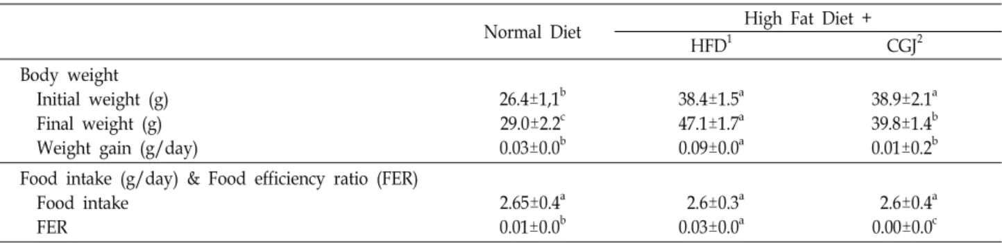
Table 1. Effect of Cheongukjang on changes in body weight, food intake and food efficiency ratio (FER) of DIO C57BL/6 mice
Normal Diet High Fat Diet +
HFD1 CGJ2
Body weight Initial weight (g) Final weight (g) Weight gain (g/day)
26.4±1,1b 29.0±2.2c 0.03±0.0b
38.4±1.5a 47.1±1.7a 0.09±0.0a
38.9±2.1a 39.8±1.4b 0.01±0.2b Food intake (g/day) & Food efficiency ratio (FER)
Food intake FER
2.65±0.4a 0.01±0.0b
2.6±0.3a 0.03±0.0a
2.6±0.4a 0.00±0.0c
1ND: Mice fed normal diet (D12451) for 26 weeks.
HFD: Mice fed High fat diet (D12450B) for 26 weeks.
CGJ: High fat diet for 13 weeks to induce obesity, followed by cheonggukjang supplemented (10%, w/w) to the high fat diet for 13 weeks
2Daily weight gain/daily weight intake
a-cMeans with different letters in the same raw are significantly different (p<0.05) by Duncan's multiple range test.
Flow cytometry analysis
The spleen, liver and kidney were collected. After lysis of the erythrocytes, the splenocyte, leukocytes of Liver and leukocytes of kidney were in a blocking buffer [PBS contain- ing 2.4G2 mAb/0.2% BSA (Bovine serum albumin)/0.1% so- dium azide], and then incubated with the relevant mAbs for 30 min at 4℃. Finally, they were washed twice with staining buffer (PBS containing 0.2% BSA/0.1% sodium azide) and analyzed by FACscan (BD: Biosciences Pharmingen, Mountain View, CA).
Statistical analysis
All results obtained were analyzed in time design using the general linear model procedure of the SAS System (SAS, 1996). Data are expressed as mean ± standard deviations values. Means with different letters are significantly differ- ent (p<0.05) by Duncan’s multiple range tests.
Results
Effects of CGJ on body weight and organ weight of diet induced obesity models
The beneficial effects of CGJ against obesity in diet in- duced obesity mice were investigated. To induce the obesity, high-fat diet was fed to C57BL/6 mice for 13 weeks. After obesity has been induced, mice in the CGJ group were fed CGJ containing high fat diet for another 13 weeks while mice in the ND group and HFD group were fed normal diet and high fat diet from the beginning of the experiment, respectively. Body weight gain and Food intakes of mice were shown in Table 1. Body weight of ND group was 26.4
g and that of HFD was 38.4 g after 13 weeks. Body weight gain for the ND, HFD, and CGJ group were 0.03, 0.09, and 0.01 g/day, respectively (p<0.05). Despite food intakes of mice were similar among 3 groups were not different, body weight gain (g/day) and final body weight of mice were significantly different (p<0.05). Organ weights of C57BL/6 mice fed CGJ supplemented high fat diet for 13 weeks after diet induced obesity (13 weeks with high fat diet) were shown in Table 2. The CGJ group significantly decreased liver weight than HFD group (p<0.05). The perirenal fat weight of ND, HFD, and CGJ group were 1.8, 2.6 and 0.2 (g/100 g body weight), respectively. But no differences were found in epididymal fat size among 3 groups.
Effects of CGJ on the plasma lipid profiles
C57BL/6 mice fed normal diet (ND) and high fat diet
(HFD) for 26 weeks. High fat diet induced obesity followed
a course of development which could be divided into 13
weeks and 26 weeks. This study was shown that after 13
weeks feeding, the contents of plasma TG of ND and HFD
group were 88, 119 mg/dl, respectively. After 13 weeks feed-
ing, the concentrations of plasma TC of ND and HFD group
were 93, 126 mg/dl, respectively. After obesity has been in-
duced, mice in the CGJ group were fed CGJ supplemented
high fat diet for another 13 weeks. The concentrations of
plasma TG and TC of C57BL/6 mice fed ND, HFD, and CGJ
supplemented HFD diet were shown in Table 3. The TG con-
centrations of in the ND, HFD and CGJ group were 99
mg/dl, 184 mg/dl and 117 mg/dl. The TC concentrations
of the ND, HFD, and CGJ group were 107, 199, and 118
mg/dl. Plasma TG and TC concentrations for the CGJ group
Table 2. Effect of cheongukjang on relative organ weight of liver, spleen, kidney, and adipose tissue in DIO C57BL/6 mice Relative weight
(g/100 g body weight) ND High fat diet
HFD1 CGJ2
Liver Kidney Spleen
4.5±0.5b 1.3±0.1NS 0.3±0.04a
5.6±0.8a 0.9±0.07 0.3±0.04ab
4.1±0.5b 1.0±0.14 0.3±0.1b Fat pad
Epididymal fat Perirenal fat
3.3±1.0NS 1.8±0.3b
4.0±0.3 2.60±0.2a
3.4±0.2 0.192±0.3b HFD: Mice fed High fat diet (D12450B) for 26 weeks.
CGJ: High fat diet for 13 weeks to induce obesity, and followed by cheonggukjang supplemented (10%, w/w) to the high fat diet for 13 weeks;
a-bMeans with different letters in the same raw are significantly different (p<0.05) by Duncan's multiple range test.
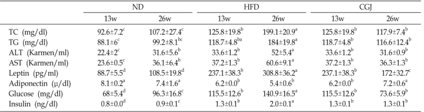
Table 3. Effect of cheonggukjang on plasma lipid levels in DIO mice.
ND HFD CGJ
13w 26w 13w 26w 13w 26w
TC (mg/dl) TG (mg/dl) ALT (Karmen/ml) AST (Karmen/ml) Leptin (pg/ml) Adiponectin (μ/dl) Glucose (mg/dl) Insulin (ng/dl)
92.6±7.2c 88.1±6c 22.4±2c 23.6±0.5c 88.7±5.5d 8.1±0.2a 68±5.4d 0.8±0.0d
107.2±27.4c 99.2±8.1bc 31.6±5.6b 36.1±6.4b 108.5±19.8d 7.4±1.6a 96.3±16.8c 0.9±0.1c
125.8±19.8b 118.7±4.8ba 33.6±1.2b 37.2±1.3b 237.1±38.3b 6.2±0.0b 115.5±12.6b 1.3±0.1b
199.1±20.9a 184±19.8a 52±5.4a 60.6±9.1a 308.8±36.2a 5.4±0.6b 140.9±16.5a 2.0±0.1a
125.8±19.8b 118.7±4.8b 33.6±1.2b 37.2±1.3b 237.1±38.3b 6.2±0.0b 115.5±12.6b 1.3±0.1b
117.9±7.4b 116.6±12.4b 31.6±0.9b 36.3±1.3b 172±32.7c 7.2±0.6a 73.6±5.9b 1.3±0.1b 13w; ND: Mice fed normal diet (D12451) for 13 weeks, HFD: Mice fed High fat diet (D12450B) for 13 weeks
26w; ND: Mice fed normal diet (D12451) for 26 weeks, HFD: Mice fed High fat diet (D12450B) for 26 weeks
CGJ: High fat diet for 13 weeks to induce obesity, and followed by cheonggukjang supplemented (10%, w/w) to the high fat diet for 13 weeks;
a-dMeans with different letters in the same raw are significantly different (p<0.05) by Duncan's multiple range test.
were significantly lower than that for the HFD group (p<0.05). After 13 weeks feeding, the levels of plasma AST of ND and HFD group were 24, 37 karmen/ml. The levels of plasma ALT of ND and HFD group were 22 and 34 kar- men/ml. After 26 weeks feeding, the AST and ALT levels of CGJ (36, 32 Karmen/ml) were significantly lower than the HFD group (61, 52 Karmen/ml). Measured values at the end of experimental period for plasma leptin, adiponectin, glucose and insulin in C57BL/6 mice fed high fat diets con- taining cheonggukjang for 13 weeks after diet induce obesity were given in Fig. 2. The secretion of plasma leptin of ND and HFD group were 89, 237 pg/ml. After 26 weeks feeding, the leptin secretions in the ND, HFD and CGJ group were 108, 309 and 172 pg/ml, respectively (p<0.05). After 13 weeks feeding, the secretion of plasma adiponectin of ND and HFD group were 8.1, 6.2 μg/ml. After 26 weeks feeding, the adi- ponectin secretions in the ND, HFD and CGJ group were 7.4, 5.2 and 7.2 μg/ml, respectively. The glucose levels of
diet-induced obesity in C57BL/6 mice were increased to 100 mg/dl (222), which feeding high fat diet for 13 weeks. After 13 weeks feeding, the level of plasma glucose of ND and HFD group were 68, 115.5 mg/dl. In this study, mice fed CGJ supplemented high fat diet for 13 weeks after diet-in- duced obesity for 13 weeks. After 26 weeks feeding, the glu- cose level in the ND, HFD and CGJ group were 96.3, 140.9 and 73.6 mg/dl, respectively. In the HFD group, plasma glu- cose levels increased by approximately 46% compared with the ND group. After 13 weeks feeding, the level of plasma insulin of ND and HFD group were 0.8 and 1.3 ng/ml. After 26 weeks feeding, the insulin level in the ND, HFD, and CGJ group were 0.9, 2.0 and 1.3 mg/dl, respectively.
Effect of CGJ on liver steatosis
High fat diet has known to alter hepatic lipid accumu-
lation and cause hepatic steatosis [26]. Given the significant
improvement in plasma lipid profile by CGJ (Table 3), we
A
B
C
D
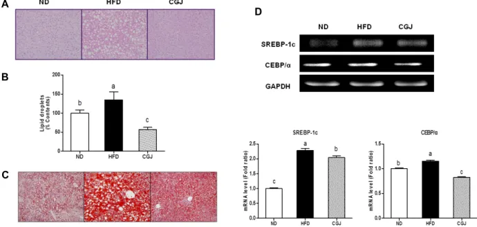
Fig. 1. Effect of resistant to steatosis of DIO C57bl/6 mice. The empty vacuole in the H&E staining and the red color droplets in Oil red O staining represent the lipid droplets in the liver. (A): Representative photomicrograph of liver sections stained with hamatoxylin and eosin(H&E). Original magnification, X200, (B): Oil Red O staining of liver. Original magnification, X200.
hypothesized that CGJ supplementation attenuates diet-in- duced obesity and hepatic steatosis. Liver of HFD group showed higher lipid droplets than those fed normal diets (ND) (Fig. 1A). Liver of HFD group were enlarged and pro- duced a yellowish color, indicating liver steatosis. On the other hand, administration of CGJ reversed the liver to re- main red and healthy. The lipid droplets were significantly lower in CGJ group than HFD group. In histology analysis, the liver of HFD mice exhibited a typical sign of fatty liver showing the accumulation of many fat droplets. To confirm the beneficial effect of cheonggukjang consumption on the lip- id droplets was demonstrated by a histological examination of liver (Fig. 1B). The contents of lipid droplets were sig- nificantly higher in the HFD mice (135%) than CGJ mice (57%). To confirm the lipid accumulation in the liver, stain- ing was performed using the Oil red O (Fig. 1C). In the Oil red O stained slides of the HFD group, many more red drop- lets within the cytoplasm of hepatocytes were present in comparison with ND and CGJ group. The induction of obe- sity with high fat diet was associated alterations in adipo- genic transcription factor. To investigate how hepatic fat ac- cumulation was regulated in ND, HFD and CGJ groups. The mRNA level of SREBP-1c and CEBP/α on the CGJ group were decreased by 10% and 29%, respectively, compared to that of the HFD (Fig. 1D).
CGJ modulate immune function of liver
The number of total hepatic leukocytes and accumulation of macrophage were investigated in C57BL/6 mice fed with cheonggukjang. (Fig. 2A). The number of CD11b
+F4/80
+T cells on ND and CGJ groups were significantly lower than the HFD group by 6 and 73%, respectively (p<0.05). To de- termine whether immature myeloid cells are crucial in medi- ating inflammatory hepatic steatosis, activation and accumu- lation of immature myeloid cells in the liver was determined (Fig. 2B, Fig. 2C). The number of Gr-1
intCD11b
highcells on ND and CGJ groups were significantly lower than the HFD group by 65 and 75%, respectively (p<0.05). The Gr-1
highCD11b
highimmature myeloid cells of ND and CGJ were significantly decreased by 45% and 90%, respectively, compared to that of the HFD group (p<0.05).
Effects of CGJ on epididymal fat pads Morphology and adipogenic transcription factor
Histological staining (H & E) revealed that the increase
in body fat in HFD. The effects of cheonggukjang con-
sumption on the adipocyte size was demonstrated by a his-
tological examination of adipose tissue. The size of adipocyte
in CGJ group were markedly reduced by 13.5% compared
to the HFD group (Fig. 3A). We measured relative levels
using cDNA synthesized with total RNA of liver by RT-PCR
(Fig. 3B). The mRNA expression of PPAR-γ on ND and CGJ
Fig. 2. Effect of cheonggukjang on macrophages and immature myeloid precursor cells in the liver of DIO C57Bl/6 mice. a-cMeans with different letters in the same raw are significantly different (p<0.05) by Duncan's multiple range test.
Fig. 3. Effect of cheonggukjang on inflammation and lipogenesis of adipose tissue in DIO C57Bl/6 mice. (A): Percent frequency distribution of adipocyte sizes indicates a shift in the size of the adipocyte population toward larger hypertrophied cells.
(B): Fold ratio : Gene expression / GAPDH × ND numerical value (ND fold ration :1).
were significantly decreased by 12% and 37%, respectively, compared to that of the HFD group (p<0.05). The mRNA expression level of SREBP-1c on ND and CGJ were sig- nificantly downregulated by 35% and 37%, respectively, compared to that of the HFD group (p<0.05). Histology on epididymal fat pads from C57BL/6 mice in macrophage clusters surrounding adipocytes. To investigate whether cheonggukjang could decrease the expression of pro-in- flammatory mediators in C57BL/6 mice. The expression of MCP-1 and TNF-α mRNA was significantly elevated in HFD group compared with those of the ND and CGJ group.
Compared with the HFD group, CGJ group significantly de- creased the level of MCP-1 and TNF-α mRNA by 49 and 59%, respectively (p<0.05). In the CGJ group, expression of TNF-α mRNA decreased by approximately 54% compared with the ND group (p<0.05).
Dissussion
The cheonggukjang is a viable candidate to improve phys- iological function due to its high aglycone of isoflavone. The aim of the present study was to verify the anti-obesity and anti-inflammation effects of dietary cheonggukjang in diet in- duced obese (DIO) mice. In this study, a significant reduc- tion in body weight gain with cheonggukjang supplementa- tion indicates that the cheonggukjang suppresses the HFD-in- duced increase in body weight gain and fat weight (Table 1). Despite similar food efficiency between the HFD and the CGJ groups, weight gain trends were significantly different.
CGJ group indicated a dramatic decrease in the liver weight (p<0.05) and a marked reduction in contents of plasma TC and TG (p<0.05). The change in body weight and liver weight were consistent with the change in lipid profiles (Table 2, Table 3). These results suggested that cheonggukjang supple- mentation has a substantial influence on lipid metabolism in diet induced obese mice. One report acclaims that epi- demically elevated levels of serum triglyceride (TG) and to- tal cholesterol (TC) called hyperlipidemia can be achieved by administration of high-fat/cholesterol diet [47]. These da- ta would suggest that cheonggukjang is a good source candi- date that can reduce the risk of lipid accumulation. Because of greater fat accumulation in the liver of mice from the high fat diet, we estimated hepatic dysfunction by measuring the activities of leptin, glucose, ALT and AST into the plasma in response to membrane damage. Supplement of cheongguk- jang downregulated levels of plasma insulin, glucose, ALT,
AST and leptin and upregulated the level of adiponectin, in spite of long term intake of high fat diet. Among the three biochemical measures, triglyceride levels exhibit significant correlation with both cholesterol and glucose [12]. The asso- ciation of obesity with increased plasma insulin level [19].
High-fat diet can promote insulin resistance and alteration
of insulin signaling in hepatocytes to promote increased in-
tracellular fatty acids and eventually deteriorate hepatic
steatosis [48]. Adiponectin is secreted by fat cells and circu-
lates in the blood. The hormone suppresses glucose pro-
duction in the liver and enhance glucose uptake into skeletal
muscle, which is known to result from anti-atherosclerotic
and insulin-sensitizing properties [43]. Promotion in ex-
pression of leptin is associated with release of pro-in-
flammatory cytokines [37]. Taken together, it can be proven
that intake of cheonggukjang has beneficial effects to upregu-
late insulin sensitivity due to reduction of blood glucose and
insulin level and to improve lipid metabolism. In addition,
we may infer that the intake of cheonggukjang may suppress
liver steatosis and proinflammatory cytokines by reduction
in levels of ALT, AST and leptin and upregulation of
adiponectin. Our observation of hepatic lipid droplets by
H&E staining to investigate improvement of plasma lipid
metabolism along with suppression of obesity-induced liver
steatosis, the lipid drop in CGJ group was smaller than HFD
(Fig. 1). Lipid accumulation is important mechanism in stea-
tosis [3]. CGJ mice are protected from obesity-induced
steatosis. These results would suggest that cheonggukjang has
anti-steatosis effect as it regulates lipid profiling. The lipid
droplets of liver studied was significantly correlated with
their plasma TG, TC, AST, ALT, leptin, adiponectin, glucose,
and insulin. These results demonstrated that administration
of the cheonggukjang played a role in improvement of plasma
lipid metabolism and liver lipid deposition. We expected
that such changes may result from a certain transcriptional
factor which manages regulation of lipogenesis and trigly-
ceride synthesis, so observed expression SREBP-1c and
CEBP/α. The sterol regulatory element-binding proteins
(SREBPs) are transcription factors to take part in the regu-
lation in the expression of several genes decoding enzymes
for fatty acid and glucose metabolism, and they mediate the
responses to changes in the nutritional status [20]. Moreover,
SREBP-1c is the predominant isoform in the liver; sup-
pression in activity of SREBP-1C by adiponectin via AMPK
suppresses hepatic lipogenesis. The CCAAT/enhancer bind-
ing protein (C/EBP) is the primary adipogenic transcription
factor and their activities are initiated by synergetic trans- activation in the expressions of several adipogenic effector genes [34]. We observed the mRNA expression level of SREBP-1c and CEBP/α were increase in the HFD group (p<
0.05), an effect significantly reversed by cheonggukjang. These results indicate that CGJ might have role on inhibiting the expression of transcription factors which take part in fat ac- cumulation of liver and lipid metabolism. Body weight of ND group was 26.4 g and that of HFD was 38.4g after 13 weeks. and ND group gained 2g, HFD group 9g and CGJ group 1g after another 13 weeks. As immune cells are acti- vated by obesity, we investigated changes in immune cells on liver steatosis upon administration of cheonggukjang.
Macrophages plays a significant role in the pathogenesis of obesity [49]. Inflammatory reaction which involves macro- phage infiltration include expression of macrophage-derived surface markers F4/80 and CD11b, both of which are also expressed on monocytes and neutrophils. Therefore, it is im- portant to prevent accumulation of macrophage as well as accumulation of fat during obesity. Adiponectin has a crucial role in suppressing macrophage activity in the liver. These results, including reduction of CD11b+F4/80+ T cells ob- served in CGJ, suggested that secretion of adiponectin in CGJ were related with decreased macrophage accumulation in the liver. Also, obvious decrease in Gr-1
intCD11b
highcells and Gr-1
highCD11b
highimmature myeloid cells were observed in CGJ group (Fig. 2). Neutrophils generate chemokines to recruit monocytes and dendritic cells and can determine whether macrophages can undergo differentiation to a pre- dominantly pro- or anti-inflammatory phenotype [39].
Accumulation of immature myeloid cells may be an im- portant component in deteriorating obesity-triggered in- flammation response of the hepatic tissue, which in turn worsens metabolic morbidity including steatohepatitis [7].
These results demonstrated that long term consumption of cheonggukjang is associated with immune modulation effects in liver despite the consumption of a high fat diet. Diet-in- duced obese mice were characterized by an increase in body weight, body fat weight, and plasma leptin. In contrast, the CGJ mice decrease body weight caused principally by a highly significant decrease in perirenal fat pad and liver, al- though there was a trend toward a decrease in lipid parameters. We measured the size of adipocyte to inves- tigate that such changes affect to the size of adipose tissue.
Increase in adiposity has strong association with severe health problems and with an overall reduced longevity of
lifetime [27]. Size of adipose tissue for in the CGJ group was
significantly lower than that for the HFD group (p<0.05). To
investigate how lipogenesis are regulated in cheonggukjang,
we examined expression of PPAR-γ and SREBP-1c in the
adipose tissue (Fig. 3B). The peroxisome proliferators-acti-
vated receptor-γ (PPAR-γ) and sterol regulatory element-
binding protein-1 (SREBP-1) are designated as key regu-
latory mediators of the adipogenesis process [25]. PPAR-γ
is highly expressed in both brown and white adipose tissue
and is observed to be a representative transcription factor
in differentiation of adipocyte [35]. The mRNA level of
PPAR-γ and SREBP-1c on the CGJ group was significantly
decreased compared to that of the HFD group. The anti-obe-
sity effect of CGJ might be due to inhibiting regulation pro-
moters of adipogenic genes such as PPARγ and SREBP-1
transcription factors, resulting in inhibition of lipid accumu-
lation by blocking adipogenesis. Inhibition of adipogenesis
could be related to obesity prevention. These results sug-
gested that CGJ reduce the (size of adipose tissue and adipo-
genesis transcription factor) decreases the risk of metabolic
syndrome. Macrophages which invade adipose tissue play
a constitutive role in deteriorating inflammatory activity ac-
counted for characteristics of increased filtration of macro-
phages into white adipose tissue [49]. Thus, suppressing
macrophage infiltration and accumulation induced by lipid
accumulation may be helpful to reduce inflammatory re-
action in case of obesity. Reduction in infiltration of macro-
phage by cheonggukjang may be helpful to suppress obe-
sity-induced inflammation. These data indicated that there
was an increase in macrophage infiltration in HFD mice but
not in ND and CGJ group (Fig. 3A). As adipose tissue has
elevated levels of macrophages, obesity results in local
inflammation. Upon activation, macrophages secrete num-
bers of cytokines and chemokines, such as MCP-1 and TNF-
α, that are known to cause insulin resistance in adipocytes
[9, 14, 15]. Obesity is often linked to increased plasma levels
of MCP-1 and overexpression in adipose tissue [15, 38]. In
adipose tissue in particular, it was often seen TNF-α regulat-
ing and interfering with adipocyte metabolism at numerous
sites including transcriptional regulation, glucose and fatty
acid metabolism [40]. CGJ is associated with reduce in-
flammation of adipose tissue mediated by MCP-1 and TNF-
α. This data indicated that CGJ had inhibited inflammatory
action in high fat diet induced obese mice (Fig. 3B). The ob-
jective of this study was to determine the underlying mecha-
nism by cheonggukjang inhibit adipogenesis. This study was
regulation of adipocytokine and suppress of liver steatosis demonstrated that cheonggukjang interfere with the adipo- genesis process in liver and adipose tissue by down-regulat- ing the expression of primary adipogenic transcription factors. Taken those results above, we could confirm im- provement of lipid metabolism, reduction of hepatic stea- tosis, immune function of liver, inflammation of adipocytes and synthesis of adipose transcript factors upon intake of cheonggukjang. Our results determined that cheonggukjang de- creases the lipogenic pathway and regulates immune func- tion, and this will provide attenuation of low-grade in- flammation in diet induced obese mice.
Acknowledgments
This research was financially supported by the Ministry of Education, Science Technology and Korea Industrial Technology Foundation through the Human Resource Training Project for Regional Innovation. I would like to ac- knowledge Kun-Young Park for reviewing the manuscript.
References
1. Akram, D. S. Report of WHO Consultation on Obesity, 2000, WHO: Obesity: Preventing and Managing the Global Epidemic, pp.6-8, World Health Organization, Geneva, Switzerland
2. Aoki, F. and Honda, H. 2007. Suppression by Licorice Flavonoids of Abdominal Fat Accumulation and Body Weight Gain in High-Fat Diet-Induced Obese C57BL/6 Mice. Biosci. Biotechnol. Biochem. 71, 206-214.
3. Bechmann, L. P., Hannivoort, R. A., Gerken, G., Hotamisligil, G. S., Trauner, M. and Canbay, A. 2012. The interaction of hepatic lipid and glucose metabolism in liver diseases. J.
Hepatol. 56, 952-64.
4. Chien, H. L., Huang, H. Y. and Chou, C. C. 2006.
Transformation of isoflavone phytoestrogens during the fer- mentation of soymilk with lactic acid bacteria and bifidobacteria. Food. Microbiol. 23, 772-778.
5. Chun, J., Kim, G. M., Lee, K. W., Choi, I. D., Kwon, G. H., Park, J. Y., Jeong, S. J., Kim, J. S. and Kim, J. H. 2007.
Conversion of isoflavone glucosides to aglycones in soymilk by fermentation with lactic acid bacteria. J. Food. Sci. 72, 39-44.
6. Chun, J., Kim, J. S. and Kim, J. H. 2008. Enrichment of iso- flavone aglycones in soymilk by fermentation with single and mixed cultures of Streptococcus infantries 12 and Weissella sp. 4. Food. Chem. 109, 278-284.
7. Deng, Z., Liu, Y., Liu, C., Xiang, X., Wang, J., Cheng, Z., Shah, S. V., Zhang, S., Zhang, L., Zhuang, X., Michalek, S., Grizzle, W. E. and Zhang, H. G. 2009. Immature myeloid
cells induced by a high-fat diet contribute to liver inflammation. Hepatology 50, 1412-1420.
8. Denlinger, L. C., Fisette, P. L., Garis, K. A., Kwon, G., Vazquez-Torres, A., Simon, A. D., Nguyen, B., Proctor, R.
A., Bertics, P. J. and Corbett, J. A. 1996. Regulation of in- ducible nitric oxide synthase expression by macrophage pu- rinoreceptors and calcium. J. Biol. Chem. 271, 337-342.
9. Fried, S. K., Bunkin, D. A. and Greenberg, A. S. 1998.
Omental and subcutaneous adipose tissues of obese subjects release interleukin-6: depot difference and regulation by glucocorticoid. J. Clin. Endocrinol. Metab. 83, 847-850.
10. Gotoh, T., Yamada, K., Yin, H., Ito, A., Kataoka, T. and Dohi, K. 1998. Chemoprevention of N-Nitroso-N-methylurea-in- duced rat mammary carcinogenesis by soy foods or bio- chanin A. Jpn. J. Can. Res. 89,137-142.
11. Hardie, L., Trayhurn, P., Abramovich, D. and Fowler, P.
1997. Circulating leptin in women; a longitudinal study in the menstrual cycle and during pregnancy. Clin. Endocrinol.
47, 101-106.
12. Hollister, L. E., Overall, J. E. and Snow, H. L. 1967.
Relationship of obesity to serum triglyceride, cholesterol, and uric acid, and to plasma glucose levels. Am. J. Clin.
Nutr. 20, 777-782.
13. Hongbin, Cui. 2001. Soybean bio-functional components de- velopment and application. 241-263. Beijing: China Light Industry Press House. Beijing. China
14. Hotamisligil, G. S. and Spiegelman, B. M. 1994. Tumor ne- crosis factor alpha: a key component of the obesity-diabetes link. Diabetes 43,1271-1278.
15. Hrnciar, J., Gábor, D., Hrnciarová, M., Okapcová, J., Szentiványi, M. and Kurray, P. 1999. Relation between cyto- kines (TNF-alpha, IL-1 and-6) and homocysteine in android obesity and the phenomenon of insulin resistance syndromes. Vnitr. Lek. 45, 11-16.
16. Iritani, N., Hosomi, H., Fukuda, H., Tada, K. and Ikeda, H. 1996. Soybean protein suppresses hepatic lipogenic en- zyme gene expression in Wistar fatty rats. J. Nutr. 126, 380-388.
17. Ishimi, Y., Yoshida, M., Wakimoto, S., Wu, J., Chiba, H., Wang, X., Takeda, K. and Miyaura, C. 2002. Genistein, a soybean isoflavone, affects bone marrow lymphopoiesis and prevents bone loss in castrated male mice. Bone 31, 180-185.
18. Karalis, K. P., Giannogonas, P., Kodela, E., Koutmani, Y., Zoumakis, M. and Teli, T. 2009. Mechanisms of obesity and related pathology: linking immune responses to metabolic stress. FFBS. J. 276, 5747-5745.
19. Karam, J. H., Grodsky, G. M. and Forsham, P. H. 1963.
Excessive insulin response to glucose in obese subjects measured by immunochemical assay. Diabetes 12, 197-204.
20. Kim, J. B., Sarraf, P., Wright, M., Yao, K. M., Mueller, E., Solanes, G., Lowell, B. B. and Spiegelman, B. M. 1998.
Nutritional and insulin regulation of fatty acid synthetase and leptin gene expression through ADD1/SREBP1. J. Clin.
Invest. 101, 1-9.
21. Kuo, L. C., Cheng, W. Y., Wu, R. Y., Huang, C. J. and Lee, K. T. 2006. Hydrolysis of black soybean isoflavone glyco-
sides by Bacillus subtilis natto. Appl. Microbiol. Biotechnol. 73, 314-320.
22. Lee, C. H. 2001. Fermentation technology in Korea. pp77.
Korea university press. Seoul. Korea
23. Lin, G., Wang, G., Liu, G., Yang, L. J., Chang, L. J., Lue, T. F. and Lin, C. S. 2009. Treatment of type 1 diabetes with adipose tissue–derived stem cells expressing pancreatic du- odenal homeobox 1. Stem. Cells. Dev. 18, 1399-1406 24. Liu, D., Zhen, W., Yang, Z., Carter, J. D., Si, H. and
Reynolds, K. A. 2006. Genistein acutely stimulates insulin secretion in pancreatic β-cells through a cAMP-dependent protein kinase pathway. Diabetes 55, 1043-1050.
25. MacDougald, O. A. and Lane, M. D. 1995. Transcriptional regulation of gene expression during adipocyte differentiation. Annu. Rev. Biochem. 64, 345-373.
26. Meli, R., Mattace, R. G., Irace, C., Simeoli, R., Di, P. A., Paciello, O., Pagano, T. B., Calignano, A., A. and Santamaria, R. 2013. High fat diet induces liver steatosis and early dysre- gulation of iron metabolism in rats. PLoS One 8, e66570.
27. Mokdad, A. H., Marks, J. S., Stroup, D. F. and Gerberding, J.L. 2005. Correction: actual causes of death in the United States. JAMA. 293, 293-294.
28. Must, A., Spadano, J., Coakley, E. H., Field, A. E., Colditz, G. and Dietz, W. H. 1999. The disease burden associated with overweight and obesity. JAMA. 282, 1523-1529.
29. Nakajima, N., Nozaki, N., Ishihara, K., Ishikawa, A. and Tsuji, H. 2005. Analysis of isoflavone content in tempeh, a fermented soybean, and preparation of a new isoflavone- enriched tempeh. J. Biosci. Bioeng. 100, 685-687.
30. Nathan, C. 2006. Neutrophils and immunity: challenges and opportunities. Nat. Rev. Immunol. 6, 173-182.
31. Park, K. Y., Jung, K. O., Rhee, S. H. and Choi, Y. H. 2003.
Antimutagenic effects of doenjang (Korean fermented soy- paste) and its active compounds. Mutat. Res. 523, 43-53.
32. Peterson, T. G., Ji, G. P., Kirk, M., Coward, L., Falany, C.
N. and Barnes, S. 1998. Metabolism of the isoflavones genis- tein and dichanin A in human breast cancer cell lines. Am.
J. Clin. Nutr. 68, 1505-1511.
33. Potter, S. M., Baum, J. A., Teng, H., Stillman, R. J., Shay, N. F. and Erdman, J. W. Jr. 1998. Soy protein and iso- flavones: their effects on blood lipids and bone density in postmenopausal women. Am. J. Clin. Nutr. 68,1375-1379.
34. Rosen, E.D. 2005. The transcriptional basis of adipocyte development. Prostaglandins. Leukot. Essent. Fatty Acids. 73, 31-34.
35. Rosen, E D., Sarraf, P., Troy, A. E., Bradwin, G., Moore, K., Milstone, D. S., Spiegelman, B. M. and Mortensen, R.
M. 1999. PPAR gamma is required for the differentiation of adipose tissue in vivo and in vitro. Mol. Cell. 4, 611-617.
36. Sahai, A., Malladi, P., Pan, X., Paul, R., Melin-Aldana, H.
and Green, R. M. 2004. Obese and diabetic db/db mice de- velop marked liver fibrosis in a model of nonalcoholic stea- tohepatitis: role of short-form leptin receptors and osteopontin. Am. J. Physiol. Gastrointest. Liver Physiol. 287, 1035- G1043.
37. Sarraf, P., Frederich, R. C., Turner, E. M., Ma, G., Jaskowiak,
N. T., Flier, J. S., Lowell, B. B., Fraker, D. L. and Alexander, H. R. 1997. Multiple cytokines and acute inflammation raise mouse leptin levels: potential role in inflammatory anorexia.
J. Exp. Med. 185, 171-175.
38. Sartipy, P. and Loskutoff, D. J. 2003. Monocyte chemo- attractant protein 1 in obesity and insulin resistance. Proc.
Natl. Acad. Sci. USA 100, 7265-7270.
39. Serhan, C. N. and Savill, J. 2005. Resolution of inflammation:
the beginning programs the end. Nat. Immunol. 6, 1191-1197.
40. Sethi, J. K. and Hotamisligil, G. S. 1999. The role of TNF alpha in adipocyte metabolism. Semin. Cell. Dev. Biol. 10, 19- 29.
41. Slavin, J. L., Karr, S. C., Hutchins, A. M. and Lampe, J. W.
1998. Influence of soybean processing, habitual diet and soy dose on urinary isoflavonoid excretion. Am. J. Clin. Nutr.
68, 1492S-1495S.
42. Soh, J. R., Shin, D. H., Kwon, D. Y. and Cha, Y. S. 2008.Effect of Cheonggukjang supplementation upon hepatic acyl-CoA synthase, carnitine palmitoyl transferase I, acyl-CoA oxidase and uncoupling protein 2 mRNA levels in C57BL/6J mice fed with high fat diet. Genes. Nutr. 2, 365-369.
43. Staiger, H., Tschritter, O., Machann, J., Thamer, C., Fritsche, A., Maerker, E., Schick, F., Haring, H. U. and Stumvoll, M.
2003. Relationship of serum adiponectin and leptin concen- trations with body fat distribution in humans. Obes. Res. 11, 368-376.
44. Sugano, M., Goto, S., Yamada, Y., Yoshida, K., Hashimoto, Y., Matsuno, T. and Kimoto, M. 1999. Cholesterol-lowering activity of various undigested fraction of soybean protein in rats. J. Nutr. 12, 977-985.
45. Teede, H. J., Dalais, F. S., Kotsopoulos, D., Liang, Y. L., Davis, S. and McGrath, B. P. 2001. Dietary soy has both ben- eficial and potentially adverse cardiovascular effects: a pla- cebo-controlled study in men and postmenopausal women.
J. Clin. Endocrinol. Metab. 86, 3053-3060.
46. Todoric, J., Löffler, M., Huber, J., Bilban, M., Reimers, M., Kadl, A., Zeyda, M., Waldhäusl, W. and Stulnig, T. M. 2006.
Adipose tissue inflammation induced by high-fat diet in obese diabetic mice is prevented by n−3 polyunsaturated fatty acids. Diabetologia 49, 2109-2119.
47. Tzang, B. S., Yang, S. F., Fu, S. G., Yang, H. C., Sun, H.
L. and Chen, Y. C. 2009. Effects of flaxseed oil on cholesterol metabolism of hamsters. Food Chem. 114, 1450-1455.
48. Uygun, A., Kadayifci, A., Yesilova, Z., Erdil, A., Yaman, H., Saka, M., Deveci, M. S., Bagci, S., Gulsen, M., Karaeren, N.
and Dagalp, K. 2000. Serum leptin levels in patients with nonalcoholic steatohepatitis. Am. J. Gastroenterol. 95, 3584- 3589.
49. Weisberg, S. P., McCann, D., Desai, M., Rosenbaum, M., Leibel, F. L. and Ferrante, A. W. Jr. 2003. Obesity is asso- ciated with macrophage accumulation in adipose tissue. J.
Clin. Invest. 112, 1796-1808.
50. Yun, J. W., Lee, B. S., Kim, C. W. and Kim, B. H. 2007.
Comparison with 3 high-fat diet for studying obesity in C57BL/6 mouse. Lab. Anim. Res. 23, 245-250.
초록:고지방식이로 유도된 비만 마우스에서 청국장의 항비만 및 항염증 효과
김지영*
(부산대학교 식품영양학과)