Classifying Host Susceptibility Using Porcine Circovirus Type 2 Viral Load and Antibody Titer
Kyu-Sang Lim
1,2, Eun-A Lee
1, Kyung-Tai Lee
2, Taehoon Chun
1, Ki-Chang Hong
1* and Jun-Mo Kim
2*
1College of Life Sciences and Biotechnology, Korea University, Seoul 136-713, Korea
2Division of Animal Genomics and Bioinformatics, National Institute of Animal Science, RDA, Wanju 55365, Korea Received November 8, 2016 /Revised December 7, 2016 /Accepted December 26, 2016
Porcine circovirus type 2 (PCV2) is a notorious and ubiquitous virus in the swine industry. The sus- ceptibility of the host to PCV2 infection is considered to be one factor associated with the dynamics of PCV2. The objective of this study was to verify the criteria for host susceptibility to PCV2, using blood parameters of post-weaned pigs naturally infected with the virus. The PCV2 DNA viral load, antibody titer, and leukopenia characteristics were measured in the serum extracted from the pigs at the 10th week. We classified the pigs into high (>5.0), intermediate (3.0 to 5.0), and low (<3.0) groups on the basis of the PCV2 viral load (log copies/ml), or as positive (≤0.50) and negative (>0.50) groups on the basis of antibody titer (sample-to-negative corrected ratio). Moreover, using these two catego- rized parameters, we suggested the criteria for classification into the susceptible and resistant groups.
Statistical analyses revealed that pigs in the susceptible group had a significantly higher viral load (p<0.001) and negative antibody titer (p<0.001), as well as significantly lower leukocyte counts (p=0.018) and lower amounts of several leukocyte components (p<0.05), than pigs in the resistant group. We concluded that the susceptible group could be considered to have PCV2-induced leukopenia. Therefore, we suggest that the combined classifications of viral loads and anti-PCV2 antibodies can be used to determine PCV2-induced leukopenia in the subclinical PCV2 infection of post-weaned pig populations.
Key words : Antibody titer, host susceptibility, leukopenia, pig, porcine circovirus type 2, viral load
*Corresponding authors
*Tel : +82-2-3290-3053, Fax : +82-2-925-1970 E-mail : [email protected] (Prof. Ki-Chang Hong) Tel : +82-63-238-7323, Fax : +82-63-238-7347 E-mail : [email protected] (Dr. Jun-Mo Kim)
This is an Open-Access article distributed under the terms of the Creative Commons Attribution Non-Commercial License (http://creativecommons.org/licenses/by-nc/3.0) which permits unrestricted non-commercial use, distribution, and reproduction in any medium, provided the original work is properly cited.
Journal of Life Science 2017 Vol. 27. No. 3. 283~288 DOI : https://doi.org/10.5352/JLS.2017.27.3.283
Introduction
Porcine circovirus type 2 (PCV2) has been widely docu- mented to wreak havoc in the swine industry [1, 8]. In partic- ular, PCV2 is a major etiological agent of postweaning multi- systemic wasting syndrome (PMWS), which occurs primar- ily in pigs between 8 and 12 wk of age, with clinical symp- toms such as weight loss, respiratory signs, and jaundice [15]. In recent years, several combined PCV2-related diseases have been named porcine circovirus-associated disease (PCVAD) in North America and porcine circovirus disease (PCVD) in Europe, because PCV2 infection appears to cause a wide variety of clinical syndromes [12]. To overcome the serious problems related with these diseases, management
strategy for controlling PCV2 have been recommended [8]
and various vaccines against PCV2 have been used world- wide [2, 4]. However, PCV2 is still ubiquitous in swine farms and their infection cannot be completely prevented.
Previous studies focused on the host genetic differences in relation with PCVAD incidence and severity [11, 13]. In general, postmortem evaluation methods are used for the identification of individuals susceptible to PCV2. However, sacrifice of the tested pigs is inevitable in postmortem evalu- ation, and the tested pigs can no longer be used as seed stocks. For this reason, PCV2 infections cannot be measured in many individuals and controlled easily in piglet farms.
Therefore, new evaluation methods are required that can ac- curately and immediately measure the virus in numerous live pigs.
Several measurable methodologies on living bodies that
are related to the PCV2 infection dynamics have been sug-
gested, for example, blood parameters like the serum viral
load and PCV2 specific antibody titer [12]. Moreover, vir-
emia, antibody response, and leukopenia are common fea-
tures of the pathogenesis of PCV2 infection [5]. Alternatively,
PCV2-induced leukopenia, which is related to lymphocyte
depletion in lymphoid tissue, is widely observed in PMWS-
affected piglets and is the primary characteristic for diagnos- ing PMWS [10]. However, those of previous studies had not been applied to estimate host susceptibilities using the com- binational parameters. The objective of current study was to verify the potential use of blood parameters such as viral load, antibody level, and leukocyte count for the identi- fication of individual susceptibility to PCV2 infection with- out necropsy in postweaning pigs. We used the PCV2 viral loads and antibody titers in the sera to develop criteria for the host susceptibility to PCV2 infection. The susceptibility criteria were confirmed by their associations with PCV2-in- duced leukopenia values.
Materials and Methods
Animals
A total of 155 crossbreds (82 females and 73 castrated males) generated from Yorkshire dams inseminated with se- men from Landrace were randomly selected from 46 litters (average, 3.37±1.50 piglet per dam) at a farm with animals that had naturally occurring PCV2 infection. Although the vaccine program of the source farm had included PCV2 and porcine respiratory syndrome virus, all the animals used in this study were not vaccinated against PCV2, so as to de- termine the host response against natural PCV2 infection.
The pigs were raised in four batches under the same condition. The experimental protocols and standard operat- ing procedures were approved by the Institutional Animal Care and Use Committee at the National Institute of Animal Science (Wanju, Republic of Korea) in 2016(Ethic approval number: 2016-191). At the age of around 4 weeks (28.34±3.49 d), blood samples were collected from each pig and analyzed for the presence of PCV2 DNA in the serum, and 142 pigs were identified as seronegative for PCV2. We therefore used these 142 pigs to further determine how many had been ex- posed to natural PCV2 infection up to 10 weeks of age, which is predominant onset age of PMWS. The PCV2 viral load, anti-PCV2 antibody titer, and leukocyte counts were quantified in blood samples collected from the pigs at 10 wk of age (69.25±2.17d).
PCV2 viral load and antibody
The viral DNA from the serum samples was isolated us- ing a commercial DNA extraction kit (Qiagen, Valencia, CA, USA). The PCV2 viral load was measured with TaqMan- based real-time polymerase chain reaction (PCR), with the
following primers and probe designed from the complete PCV2 genome (GenBank Accession No. FR823451.1): for- ward primer, 5'-TCGATCTCAAGGACAACGGAGT-3'; re- verse primer, 5'-TTGGTCTTCCAATCACGCTTCTGC-3'; and probe, 5'-CAGAGCAGCACCCTGTAACGTTTGTCA-3. The size of the amplified product was 173 bp. The reaction con- tained 500 nM of each primer, 250 nM of the probe, 25 μl of 2× TaqMan Universal Master Mix (Applied Biosystems, Foster City, CA, USA), and 2.5 μl of the template. Nuclease- free water was added to bring the final volume to 50 μl.
Amplification was performed under Universal cycling con- ditions (2 min at 50°C, 10 min at 95°C, and 45 cycles of 15 s at 95°C and 1 min at 58°C). The plasmid (pPCV2) used as the standard DNA was constructed by ligating a PCR fragment into the T Easy vector, according to the manu- facturer’s instructions (Promega, Madison, WI, USA). The cloned fragment included a 314 bp region of the PCV2 open reading frame 3. The plasmid was propagated in PK-15 por- cine kidney cells (ATCC), purified using a Miniprep kit (Qiagen, Valencia, CA, USA), and quantified using a spec- trophotometer (Thermo Scientific, Waltham, MA, USA).
Ten-fold dilutions were made to obtain 10
11to 10
1plasmids per 2.5 μl sample for the real-time PCR. We transformed the unit of PCV2 DNA viral load (copies/ml) into log base 10.
Anti-PCV2 antibodies in the serum were measured with a commercial PCV2 Ab Mono Blocking kit (Synbiotics, Lyon, France), according to manufacturer’s instructions. The an- ti-PCV2 antibody titers were expressed as a sample-to-neg- ative corrected (SNc) ratio. Samples were considered neg- ative if the SNc ratio was higher than 0.50 [17]. The indices for leukocytes including total count, neutrophils, lympho- cytes, monocytes, eosinophils, and basophils were counted using a fully automatic hematology analyzer for animals (Drew Scientific Inc., Dallas, TX, USA) in accordance with the manufacturer’s recommendations.
Statistical analysis
A total of 142 pigs at 10 wk of age were classified into
three groups on the basis of the degree of viral load (high,
intermediate, low), using the FASTCLUS procedure (SAS
software). The pigs were also separated into two groups on
the basis of the SNc ratio of the antibody titer. The SNc ratio
of the positive group was ≤0.50, whereas that of the neg-
ative group was >0.50 (Fig. 1). Finally, we classified the sus-
ceptible and resistant groups among the studied population
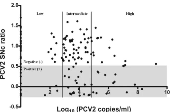
Fig. 1. Scatterplot representing the distribution of porcine circo- virus type 2 (PCV2) viral loads and anti-PCV2 antibodies in 10-wk-old naturally infected pigs (n=142). The PCV2 viral loads in the sera were expressed as the logarithm of the PCV2 DNA copies/ml. The two vertical solid lines divide the PCV2 viral loads (log copies/ml) into the high (>5.0), intermediate (3.0 to 5.0), and low (<3.0) groups.
PCV2 antibody titer levels were classified according to the sample-to-negative corrected (SNc) ratio (positive, ≤ 0.50; negative, >0.50). The positive (+) range of PCV2 antibody titer is represented by the gray background.
on the basis of the combined viral loads (log PCV2 cop- ies/ml) and antibody titers (SNc ratio). Pigs were classified as susceptible if they showed a high PCV2 viral load (>5.0 log PCV2 copies/ml) with negative PCV2 antibody titer, whereas they were grouped as resistant if they showed a low PCV2 viral load (<3.0 log PCV2 copies/ml) with PCV2 antibody level.
The mixed model (SAS procedure MIXED; SAS software) was used to determine the relationships between the groups and measured traits. The model used was as follows: y
ijklm= μ + G
i+ S
j+ B
k+ M
l+ e
ijklm, where y
ijklmis the observation of the traits, μ is the general mean, G
iis the fixed effect of group i, S
j is the fixed effect of sex j, Bkis the fixed effect of batch j, M
l is the random effect of mother l, and ejjklmis the random error. When significant differences were de- termined, the mean values were separated by the probability difference (PDIFF) option.
Results and Discussion
In general, piglets are exposed through horizontal PCV2 infection, by direct or nose-to-nose contact with pigs that are already infected, after weaning and mingling in the post- weaning house [14]. Following PCV2 infection, viremia and seroconversion are noted in the serum. Along with the PCV2
viral load, the PCV2 specific antibody titer is generally con- sidered to be informative of the immune status toward PCV2 infection. Therefore, the presence of PCV2 genomic DNA and PCV2 specific antibodies in the serum indicates that the tested pigs have an infectious level of PCV2 [7].
The distributions of the PCV2 viral loads (log PCV2 cop- ies/ml) and anti-PCV2 antibody titers (SNc ratio) in the 142 pigs at 10 wk of age are shown in Fig. 1. The frequency of PCV2 DNA detected in the sera was 77% (n=110), and the average viral load was 4.22±1.43 log copies/ml. The viral loads ranged from 1.66 to 9.24 log copies/ml. Although the PCV2 viral loads were widely distributed in the study pop- ulation, a relatively lower number of pigs (32%, n=46) showed a positive antibody titer against PCV2 (SNc ratio
≤0.50).
In a previous study, PCV2 genomic DNA and antibodies were detected in sera from both healthy pigs and PCVAD- diagnosed pigs [15]. However, other previous studies re- ported that the PCV2 viral loads in the case of PMWS in PCVAD-diagnosed pigs were higher (>7 log PCV2 cop- ies/ml in the case of PMWS) than those in healthy pigs (<4 log PCV2 copies/ml) [3, 16]. In our population, eight pigs showed upper levels of PCV2 viral load (i.e., >7.0 log cop- ies/ml). However, only three of these eight pigs showed positive PCV2 antibodies (SNc ratio ≤0.50). Moreover, we observed a weak negative correlation between the PCV2 vi- ral load and antibody titer in our experimental population (r=-0.17, p=0.035; data was not shown). Based on the two-di- mensional spread of PCV2 viral load and antibody titer, we postulate that the serum PCV2 viral load and antibody titer during the post-weaning period could be challenged as be- ing useful parameters for the evaluation of host suscepti- bility to PCV2 infection, and there were high variations of host susceptibility in the population.
Classifications based on the PCV2 viral load and antibody titer were conducted to investigate the host susceptibility against PCV2 infection under the living conditions used (Table 1). The 10-wk-old pigs were classified by cluster anal- ysis into three groups on the basis of PCV2 viral load (high,
>10
5copies/ml; intermediate, 10
5to 10
3copies/ml; and low,
<10
3copies/ml). The PCV2 viral loads were significantly dif-
ferent among the groups (p<0.001). However, no significant
difference was observed in the SNc ratio of anti-PCV2 anti-
bodies (p=0.908). Moreover, the leukocyte counts and all leu-
kocyte components were not significantly different among
the three groups.
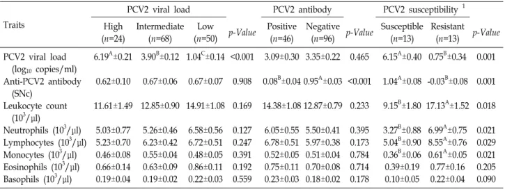
Table 1. Classification of pigs according to the blood parameters of porcine circovirus type 2 (PCV2), viral load, antibody, and susceptibility and their associations
Traits
PCV2 viral load PCV2 antibody PCV2 susceptibility 1
High (n=24)
Intermediate (n=68)
Low
(n=50) p-Value Positive (n=46)
Negative
(n=96) p-Value Susceptible (n=13)
Resistant
(n=13) p-Value PCV2 viral load
(log10 copies/ml) Anti-PCV2 antibody (SNc)
Leukocyte count (103/μl)
Neutrophils (103/μl) Lymphocytes (103/μl) Monocytes (103/μl) Eosinophils (103/μl) Basophils (103/μl)
6.19A±0.21 0.62±0.10 11.61±1.49
5.03±0.77 5.23±0.70 0.46±0.08 0.66±0.14 0.19±0.04
3.90B±0.12 0.67±0.06 12.85±0.90
5.26±0.46 6.23±0.42 0.55±0.04 0.63±0.09 0.19±0.02
1.04C±0.14 0.67±0.07 14.91±1.08
6.58±0.56 6.72±0.51 0.48±0.05 0.86±0.11 0.22±0.03
<0.001 0.908 0.169
0.127 0.247 0.391 0.192 0.559
3.09±0.30 0.08B±0.04 14.38±1.08
6.05±0.55 6.78±0.51 0.52±0.05 0.75±0.11 0.23±0.03
3.35±0.22 0.95A±0.03 12.87±0.79
5.50±0.41 5.97±0.38 0.51±0.04 0.70±0.08 0.18±0.02
0.465
<0.001 0.233
0.395 0.173 0.784 0.714 0.178
6.15A±0.40 1.04A±0.08 9.15B±1.80
3.27B±0.88 5.04B±0.90 0.36B±0.06 0.39±0.19 0.10±0.05
0.75B±0.34 -0.03B±0.08 17.13A±1.52
6.99A±0.75 8.55A±0.76 0.61A±0.05 0.77±0.16 0.22±0.04
0.001 0.001 0.018
0.021 0.029 0.021 0.205 0.090
A,B,C
Different superscript letters within the row indicate significant differences (p<0.05).
1The susceptible group has a high PCV2 viral load with negative PCV2 antibody level, whereas the resistant group has a low PCV2 viral load with positive PCV2 antibody level.
SNc, sample-to-negative corrected ratio.
The alternative grouping was based on the antibody sta- tus as expressed by the SNc ratio (positive, ≤0.50; negative,
>0.50). The associations of the groups were not significant with all of the measured traits, except for the anti-PCV2 anti- body levels (p<0.001). Therefore, each classification had no significant effect on each of the other quantified parameters, as well as on any of the leukocyte characteristics. Based on each classification result, we concluded that either the PCV2 viral load or the antibody titer alone cannot be used to eval- uate the host’s susceptibility to PCV2 infection.
Next, the pigs were classified into PCV2-susceptible and PCV2-resistant groups by combining the two kinds of classi- fication used above. The susceptible group had a high PCV2 viral load with negative PCV2 antibody level, whereas the resistant group had a low PCV2 viral load with positive PCV2 antibody level (Fig 1). As shown in Table 1, the associ- ation results were highly significant between the two groups with respect to the quantifiable parameters and leukocyte characteristics. The susceptible group showed a significantly higher viral load (p<0.001) and PCV2 SNc ratio (p<0.001) than did the resistant group. In addition, the total leukocyte counts were considerably lower in the susceptible group (p=0.018). Specifically, the mean value of leukocyte counts for the susceptible group was lower than the reference range (11 to 22 10
3/μl) for healthy pigs (Duncan et al., 1994).
Among the leukocyte components, the neutrophils, lympho- cytes, and monocytes were also significantly lower in the
susceptible group (p=0.021, p=0.029, and p=0.021, respectively).
Based on the significantly low levels of these leukocyte com- ponents, the susceptible group could be considered as hav- ing PCV2-induced leukopenia. Considering the association of leukopenia with PCV2, our criteria using the PCV2 viral load and antibody titer may be applicable for evaluating sus- ceptibility against PCV2 infection.
Previous studies have also attempted challenge experi-
ments using the PCV2 viral load and antibody titer as the
blood parameters related with PCV2 infection, respectively
[6, 9]. However, those studies used experimental pigs that
were inoculated at the same time point, and they could
measure the values under the control of the exact points of
post day of infection. In the current study, a naturally
PCV2-infected pig population was used. Consequently, it
was difficult to control not only the point of PCV2 infection
in each individual but also to control various concurrent
co-infections by other pathogens. Therefore, the current
study’s suggested criteria of serum PCV2 viral load and anti-
body titer had limitations with respect to identifying the pre-
cise susceptibility at the individual level and having a direct
relationship with the clinical PCVAD diagnosis (e.g.,
PMWS). However, we measured the leukopenia character-
istics in the serum instead of making a simple PCVAD diag-
nosis and observations of clinical symptoms. Our results
suggest that the optimum levels of the two parameters can
be applicable to verifying PCV2-induced leukopenia in the
field, with a naturally PCV2-infected population. Moreover, it could be easily evaluated in the serum without the need to sacrifice the animals, so these parameters can be used as phenotypes for host susceptibility against PCV2 in further studies. In conclusion, we suggest that the combined criteria of a PCV2 viral load of >5.0 copies/ml and negative results for antibodies against PCV2 can be useful for determining PCV2-induced leukopenia in subclinical PCV2 infection of pig populations.
Acknowledgements
This work was supported by a grant from the Next- Generation BioGreen 21 Programs (PJ01113002: Develop- ment of genetic markers related to the resistance to PMWS by dissecting the molecular mechanism of PCV2 and host cell interactions, and PJ01181602: Investigation of genetic markers related to PRRSV resistance), Rural Development Administration, Republic of Korea) and by 2016 Postdoctoral Fellowship Program of National Institute of Animal Science, Rural Development Administration, Republic of Korea.
References
1. Alarcon, P., Rushton, J. and Wieland, B. 2013. Cost of post- weaning multi-systemic wasting syndrome and porcine cir- covirus type-2 subclinical infection in England - an econom- ic disease model. Prev. Vet Med. 110, 88-102.
2. Beach, N. M. and Meng, X. J. 2012. Efficacy and future pros- pects of commercially available and experimental vaccines against porcine circovirus type 2 (PCV2). Virus Res. 164, 33-42.
3. Brunborg, I. M., Moldal, T. and Jonassen, C. M. 2004.
Quantitation of porcine circovirus type 2 isolated from se- rum/plasma and tissue samples of healthy pigs and pigs with postweaning multisystemic wasting syndrome using a TaqMan-based real-time PCR. J. Virol. Methods 122, 171- 178.
4. Chae, C. 2012. Commercial porcine circovirus type 2 vac- cines: efficacy and clinical application. Vet. J. 194, 151-157.
5. Darwich, L., Segales, J., Domingo, M. and Mateu, E. 2002.
Changes in CD4(+), CD8(+), CD4(+) CD8(+), and im- munoglobulin M-positive peripheral blood mononuclear cells of postweaning multisystemic wasting syndrome-af- fected pigs and age-matched uninfected wasted and healthy pigs correlate with lesions and porcine circovirus type 2 load in lymphoid tissues. Clin. Diagn. Lab. Immunol. 9, 236- 242.
6. Engle, T. B., Jobman, E. E., Moural, T. W., McKnite, A. M, Bundy, J. W., Barnes, S. Y., Davis, E. H., Galeota, J. A., Burkey, T. E., Plastow, G. S., Kachman, S. D. and Ciobanu,
D.C. 2014. Variation in time and magnitude of immune re- sponse and viremia in experimental challenges with Porcine circovirus 2b. BMC Vet. Res. 10, 286.
7. Krakowka, S., Ellis, J., McNeilly, F., Waldner, C. and Allan, G. 2005. Features of porcine circovirus-2 disease: correla- tions between lesions, amount and distribution of virus, and clinical outcome. J. Vet. Diagn. Invest. 17, 213-222.
8. Madec, F., Rose, N., Grasland, B., Cariolet, R. and Jestin, A. 2008. Post-weaning multisystemic wasting syndrome and other PCV2-related problems in pigs: a 12-year experience.
Transbound Emerg. Dis. 55, 273-283.
9. McKnite, A. M., Bundy, J. W., Moural, T. W., Tart, J. K., Johnson, T. P., Jobman, E. E., Barnes, S. Y., Qiu, J. K, Peterson, D. A., Harris, S. P., Rothschild, M. F., Galeota, J.
A., Johnson, R. K., Kachman, S. D. and Ciobanu, D. C. 2014.
Genomic analysis of the differential response to ex- perimental infection with porcine circovirus 2b. Anim. Genet.
45, 205-214.
10. Nielsen, J., Vincent, I. E., Botner, A., Ladekaer-Mikkelsen, A. S., Allan, G., Summerfield, A. and McCullough, K. C.
2003. Association of lymphopenia with porcine circovirus type 2 induced postweaning multisystemic wasting syn- drome (PMWS). Vet. Immunol. Immunopathol. 92, 97-111.
11. Opriessnig, T., Fenaux, M., Thomas, P., Hoogland, M. J., Rothschild, M. F., Meng, X. J. and Halbur, P. G. 2006.
Evidence of breed-dependent differences in susceptibility to porcine circovirus type-2-associated disease and lesions. Vet.
Pathol. 43, 281-293.
12. Opriessnig, T., Meng, X. J. and Halbur, P. G. 2007. Porcine circovirus type 2 associated disease: update on current ter- minology, clinical manifestations, pathogenesis, diagnosis, and intervention strategies. J. Vet. Diagn. Invest. 19, 591-615.
13. Opriessnig, T., Patterson, A. R., Madson, D. M., Pal, N., Rothschild, M., Kuhar, D., Lunney, J. K., Juhan, N. M., Meng, X. J. and Halbur, P. G. 2009. Difference in severity of porcine circovirus type two-induced pathological lesions between Landrace and Pietrain pigs. J. Anim. Sci. 87, 1582- 1590.
14. Patterson, A. R. and Opriessnig, T. 2010. Epidemiology and horizontal transmission of porcine circovirus type 2 (PCV2).
Anim. Health. Res. Rev. 11, 217-234.
15. Segales, J., Allan, G. M. and Domingo, M. 2005. Porcine cir- covirus diseases. Anim. Health. Res. Rev. 6, 119-142.
16. Segales, J., Calsamiglia, M., Olvera, A., Sibila, M., Badiella, L. and Domingo, M. 2005. Quantification of porcine circovi- rus type 2 (PCV2) DNA in serum and tonsillar, nasal, tra- cheo-bronchial, urinary and faecal swabs of pigs with and without postweaning multisystemic wasting syndrome (PMWS). Vet. Microbiol. 111, 223-229.
17. Sinha, A., Shen, H. G., Schalk, S., Beach, N. M., Huang, Y.
W., Halbur, P. G., Meng, X. J. and Opriessnig, T. 2010.
Porcine reproductive and respiratory syndrome virus in- fection at the time of porcine circovirus type 2 vaccination has no impact on vaccine efficacy. Clin. Vaccine. Immunol.
17, 1940-1945.
초록:돼지 써코바이러스 2형 감염량과 항체가를 이용한 자돈의 저항성군 선발법
임규상
1,2․이은아
1․이경태
2․전태훈
1․홍기창
1*․김준모
2*
(1고려대학교 생명과학대학 생명공학부, 2농촌진흥청 국립축산과학원 동물유전체과)