Received: 9 November, 2013 Revised: 12 December, 2013 Accepted: 15 December, 2013 Corresponding author: Eun-Jung Kim
Department of Physical Therapy, Nambu University, 23 Cheomdanjungang-ro, Gwangsan-gu, Gwangju 506-706, Republic of Korea Tel: 82-61-970-0235 Fax: 82-62-970-0492 E-mail: [email protected]
This is an Open-Access article distributed under the terms of the Creative Commons Attribution Non-Commercial License (http://creativecommons.org/licens es/by-nc/3.0) which permits unrestricted non-commercial use, distribution, and reproduction in any medium, provided the original work is properly cited.
Copyright © 2013 Korean Academy of Physical Therapy Rehabilitation Science
http://dx.doi.org/10.14474/ptrs.2013.2.2.87 Phys Ther Rehabil Sci
pISSN 2287-7576 2013, 2 (2), 87-91
eISSN 2287-7584 www.jptrs.org
The effect of physical training on glutamate transporter expression in an experimental ischemic stroke rat model
Gye-Yeop Kim
a, Eun-Jung Kim
baDepartment of Physical Therapy, Dongshin University, Naju, Republic of Korea
bDepartment of Physical Therapy, Nambu University, Gwangju, Republic of Korea
Objective: The present study was aimed at determining the effect of physical training on glutamate transporter activity in a mid- dle cerebral artery occlusion (MCAO)-induced ischemia injury rat model.
Design: Randomized controlled trial.
Methods: In this study, we randomly divided them into three groups. Group I included non-occlusion sham controls (n=10), Group II included non-physical training after MCAO (n=10), and Group III included rats that were subjected to physical training after MCAO (n=10). Rats in the physical training group underwent treadmill training, which began at 24 h after MCAO and con- tinued for 14 consecutive days. The training intensity was gradually increased from 5 m/min on the first day to 12 m/min on day 3, and it was maintained until day 14. Focal cerebral ischemia was examined in adult male Sprague-Dawley rats by using the MCAO model. We determined the functional outcomes for each rat on days 1, 7, and 14. Glutamate transporter-1 (GLT-1) activity in the cortex of rats from all three groups was examined at the end of the experiment.
Results: Out result show that MCAO rats exhibited severe neurological deficits on the 1 day, and there was no statistically sig- nificant in each groups. We observed that the functional outcomes were improved at days 7 and 14 after middle cerebral artery oc- clusion, and GLT-1 activity was increased in the physical training group (p<0.05).
Conclusions: These results indicated that physical training after focal cerebral ischemia exerts neuroprotective effects against ischemic brain injury by improving motor performance and increasing the levels of GLT-1 activity.
Key Words: Glutamate transporter, Physical training, Stroke
Introduction
Stroke is a major cause of morbidity and mortality around the world, and there is an association between age and the risk of stroke [1]. Ischemic strokes, accounting for 80% of all strokes may be caused due to atherosclerotic disease, em- bolism, hypercoagulable disorders, and in certain cases, due to undetermined causes [2]. It is also one of the leading caus- es of motor dysfunction, cognitive impairment, movement and gait disorder [3]. The restoration of these main functions is therefore, a major aim in stroke rehabilitation.
Major ischemic brain repair processes during stroke re-
covery include neuralplasticity, angiogenesis, and neuro- genesis [4,5]. These brain repair processes are influenced by environmental stimulation factors such as sensory and mo- tor stimuli through the neuronal pathway. The ischemic brain is the most responsive to these alterations and interacts with the environment to modify its neural circuitry [6].
Physical exercise and training is an effective rehabilitation
method for protecting neural cells against ischemia-induced
brain injury [7,8]. Physical exercise is known to simulta-
neously promote the neuronal cell survival mechanisms,
while inhibiting the neuronal apoptotic pathways [9]. In ad-
dition, it increases capillary density by inducing angiogenic
nal circuits and increased motor function, thus ameliorating pathological conditions [11,12]. Considering the several dif- ferent type of physical training available, it is important to know which of these stroke rehabilitation strategies is the most effective in facilitating motor function recovery [13-15]. Glutamate is the main excitatory neurotransmitter, during the ischemic condition, which mediates a large ma- jority of synaptic transmission and is responsible for the ex- pression of neural plasticity and higher brain functions [16,17]. Glutamate overstimulates N-methyl-D-aspartate receptors, which upon activation mediate calcium influx and acts as the primary factor in calcium-led mechanisms of neu- ronal cell death and neuroinflammation, following ischemic stroke [18]. The inhibition of glutamate-induced neuronal excitotoxicity has been a therapeutic target for stroke for many years [19]. Several recent studies indicate that phys- ical training also plays an important role in angiogenesis, neuronal plasticity, neuroinflammation, and functional re- covery after stroke [20-22].
However, the exact mechanisms underlying the effective- ness of physical training are not yet very clear. Here, we sought to lassess the contribution of physical training in pro- moting the functional outcome and glutamate transporter ac- tivity in focal cerebral ischemic injury rats.
Methods
Rat middle cerebral artery occlusion rat model
Thirty male Sprague-Dawley rats (weight=250-260 g) were used in this study. Rats were housed at a temperature of 25.0±1.0
oC and 50±5% a humidity, with a 12-h light-dark cycle, and had free access to food and water. The rats were acclimated for one week, and were randomized into three groups. Group I included the non-occlusion sham control rats (n=10), Group II included the rats subjected to non-physical training after middle cerebral artery occlusion (MCAO) (n=10), and Group III included rats that underwent physical training after MCAO (n=10). All animal ex- perimental protocols were performed in accordance with the guidelines of the institution’s animal care and use committee of Dongshin University. Focal cerebral ischemia was in- duced with a modified intraluminal suture method, which was previously described in detail [23]. Briefly, the left com-
common carotid arteries, a 3-0 silicon rubber-coated mono- filament was inserted through the common carotid artery in- to the internal carotid artery to a depth of 18-20 mm beyond the carotid bifurcation at the base of the middle cerebral artery. An atraumatic aneurysm clip was placed on the in- ternal carotid artery to prevent bleeding. The clip and the monofilament were removed after 1 h to stimulate transient ischemia, and after 24 h to stimulate permanent ischemia.
Finally, the incision was sutured once the clip and the mono- filament were removed. In the Group I, all steps were in- cluded except for MCAO procedure.
Treadmill exercise and functional outcomes
Rats in the physical training group underwent treadmill training 24 h after MCAO for 14 consecutive days. The training intensity was gradually increased from 5 m/min on the first day to 12 m/min on day 3, and it was maintained un- til day 14, as described in detail in our previous study [24].
On days 1, 7, and 14 after MCAO, neurological deficit scores were tested as previously described, and all rats were scored by an observer blinded to the experiment design with the following criteria: score 0, no neurological symptoms;
score 1, unable to extend right forepaw fully; score 2, re- duced grip of the right forelimb; score 3, torso turning to the right side when held by tail; score 4, circling or walking to the right; score 5, failure to walk without help; score 6, no spontaneous activity or narcosis; and score 7, dead.
Western blotting analysis
About 10μg of protein was separated using 12% sodium
dodecylsulfate-polyacrylamide gel electrophoresis after
brain tissues were collected, transferred onto a polyvinyl-
idene difluoride membrane, and blocked in 5% non-fat milk
at room temperature for 2 h. Resultant protein was incubated
in rabbit anti-GLT-1 (Santa Cruz Biotechnology, Santa
Cruz, CA, USA) and β-actin (Santa Cruz Biotechnology),
followed by incubation in horseradish peroxidase-labeled
goat anti-rabbit immunoglobin (1:5,000, Zymed, San Fran-
cisco, CA, USA) at room temperature for 3 h. Membranes
were finally incubated with a chemiluminescent reagent
(PerkinElmer Life Sciences, Boston, MA, USA) and the sig-
nals produced were recorded on X-ray film (BIOMAX XAR
Film, Rochester, NY, USA) for a densitometric analysis.
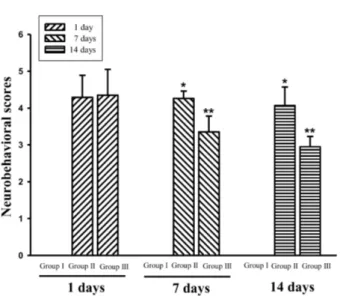
Figure 1. The effect of physical training improved neurobehavioral recovery. Values are presented as mean (SD) of three independent experiments. *p<0.05 compared to Group I, **p<0.05 compared to Group II.
Figure 2. The effect of physical exercise increased expression of glutamate transporter-1 (GLT-1). (A) Representative images of Western blotting for GLT-1 and glyceraldehyde 3-phosphate de- hydrogenase (GAPDH). (B) Quantification of the optical density for GLT-1. Values are presented as mean (SD) of three independent experiments. *p<0.05 compared to Group I, **p<0.05 compared to Group II.
Data analysis
Data analysis was performed with PASW Statistics 18.0 (IBM Co., Armonk, NY, USA). All of the data were ex- pressed as mean±standard deviation (SD). Differences in limb placement test scores were examined with a parametric one-way ANOVA and Scheffe’s post hoc procedure at 95%
significance levels. For test based on scoring systems, the nonparametric Kruskal-Wallis test was used with the multi- ple-comparison post hoc test to determine the number and relation of the group differences at a 95% significance level.
Results
All rats with MCAO exhibited severe neurological defi- cits on the first day, and there was no statistically significant difference between the non-physical training group and the physical training group (4.29±0.60 vs. 4.35±0.70 in Group II and Group III, respectively). The effect of physical train- ing on the recovery of function was evaluated at on days 7 (4.26±0.20 vs. 3.35±0.43 in Group II and Group III, re- spectively) and 14 (4.07±0.50 vs. 2.95±0.28 in Group II and Group III, respectively) after MCAO (p<0.05; Figure 1).
We observed that physical training significantly promoted functional outcomes on the 7th and 14th day after MCAO (p
<0.05). All rats in the Group I evaluated on days 1, 7, and 14, exhibited no neurological deficits. We analyzed brain fraction extracts from the rats in all three groups at the end of
the experiment to determine glutamate transporter-1 (GLT-1) activity levels. Compared to the Group II, the Group III ex- hibited an increasing trend in GLT-1 activity. After MCAO, the GLT-1 activity significantly changed by 83.56 % in the Group II and by 134.84 % in Group III (p<0.05; Figure 2).
Discussion
About of the strokes is ischemic, and 20% is hemorrhagic [25]. Patients with stroke experience various symptoms that limit their daily activities, which include physical disability, neglect, memory, cognitive, and sensory impairment [26,27].
Brain ischemia is one of the leading causes for severe dis- abilities during their lifetime and often leads to irreversible brain damage described by characteristic adenosine tri- phosphate depletion, neuronal oxidative stress, release of in- flammation cytokines and abnormal release of neuro- transmitters [28,29].
Physical activity enhanced the promotion of cell survival
mechanisms. A number of previous studies have inves-
tigated the role of exercise in promoting enhanced cell sur-
vival against brain ischemia and improved functional recov-
ery [30,31]. Our result show that physical training sig-
nificantly promoted the functional outcomes on day 14 after
MCAO. The results indicated that exercise improved the re-
However, whether physical training for two weeks could in- fluence the excessive release of glutamate caused by cere- bral ischemia is still unknown.
After MCAO, excessive glutamate release could evoke postsynaptic depolarization and cause influx of cation such as calcium and sodium into the cells, thereby inducing neu- ronal excitotoxicity [33,34]. The GLT-1 is a major glutamate transporter that has been shown to exert neuroprotection in various animal models of ischemic injury and motor neuron degeneration [35-37]. In our study, there were significant in- hibitory effects on GLT-1 activity observed in the ischemic stroke model, and physical training led to an increased in GLT-1 activity and improvements in motor function.
Changes in GLT-1 activity might reflect neuroprotective characteristics that promote the survival of the cerebral cor- tex, as has been shown in experimental experiments models of focal cerebral ischemia [34]. Improvements in neuro- logical functions indicated that alterations in GLT-1 ex- pression might be correlated with the neuroprotection.
Thus, our data clearly show that physical training appears to act as a major homeostatic regulator of motor function and GLT-1 upregulation. These findings suggest that physical training is important in functional recovery. The present data revealed that physical training promoted functional out- comes and improved the GLT-1 expression in the ischemic region after MCAO, and this study confirms that physical activity is an early physical therapeutic strategy in the re- habilitation of ischemic stroke.
References
1. Barker-Collo S, Starkey N, Lawes CM, Feigin V, Senior H, Parag V. Neuropsychological profiles of 5-year ischemic stroke survi- vors by Oxfordshire stroke classification and hemisphere of lesion. Stroke 2012;43:50-5.
2. Ashjazadeh N, Fathi M, Shariat A. Evaluation of homocysteine level as a risk factor among patients with ischemic stroke and its subtypes. Iran J Med Sci 2013;38:233-9.
3. Singh P, Kaur R, Kaur A. Endovascular treatment of acute ische- mic stroke. J Neurosci Rural Pract 2013;4:298-303.
4. Yin X, Meng F, Wei W, Li A, Wang Y, Chai Y, et al. Role of mouse nerve growth factor in neural recovery following hy- poxic-ischemic brain damage. Int J Clin Exp Med 2013;6:951-5.
5. Font MA, Arboix A, Krupinski J. Angiogenesis, neurogenesis and neuroplasticity in ischemic stroke. Curr Cardiol Rev 2010;
6:238-44.
physical exercise. Curr Drug Targets 2012;13:247-62.
8. Dimyan MA, Cohen LG. Neuroplasticity in the context of motor rehabilitation after stroke. Nat Rev Neurol 2011;7:76-85.
9. Hötting K, Röder B. Beneficial effects of physical exercise on neuroplasticity and cognition. Neurosci Biobehav Rev 2013;37:
2243-57.
10. Ding YH, Luan XD, Li J, Rafols JA, Guthinkonda M, Diaz FG, et al. Exercise-induced overexpression of angiogenic factors and reduction of ischemia/reperfusion injury in stroke. Curr Neuro- vasc Res 2004;1:411-20.
11. Quaney BM, Boyd LA, McDowd JM, Zahner LH, He J, Mayo MS, et al. Aerobic exercise improves cognition and motor func- tion poststroke. Neurorehabil Neural Repair 2009;23:879-85.
12. Harris JE, Eng JJ. Strength training improves upper-limb func- tion in individuals with stroke: a meta-analysis. Stroke 2010;41:
136-40.
13. Ribeiro T, Britto H, Oliveira D, Silva E, Galvão E, Lindquist A.
Effects of treadmill training with partial body weight support and the proprioceptive neuromuscular facilitation method on hemi- paretic gait: a randomized controlled study. Eur J Phys Rehabil Med 2013;49:451-61.
14. Stoller O, de Bruin ED, Knols RH, Hunt KJ. Effects of car- diovascular exercise early after stroke: systematic review and meta-analysis. BMC Neurol 2012;12:45.
15. Shaughnessy M, Michael K, Resnick B. Impact of treadmill ex- ercise on efficacy expectations, physical activity, and stroke recovery. J Neurosci Nurs 2012;44:27-35.
16. Foo K, Blumenthal L, Man HY. Regulation of neuronal bio- energy homeostasis by glutamate. Neurochem Int 2012;61:389- 96.
17. Lee JM, Grabb MC, Zipfel GJ, Choi DW. Brain tissue responses to ischemia. J Clin Invest 2000;106:723-31.
18. Kristián T, Siesjö BK. Calcium in ischemic cell death. Stroke 1998;29:705-18.
19. Garber K. Stroke treatment--light at the end of the tunnel? Nat Biotechnol 2007;25:838-40.
20. Zhang P, Yu H, Zhou N, Zhang J, Wu Y, Zhang Y, et al. Early ex- ercise improves cerebral blood flow through increased angio- genesis in experimental stroke rat model. J Neuroeng Rehabil 2013;10:43.
21. Forrester LW, Wheaton LA, Luft AR. Exercise-mediated loco- motor recovery and lower-limb neuroplasticity after stroke. J Rehabil Res Dev 2008;45:205-20.
22. Mattson MP. Glutamate and neurotrophic factors in neuronal plasticity and disease. Ann N Y Acad Sci 2008;1144:97-112.
23. Longa EZ, Weinstein PR, Carlson S, Cummins R. Reversible middle cerebral artery occlusion without craniectomy in rats.
Stroke 1989;20:84-91.
24. Zhang P, Zhang Q, Pu H, Wu Y, Bai Y, Vosler PS, et al. Very ear- ly-initiated physical rehabilitation protects against ischemic brain injury. Front Biosci (Elite Ed) 2012;4:2476-89.
25. Runchey S, McGee S. Does this patient have a hemorrhagic stroke?: clinical findings distinguishing hemorrhagic stroke from ischemic stroke. JAMA 2010;303:2280-6.
26. Jehkonen M, Yliranta A, Rasimus S, Saunamäki T. Neglect re- habilitation after stroke. Duodecim 2013;129:506-13.
27. Li W, Huang R, Shetty RA, Thangthaeng N, Liu R, Chen Z, et al.
Transient focal cerebral ischemia induces long-term cognitive function deficit in an experimental ischemic stroke model.
Neurobiol Dis 2013;59:18-25.
28. Sánchez-Mendoza E, Bellver-Landete V, González MP, Merino JJ, Martínez-Murillo R, Oset-Gasque MJ. Brain repair after is- chemic stroke: role of neurotransmitters in post-ischemic neurogenesis. Rev Neurol 2012;55:533-42.
29. Namura S, Ooboshi H, Liu J, Yenari MA. Neuroprotection after cerebral ischemia. Ann N Y Acad Sci 2013;1278:25-32.
30. Arrick DM, Sun H, Mayhan WG. Influence of exercise training on ischemic brain injury in type 1 diabetic rats. J Appl Physiol (1985) 2012;113:1121-7.
31. Enzinger C, Dawes H, Johansen-Berg H, Wade D, Bogdanovic M, Collett J, et al. Brain activity changes associated with tread- mill training after stroke. Stroke 2009;40:2460-7.
32. Cechetti F, Rhod A, Simão F, Santin K, Salbego C, Netto CA, et
al. Effect of treadmill exercise on cell damage in rat hippocampal slices submitted to oxygen and glucose deprivation. Brain Res 2007;1157:121-5.
33. Smith WS. Pathophysiology of focal cerebral ischemia: a ther- apeutic perspective. J Vasc Interv Radiol 2004;15:S3-12.
34. Shen H, Chen GJ, Harvey BK, Bickford PC, Wang Y. Inosine re- duces ischemic brain injury in rats. Stroke 2005;36:654-9.
35. Ketheeswaranathan P, Turner NA, Spary EJ, Batten TF, McColl BW, Saha S. Changes in glutamate transporter expression in mouse forebrain areas following focal ischemia. Brain Res 2011;
1418:93-103.
36. Namura S, Maeno H, Takami S, Jiang XF, Kamichi S, Wada K, et al. Inhibition of glial glutamate transporter GLT-1 augments brain edema after transient focal cerebral ischemia in mice.
Neurosci Lett 2002;324:117-20.
37. Verma R, Mishra V, Sasmal D, Raghubir R. Pharmacological evaluation of glutamate transporter 1 (GLT-1) mediated neuro- protection following cerebral ischemia/reperfusion injury. Eur J Pharmacol 2010;638:65-71.