http://dx.doi.org/10.12925/jkocs.2015.32.1.165
N-propyl–N,N-dimethylethanolamine 반응에서 유동성 변화와 상전이에 관한 연구
김기준
✝⋅ 성완모⋅이주엽
대진대학교 공과대학 화학공학과
(2015년 2월 17일 접수; 2015년 3월 10일 수정; 2015년 3월 25일 채택)
A Study on Mobility Gradients and Phase Transitions in N-propyl–N,N-dimethylethanolamine Reaction
Ki-Jun Kim✝⋅Wan-Mo Sung⋅Joo-Youb Lee*
Dept. of Chemical Engineering, DaeJin University, Pochun 487-711, Korea
*
Dept of Disaster Management and Safety Engineering, Junwon University, Chungbuk 361-763, Korea
(Received February 17, 2015; Revised March 10, 2015; Accepted March 25, 2015)
요 약 : N-propyl-N,N-dimethylethanolamine의 용액을 만들기 위해 산성 조건 하에서 6분간 초음 파처리하여 제조하였다. N-propyl-N,N-dimethylethanolamine 의 촉매 가수분해는 온도 30~55 ℃에 서 uni-lamellar vesicle과 uni-lamellar와 multi-lamellar가 뭉쳐진 혼합물에서 연구되었다. 이들의 차 이는 촉매효과에 대해 multi-lamellar보다 uni-lamellar에서 영향이 크게 나타났다. Vesicle의 상전이 온 도는 37~44 ℃이며, multi-lamellar의 분자 입자의 크기는 uni-lamellar의 입자의 크기보다 크게 나타 났다.
주제어 : N-propyl-N,N-dimethylethanolamine, 상전이, 가수분해, 입자 분포크기
Abstract : N-propyl-N,N-dimethylethanolamine was directly ultrasonicated in acidic water for 6 minute to give clear stock solutions. The catalytic hydrolysis of N-propyl-N,N- dimethylethanolamine was studied at 30~55 ℃ in the presence of uni-lamellar vesicle and mixture of uni- and multi-lamellar aggregates. The difference of rate between uni- and mixture was observed, where uni-lamellar reaction was more catalytic effect. The phase transition temperature of vesicle was 37~44 ℃. The particle size of multi-lamellar than that of uni-lamellar of biological membrane was measured more largely.
Keywords : N-propyl-N,N-dimethylethanolamine, phase transition, hydrolysis, particle size
✝
Corresponding author
(E-mail: [email protected])
1. 서 론
인지질(phospholipid)은 생체막(biomembrane) 의 중요한 구성 성분으로서 그 생물학적인 특성 이 많이 보고되어 있으며1-4), 이들 여러 특성 중 에 두개의 긴 알킬사슬의 길이와 불포화도에 따 른 물리적인 현상이 중요하여 활발하게 연구되어 의학 및 유전학에 응용되고 있다5~6). 또한 천연지 질과 유사한 합성지질의 이중층막에 대한 연구도 활발하였으며, 3중 알킬사슬을 함유한 암모늄염 들도 이중층막이 형성됨이 보고되었다7~9). 따라서 본 연구에서는 vesicle-protein으로서 N-propyl-N,N-dimethylethanolamine(PDMEE A)를 사용하고 biological membrane으로 이루어 진 이중층막(biological membrane) vesicle을 초 음파 처리법으로 제조하였고, sepharose-4B로 충 진된 컬럼을 사용하여 single uni-lamellar vesicle 을 분리하였다. 이때 염기 촉매(sodium dodecyl sulphate, SDS)에 의한 PDMEEA의 가수분해 반 응(hydrolysis reaction)을 상전이 효과, vesicle-protein과 vesicle의 특성 연구를 위해 uni-lamellar와 multi-lamellar를 비교 검토하였 다.
2. 실 험
2.1. 시약 및 기기
PDMEEA와 SDS는 Sigma 사 제품을 정제하지 않고 사용했으며, HCl과 sodium borax는 Wako 사의 GR급을, sepharose-4B는 Aldrich 사 제품 을 사용했다. 모든 수용액은 이차증류수를 사용하 여 제조하였다. 흡광도의 측정은 Pye-Unicam 사 의 uv-visible spectrophotometer를 사용하였고, pH meter는 Orion 사의 제품을 사용하였다. 입 자크기 분포(particle size distribution)는 레이저 산란의 Coulter(Miami FL Co., model No.
LS230, USA)로 측정하였으며, 초음파 처리는 태 금초음파사의 20 kHz ultrasonicator을 사용하였 다.
2.2. Vesicle-protein 반응속도 측정
pH = 2.32의 sodium borax-HCl 완충용액에 2×10⁻²M와 2×10⁻³M PDMEEA 용액 5 mL을 vial에 취해 5 ℃ 이하의 항온상태에서 5분간 초 음파 처리하여 vesicle-protein을 제조하였다. 이
투명한 용액 2 mL을 직경 1.5 cm 길이 25 cm 인 sepharose-4B로 채운 컬럼에 분당 0.6 mL 의 속도로 용출시켰다. 이때 275 nm 파장에서 각 분액의 혼탁도(turbidity)로부터 단일층 (uni-lamellar)과 다중층(multi-lamellar)의 vesicle-protein들을 분리했다. 분리한 보관용액을 완충액으로 희석시킨 다음, 희석된 시료 2 mL을 quartz cell에 취해 항온으로 유지시킨 후 진한 SDS용액을 micro-syringe로 주입하여 가수분해 되어 나오는 물질을 400 nm에서 흡광도의 증가 량으로 반응속도를 측정하였다. Pseudo-first order rate constant(ko)는 시간(t)에 대한 ln(A∞
-At)를 도시해 기울기로서 구했다.
3. 결과 및 고찰
컬럼 용출시의 turbidity10)는 Fig. 1과 같다.
Fig. 1. Turbidity of the each fraction rate of PDMEA at 275nm wave length.
염기촉매하의 PDMEEA의 가수분해 반응식은 다음과 같으며, 30℃, pH = 10.07, Vesicle- protein 이중층막 안에서 PDMEEA의 가수분해 과정의 스펙트럼 변화가 발생하는 것은 다음과 같은 분리현상이 생긴 원인으로 추정한다.
NO2-⌬-O2C-(CH)14-CH3 + H2O → NO2-⌬-OH + HO2C-(CH)14-CH3
TEMP(℃) 30 35 38 40 42 45 47 50
kobs(mim-1) 0.00126 0.00185 0.00224 0.00424 0.00601 0.00916 0.0105 0.0111 Table 2. The observed pseudo first order rate constants on PDMEEA catalyzed hydrolysis at the
various temperature ; no column elution, pH = 10.07, 0.01M borax buffer
TEMP(℃) 30 33 35 36 37 38 39 kobs(mim-1) 0.00312 0.00365 0.00418 0.00435 0.00553 0.00689 0.00803 TEMP(℃) 40 41 42 43 44 45 47 kobs(mim-1) 0.00913 0.0102 0.0112 0.0116 0.0129 0.0132 0.0134 Table 3. The observed pseudo first order rate constants on the PDMEEA catalyzed hydrolysis at
the various temperature ; column elution, pH = 10.07, 0.01M borax buffer.
Fig. 2. Time dependence absorption spectra during the vesicle-protein(PDMEA) catalyzed hydrolysis at the pH 10.07 ; dashed line : acidic medium, others line : alkaline medium, 30℃.
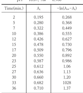
PDMEEA 가수분해 반응시 반응물의 최대흡수 파장은 267 nm이었고, 생성물의 최대흡수 파장 은 400 nm로 나타났다. Table 1에 33 ℃에서 시 간에 따른 vesicle-protein의 스펙트럼의 변화를 나타내었다.
Table 1. The observed absorbance for PDMEA catalyzed hydrolysis by SDS at 33 ℃, pH = 10.07, A∞ = 0.960
Time(min.) At -ln(A∞-At) 2
5 7 10 12 15 17 20 23 25 27 30 33 36
0.195 0.280 0.322 0.386 0.426 0.478 0.509 0.550 0.587 0.612 0.636 0.660 0.682 0.710
0.268 0.368 0.449 0.555 0.627 0.730 0.796 0.892 0.986 1.06 1.13 1.20 1028 1.37
Table 2와 Table 3에 컬럼을 통과시키지 않은 경우와 컬럼을 통과시킨 경우의 가수분해 속도상 수를 각각 나타내었다.
Fig. 3. Temperature dependence on PDMEEA catalyzed hydrolysis ; ∆ : no column elution, ⃞ : column elution.
Fig. 3에서 컬럼을 통과시킨 경우에 더 큰 속 도상수 값을 얻었는데, 이것은 uni-lamellar 구 조인 vesicle이 더욱 큰 소수성을 같기 때문에11) multi-lamellar 구조인 경우보다 큰 촉매 효과를 나타난다12). PDMEEA의 상전이 온도는 42℃로 알려져 있으나13), 본 실험의 경우는 비교적 넓게 관측되었다. 이는 PDMEA의 긴 알킬사슬에 기인 한 것으로 생각된다14).
Fig. 4. Particles size distribution of coating on PDMEA catalyzed hydrolysis by SDS at 33 ℃, pH = 10.07, (a) : no column elution, (b) : column elution.
Fig. 4에서 PDMEEA에 관한 SDS촉매효과의 유동성과 분자의 입자크기를 확인코자 초음파 처
리를 10분 동안 수행한 후에 칼럼을 통과한 것과 통과하지 않은 것을 비교하였다. (a)는 칼럼을 통 과하지 않은 PDMEEA의 multi-lamellar로서 vesicle + protein 혼합물로서 분자의 입자크기가 72.3㎛ 로 나타났으나, (b)는 칼럼을 통과하여 분 리가 이루어진 상태로 uni-lamellar micells로서 분자의 입자크기가 27.3㎛ 로서 작게 측정되었다 고 사료된다.
4. 결 론
Vesicle과 Protein의 화합물, PDMEEA로 이루 어진 이중층 vesicle-protein에서 PDMEEA의 가 수분해 반응은 지질-단백질의 상전이에 크게 영 향을 받는 것으로 나타났다. PDMEEA의 고체상 에서 액정상(liquid crystal phase)으로의 상전이 온도(Tc)는 37~44℃로 나타났으나 순수한 PDMEEA만의 vesicle-protein에서의 상전이 온 도와 비교할 때 전이 온도가 조금 높고 넓게 분 포됨이 관측되었으며, 이것은 impurity 효과에 기 인한 것으로 사료되며, vesicle-protein의 PDMEA에 관한 SDS촉매효과는 uni-lamellar PDMEEA인 경우가 uni-와 multi-lamellar 혼합 물의 시료 PDMEEA의 경우 보다 크게 나타났으 나, 입자 분포크기는 반대로 작게 나타났다. 이것 은 uni-lamellar 구조인 경우가 hydrophobic effect가 크기 때문에 hydrophilic site에서 일어나 는 가수분해 반응이 증가된 것으로 생각된다.
References
1. E. A. Dennis, J. Cao, Y. H. Hsu, V.
Magrioti, G. Kokotos, Phospholipase A2 enzymes: physical structure, biological function, disease implication, chemical inhibition, and therapeutic intervention, Chem. Rev. 111, 6131, (2011).
2. D. L. Scott, S. P. White, Z. Otwinowski, W. Yuan, M. H. Gelb, P. B. Sigler, Interfacial catalysis — the mechanism of phospholipase A2, Science 250, 1541, (1990).
3. K. Wagner, G. Brezesinski, Phospholipases to recognize modelmembrane structures on
a molecular length scale, Curr. Opin.
Colloid Interface Sci. 13 47, (2008).
4. J. E. Burke, E. A. Dennis, Phospholipase A2 structure/function, mechanism, and signaling, J. Lipid Res. 50, S237, (2009).
5. O. G. Mouritsen, T. L. Andresen, A.
Halperin, P. L. Hansen, A. F. Jakobsen, U.B. Jensen, M. O. Jensen, K. Jorgensen, T. Kaasgaard, C. Leidy, A. C. Simonsen, G. H. Peters, M. Weiss, Activation of interfacial enzymes at membrane surfaces, J. Phys. Condens. Matter 18, S1293, (2006).
6. M. H. Gelb, M. K. Jain, A. M. Hanel, O.
G. Berg, Interfacial enzymology of glycerolipid hydrolases: lessons from secreted phospholipases A2, Annu. Rev.
Biochem. 64, 653, (1995).
7. O. Berg, M. Gelb, M.-D. Tsai, M. K.
Jain, Interfacial enzymology: the secreted phospholipase paradigm, Chem. Rev. 101 2613, (2001).
8. O. Berg, B. Z. Yu, J. Rogers, M. K. Jain, Interfacial catalysis by phospholipase A2:
determination of the interfacial kinetic rate constants, Biochemistry 30, 7283, (1991).
9. W. R. Burack, Q. Yuan, R. L. Biltonen, Role of lateral phase separation in the modulation of phospholipase A2 activity, Biochemistry 32, 583, (1993).
10. U. Dahmen-Levison, G. Brezesinski, H.
Mohwald, Specific adsorption of PLA2 at monolayers, Thin Solid Films 327(5), 616, (1998).
11. D. W. Grainger, A. Reichert, H.
Ringsdorf, C. Salesse, An enzyme caught in action: direct imaging of hydrolytic function and domain formation of phospholipase A2 in phosphatidylcholine monolayers, FEBS Lett. 252, 73, (1989).
12. A. Chonn, S. C. Semple, P. R. Cullis, Association of blood proteins with large unilamellar liposomes in vivo. Relation to circulation lifetimes, J. Biol. Chem. 267, 18759, (1992).
13. H. Maeda, The enhanced permeability and retention (EPR) effect in tumor vasculature: the key role of tumor-selective macromolecular drug targeting, Adv.
Enzyme Regul. 41, 189, (2001).
14. A. A. Gabizon, Liposomal drug carrier systems in cancer chemotherapy: current status and future prospects, J. Drug Target.
10, 535, (2002).
15. G. Batist, K. A. Gelmon, K. N. Chi, W.
H. Miller, S. K. L. Chia, L. D. Mayer, C.
E. Swenson, Safety, pharmacokinetics, and efficacy of CPX-1 liposome injection in patients with advanced solid tumors, Clin.
Cancer Res. 15 , 692, (2009).