․Correspondence to : Ki-ho Cho Hoegi-dong 1, Dongdaemun-gu, Seoul, Korea
Dept. of Cardiovascular and Neurologic Diseases(Stroke center), College of Oriental Medicine, Kyung-Hee University TEL: 02-958-9134
E-mail: [email protected]
․이 논문은 2006년도 경희대학 대학원 한의학 석사학위 논문임.
Effect of Acupuncture on the Expressions of Neuropeptide Y and Leptin Receptor in the Hypothalamus of Food-deprived Rats
Mi-a Kim1, Woo-sang Jung1, Sang-kwan Moon1, Young-suk Kim1, Chang-ju Kim2, Ki-ho Cho1
1Dept. of Cardiovascular and Neurologic Diseases(Stroke center), College of Oriental Medicine, Kyung-Hee University
2Dept. of physiology, college of Medicine, Kyung-Hee University
ABSTRACT
Objectives :
This study aimed to find out whether acupuncture at various acupoints shows any effects on appetite by regulating neurotransmitters in the hypothalamus through the expression of NPY and LR in the PVN via immunohistochemistry.
Methods :
Male Sprague-Dawley rats were divided into eight groups of five mice each. The rats in the acupuncture groups were treated with acupuncture at respective acupoints, twice per a day for 3 days. The animals were sacrificed 72 h after commencement of the experiment, the brains being dissected into serial coronal sections. Expressions of neuropeptide Y and leptin receptor in the hypothalamus were assessed by immunohistochemistry.
Results :
NPY expression in the PVN was enhanced and LR expression in the PVN was decreased by food deprivation.
NPY immunoreactivity in the PVN of the food-derived rats was decreased by acupuncture at the auricular acupoint, Zusanli-acupoint, a and non-acupoint. However, acupuncture at the auricular acupoint showed most potent suppressing effect on NPY expression in the PVN of food-deprived rats. LR expression in the PVN decreased following food deprivation, and auricular acupuncture increased LR expression in the PVN of food-deprived rats. In normal conditions (fed state), LR expression in the PVN was not changed by acupuncture treatment at several sites.
Conclusions :
From this study, we have shown that acupuncture at the auricular acupoint exerts the most potent appetite suppressing effect on the food restriction state.
Key words : acupuncture, appetite, neuropeptide Y and leptin R in the hypothalamus
Ⅰ. Introduction
The prevalence of obesity has been increasing in
Korea due to the influence of westernized diet and
lack of exercise. Obesity has been accepted to be a
strong predictor of metabolic syndrome, which is a
cluster of abnormal energy metabolic condition such
as dyslipidemia, hyperglycemia and hypertension related with coronary heart disease and cerebrovascular disease.
Obesity results from over-accumulation of energy caused by imbalance between energy intake and energy expenditure. Positive caloric balance and storage of energy in adipose tissue cause adipocyte hypertrophy and visceral adipose tissue accumulation.
Free fatty acids released from the visceral fat increase hepatic lipase activity, which leads to the removal of lipid from LDL (low density lipoprotein) and HDL (high density lipoprotein), making them smaller, more dense and more susceptible to oxidation. Esterification of free fatty acids and adipocyte lipolysis activate TLR4/NF-κB pathway, inducing cytokines such as TNFα, IL-6, which produce chronic inflammation and inhibit insulin signaling. Thus, obesity leads to a state of insulin resistance, increased inflammatory response, atherogenic dyslipidemia, and significantly enhanced lipid oxidation. As a result, it is required to put much emphasis on the prevention and treatment against obesity.
One way of treating obesity is to control energy intake and expenditure. The drugs widely used now are developed to increase energy expenditure, but many side-effects have been reported. One of methods to regulate energy intake is to control appetite. In Oriental medicine, acupuncture has been used to suppress appetite in obesity patients.
Acupuncture has been applied as a clinical treatment for various diseases in Oriental medicine.
It is known to possess various effects, such as analgesia, promotion of homeostasis, improvement in brain circulation, neuromodulatory function in the central nervous system
1-3. In terms of oriental medicine, obesity and excess appetite result from
stagnation of Qi in the spleen and stomach and the development of heat in the stomach and intestine.
Many studies suggested that acupuncture may regulate appetite. Previous studies have shown that neuronal activity of lateral hypothalamus (LHA), as a feeding center, was significantly inhibited and neuronal activity of ventromedial hypothalamus (VMH), as a satiety center, was significantly increased by auricular acupuncture
4. Recently, it has been reported that eletroacupuncture upregulated the expression of melanocyte-stimulating hormone (MSH) and cocaine and amphetamine -regulated transcript peptide (CART) in the hypothalamic arcuate nucleus of diet-induced obese rats
5,6.
Among the various neuropeptides and hormones involved in appetite regulation, of particular interest are neuropeptide Y (NPY), an appetite- inducing factor, and leptin, an appetite suppressing factor. NPY is a 36-amino-acid peptide thought to be a key neurotransmitter in the regulation of food intake. Within the hypothalamus, NPY is synthesized largely in neurons whose cell bodies lie in the arcuate nucleus (ARN) and which project to the paraventricular nucleus (PVN), where NPY is released from nerve terminals
7-10.
Increases in both biosynthesis and release of NPY along the discrete neuronal pathways constitute a specific hypothalamic response to starvation
11. Central administration of NPY into the cerebral ventricles or directly into the PVN increases food intake, decreases energy expenditure, reduces sympathetic outflow to brown adipose tissue
12. Thus, NPY administration leads to a state of positive energy balance and increases fat storage.
Leptin, which is produced from the ob (obese)
gene
13, provides a feedback signal from fat stores to influence overall energy balance. A major site of action for inhibitory effect of leptin on feeding is the hypothalamic arcuate nucleus. Leptin interacts with two distinct cell types within the arcuate:
the medial neurons that contain the orexigenic peptides neuropeptide Y (NPY) and agouti-related peptide, and the lateral neurons that express proopiomelanocortin (POMC), the precursor for the anorexigenic peptide
14-16. Increasing leptin level causes to decrease NPY and agouti-related peptide expression and to increase POMC expression.
These changes are thought to mediate the effect of leptin on food intake.
Leptin exerts its actions by activation of the leptin receptor which is the product of the diabetes (db) gene
17. The leptin receptor (LR), which possesses only one transmembrane domain, is known to be presented in the choroids plexus, cerebral cortex, hippocampus, thalamus, and hypothalamus
17. Neurons with particularly strong LR-like immunoreactivity are found in the ARN and PVN of the hypothalamus
18. NPY is the key component of feeding drive systems and its secretion and action are regulated negatively by neuropeptides such as melanocortin, corticotropin-releasing factor and glucagon like peptide 1, which are also regulated by leptin.
Therefore NPY and leptin are important factors to control appetite.
This study is designed to find out whether acupuncture at various acupoints exerts any effects on appetite by regulating neurotransmitters in the hypothalamus through the expression of NPY and LR in the PVN via immunohistochemistry.
Ⅱ. Materials and methods
1. Experimental animals and acupuncture treatment Male Sprague-Dawley rats weighing 280±20 g (6 weeks old) were used for the experiment. Each animals was housed at a controlled temperature (20±2 ℃) and maintained under light-dark cycles, each consisting of 12 h of light and 12 h of darkness with food and water available for 7 days prior to the starting of experiment.
The animals were divided into eight groups of five mice each: the fed group, the fed and auricular acupunctured group, the fed and Zusanli - acupunctured group, the fed and nonacupoint- acupunctured group, the food-deprived group, the food-deprived and auricular acupunctured group, the food-deprived and Zusanli -acupunctured group, the food-deprived and nonacupoint-acupunctured group. The animals in the fed groups received abundant food and water, while food was withheld from those in the food-deprived groups for 72 h.
The rats in the acupunctured groups were
treated with acupuncture at respective acupoint,
twice per day at 10:00h and at 17:00h for three
days. Auricular point, supplied by the auricular
branch of the vagus nerve, represents ‘stomach
point' in Oriental medicine. Zusanli acupoint (ST
36) located 5 mm lateral and distal to the
anterior tubercle of the tibia. Zusanli is one of the
main acupuncture points of stomach channel of
foot yangming. The reason Zusanli acupoint is
selected is that Zusanli streamlines the stagnation
of stomach Qi and disperses the heat in the
stomach and intestine. Both hip sides were used
as a nonacupoint. For acupuncture stimulation,
stainless steel needles of 0.3 mm diameter and 25 mm
length, were inserted. After inserting acupuncture
needles into the auricular acupoint, Zusanli acupoint, or nonacupoint, left in place for 20 min, no noticeable change in behavior and movement indicative of stress was observed.
2. Tissue preparation
The animals were sacrificed at 72 h after commencement of the experiment, anesthetized with sodium pentobarbital. After a complete lack of response was observed, the rats were transcardially perfused with 50 mM phosphate-buffered saline (PBS) and then fixed with 4% paraformaldehyde in 100m M phosphate buffer at pH 7.4. The brains were dissected, postfixed in the same fixative overnight, and transferred into a 30% sucrose for cryoprotection. Serial coronal sections of 40 mm thickness were made with a freezing microtome (Leica, Nussloch, Germany).
3. Immunohistochemistry
Free-floating tissue sections were washed twice for 15 min in 50mM PBS, and then permeabilized in 0.5 % Triton X-100 for 30 min. After washing twice with PBS, sections were incubated overnight with rabbit anti-NPY antibody (DiaSorin, Stillwater, MN, USA) at a dilution of 1:4000 for visualization of NPY distribution or with goat anti-leptin receptor antibody (Santa Cruz Biotechnology, Santa Cruz, CA, USA) and at a dilution of 1:1000 for visualization of leptin receptor distribution.
The sections were then washed three times with PBS and incubated for 1 h with biotinylated anti-rabbit secondary antibody for anti-NPY immunohistochemistry or with anti-goat secondary antibody (Vector Laboratories, Burlingame, CA, USA).
The sections were subsequently incubated with an
avidin-biotin-peroxidasecomplex (Vector Laboratories) for 1 h at room temperature.
Immunoreactivity was visualized by incubation with the sections in a solution consisting of 0.02%
3,3-diaminobenzidine (DAB) containing nickel chloride and 0.01% H
2O
2in 50 μM Tris -buffer (pH 7.6) for approximately 5 min.
4. Data analysis
The intensity of NPY-specific staining was assessed in a quantitative fashion according to a microdensitometrical method based on optical density (mean gray scale) using an image analyzer (Multiscan, Fullerton, CA, USA). For assessment of LR-specific immunoreactivity, the number of LR- immunoreactive neurons was counted hemilaterally in each section through a lightmicroscope (Olympus, Tokyo, Japan).
Statistical analysis was performed using one-way ANOVA followed by Scheffe`s post-hoc test. The results are presented as the mean±standard error of the mean (S.E.M.). Differences were considered significant at p< 0.05.
Ⅲ. Results
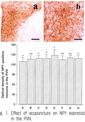
1. Effect of acupuncture on NPY expression in the PVN
The intensity of NPY immunoreactivity in the PVN of the hypothalamus was about 129.41±2.32 in the fed group, 136.84±2.71 in the fed and auricular acupunctured group, 139.16±1.85 in the fed and Zusanli -acupunctured group, 139.35±3.04 in the fed and nonacupoint acupunctured group, 148.77
±4.76 in the food-deprived group, 135.71±3.17 in
the food-deprived and auricular acupunctured group,
142.01±1.38 in the food-deprived and Zusanli -
acupunctured group, 140.22±1.89 in the food-deprived and nonacupoint acupunctured group.
The present results show that NPY expressions in the PVN was increased following food deprivation (E), and auricular acupoint, Zusanli acupoint and nonacupoint acupuncture suppressed NPY expression at food deprivation (F,G,H) ( p< 0.05).
Significant difference of NPY expression among auricular acupoint, Zusanli acupoint, nonacupoint acupuncture was not shown, but acupuncture at auricular acupoint showed most potent suppressing effect on NPY expression in the PVN of food- deprived rats (F). Under normal condition (fed state), acupuncture enhanced NPY expression (B, C, D).
Fig. 1. Effect of acupuncture on NPY expression in the PVN.
Upper : Photomicrographs of NPY-immuno reactive neurons in the PVN. The scale bar represents 100 μm in a and 50μm in b.
Lower : NPY-immunoreactive neurons staining
intensities in the PVN. The NPY levels of each group were assessed from optical density measurement. The data are represented as the mean±S.E.M.
129.41±2.32 in the fed group(A), 136.84±2.71 in the fed and auricular acupunctured group(B), 139.16±1.85 in the fed and
Zusanli
-acupunctured group(C), 139.35±3.04 in the fed and nonacupoint acupunctured group(D), 148.77±4.76 in the food- deprived group(E), 135.71±3.17 in the food-deprived and auricular acupunctured group(F), 142.01±1.38 in the food-deprived andZusanli
-acupunctured group(G), 140.22±1.89 in the food-deprived and nonacupoint acupunctured group(H).Optical density of NPY-positive neurons in the PVNa : <135 in the optical density measurement b : 135≤NPY≤143 in the optical density measurement
c : >143 in the optical density measurement
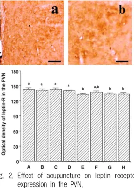
2. Effect of acupuncture on LR expression in the PVN
The number of LR-immunoreactive cells in the PVN of the hypothalamus was about 143.66±2.51 in the fed group, 142.17±1.96 in the fed and auricular acupuncture group, 144.10±1.93 in the fed and Zusanli -acupunctured group, 141.19±1.18 in the fed and nonacupoint acupunctured group, 135.00±
0.95 in the food-deprived group, 138.99±1.79 in the food-deprived and auricular acupunctured group, 135.43±1.63 in the food-deprived and Zusanli - acupunctured group, 135.19±2.20 in the food-deprived and nonacupoint acupunctured group.
The present results show that LR expression in the PVN was decreased following food deprivation.
Acupuncture at auricular acupoint enhanced food deprivation-induced decrease of LR expression.
Acupuncture at Zusanli acupoint and nonacupoint
exerted no significant effect on LR expression in
the PVN under food deprived condition. Under
normal condition (fed state), acupuncture exerted
no significant effect on LR expression in the PVN.
Fig. 2. Effect of acupuncture on leptin receptor expression in the PVN.
Upper : Photomicrographs of leptin receptor- immunoreactive neurons in the PVN. The scale bar represents 100 μm in a and 50μm in b.
Lower : number of leptin receptor-positive cells in the PVN of each group. The data are represented as the mean±S.E.M.
143.66±2.51 in the fed group(A), 142.17±1.96 in the fed and auricular acupuncture group(B), 144.10±1.93 in the fed and
Zusanli
-acupunctured group(C), 141.19±1.18 in the fed and nonacupoint acupunctured group(D), 135.00±0.95 in the food- deprived group(E), 138.99±1.79 in the food-deprived and auricular acupunctured group(F), 135.43±1.63 in the food-deprived and
Zusanli
-acupunctured group(G), 135.19±2.20 in the food-deprived and nonacupoint acupunctured group(H).Optical density of Leptin-R in the PVN a : ≥140 in the optical density measurement b : <140 in the optical density measurement
Ⅳ. Discussion
NPY expression on the PVN was enhanced and LR expression on the PVN was decreasd by food
deprivation in this study. The primary site regulating appetite and energy balance is hypothalamus, which integrate a wide range of humoral and neural signals (peptides released from the gastrointestinal tract and gastric vagal stimuli) and regulate appetite. Several feeding regulating peptides, such as neuropeptide Y (NPY) and proopiomelanocortin (POMC), are presented in neurons of the hypothalamic PVN, which also serves as a primary targeting site for leptin. Leptin, a feeding inhibitory hormone, is secreted by adipocytes in response to taking food. During food deprivation, plasma leptin levels rapidly decline, which in turn stimulates food intake by increasing NPY and agouti-related protein (AgRP) in the arcuate nucleus
14-16. Leptin can also influence neuronal activity by opening ATP- sensitive potassium channels, thus hyperpolarizing glucose-responsive neurons in the hypothalamus
19,20. The decrease in food intake by leptin is associated with inhibition of glucose-responsive neurons in the LH, and stimulation of glucose-responsive neurons in the VMH and PVN
21.
The combination of peripheral meal-related stimuli and central leptin have increased the number of neurons expressing c- fos (marker of neuronal activity in hypothalamic areas of the gut-brain axis)
22.
In the present study, NPY immunoreactivity in the PVN of the food-derived rats was decreased by acupuncture at auricular acupoint, Zusanli -acupoint and nonacupoint. However, acupuncture at auricular acupoint showed most potent suppressing effect on NPY expression in the PVN of food-deprived rats.
It was reported that acupuncture decreases
NPY expression in the hypothalamus of rats with
streptozotocin induced diabetes, suggesting that
acupuncture treatment may be effective in curbing
the hyperphagia of diabetes
23.
Under normal condition (fed state), acupuncture at auricular acupoint, Zusanli acupoint and nonacupoint increased NPY expression in the PVN. The present results consist with other reports that acupuncture acts as a stress under normal condition
24.
The present study also showed that LR expression in the PVN was decreased following food deprivation, and auricular acupuncture increased LR expression in the PVN of food-deprived rats.
In normal conditions (fed state), LR expression in the PVN was not changed by acupuncture treatment at several sites.
Shiraishi et al.
21reported that auricular acupuncture stimulation reduced body weight by enhancing lipid metabolism in both non-obese and mildly obese humans. In experimentally induced obese rats, acupuncture increased the excitability of the VMH (60.5%, n=18, p <0.01) and decreased the excitability of the LHA(45.6%, n=12, p <0.01)
25,4,26. Recently, it has been reported that eletroacupuncture upregulated the expression of melanocyte-stimulating hormone (MSH) and cocaine and amphetamine- regulated transcript peptide (CART) in the hypothalamic arcuate nucleus of diet-induced obese rats
5,6.
Stimulation of vagal branch at auricle by acupuncture decreased NPY expression in the PVN under starvation state, which is thought to be the result of augmentated gut-brain reflex by stimulating vagal afferent neurons. Moreover, auricular acupuncture increased LR expression in the PVN under starvation state. In the present study, we have shown that acupuncture at auricular acupoint exert most potent appetite suppressing effect on
food restriction state.
The relation of acupuncture with the peptides released from gastrointestinal tract such as CCK, ghrelin, and whether auricular acupuncture affects directly on the leptin or leptin receptois are needed to be clarified in next studies.
Ⅴ. Conclusion
The most important factor regulating energy intake is appetite and the primary site regulating appetite and energy balance is hypothalamus. In this study, NPY expression in the PVN was enhanced and LR expression in the PVN was decreasd by food deprivation. NPY immunoreactivity in the PVN of the food-derived rats was decreased by acupuncture at auricular acupoint, Zusanli -acupoint, and nonacupoint. However, acupuncture at auricular acupoint showed most potent suppressing effect on NPY expression in the PVN of food-deprived rats.
Under normal condition(fed state), acupuncture at
auricular acupoint, Zusanli acupoint, and nonacupoint
increased NPY expression in the PVN. The present
results consist with other reports that acupuncture
acts as a stress under normal condition
24. The
present study also showed that LR expression in
the PVN was decreased following food deprivation,
and auricular acupuncture increased LR expression
in the PVN of food-deprived rats. In normal
conditions (fed state), LR expression in the PVN
was not changed by acupuncture treatment at
several sites. From this study, we have shown
that acupuncture at auricular acupoint exert most
potent appetite suppressing effect on food restriction
state.
침치료가 굶긴 쥐 시상하부에서 neuropeptide Y(NPY) 와 leptin receptor(LR)의 발현에 미치는 영향에 대한 실험적 연구
김미아1, 정우상1, 문상관1, 김영석1, 김창주2, 조기호1
1경희대학교 한의과대학 한방순환신경내과학 교실,2경희대학교 의과대학 생리학교실
초 록목 적 :
본 연구에서는 침치료가 비만치료의 주요방법인 식욕억제에 효과적인지 알아보기 위하여 굶긴 쥐 시상하부에서 NPY와 LR의 발현 변화를 실험을 통하여 연구하였다.
방 법 :
생후 6주 Sprague-Dawley rats를 한 군에 다섯 마리씩 배정하여 대조군-무치료, 대조군-이침치료, 대조군-족삼리 치료, 대조군-임의혈치료, 실험군-무치료, 실험군-이침치료, 실험군-족삼리치료, 실험군-임의혈치료의 8군으로 분류하여 실험하 였다. 실험군은 3일간 물만 공급하고 사료를 금식시켰고, 침자극은 하루 2회 3일간 시행하였다.
침은 직경 0.3 mm 이었고, 양측혈을 사용하였고, 2-4 mm 깊이로 자입 하였으며, 20분 유침하였다. 3일 후 pentobarbital로 마취하여 사망시켰고, 4% paraformaldehyde로 조직을 고정한 후 뇌조직을 관상으로 40 μm두께로 잘랐다. 식욕조절중후인 시상 하부의 paraventricular nucleus(PVN)에서 NPY와 LR의 발현을 면역조직법으로 보았다.
결 과 :
굶기지 않은 대조군에 비하여 굶긴 실험군에서 시상하부 PVN 에서 NPY의 증가와 LR 감소가 나타났다. 굶긴 실 험군에 이침, 족삼리, 임의혈 자침시 시상하부 PVN 에서 NPY의 감소가 나타났으며, 이침이 타침 자침에 비하여 NPY의 감소 를 더욱 크게 나타냈다. 굶기지 않은 정상상태에서 이침, 족삼리, 임의혈 자침시 NPY의 증가가 있었으며 이는 자침에 의한 스 트레스로 인한 것으로 보여진다. 굶긴 실험군에 이침 자침 시에만 시상하부 PVN에서 LR의 증가가 있었으며 정상상태에서 자 침시 LR 발현의 변화는 없었다.
결 론 :