Cavernous Hemangioma of the Uterus in a Postmenopausal Woman - A Case Report -
3
0
0
전체 글
(2)
(3)
수치
관련 문서
Known typical imaging findings of intracranial extracerebral cavernous hemangiomas are a well-defined contour-bulging mass, with marked homogeneous enhancement or a filling-in
In conclusion, tracheal cavernous hemangioma that may produce massive hemoptysis, can be controlled efficiently and definitively with laser by rigid laryngoscopy, if located in
Herein, we report a rare case of intramuscular hemangioma located in the zygomaticus minor muscle, which is related to smiling and usually runs along the orbicularis oculi
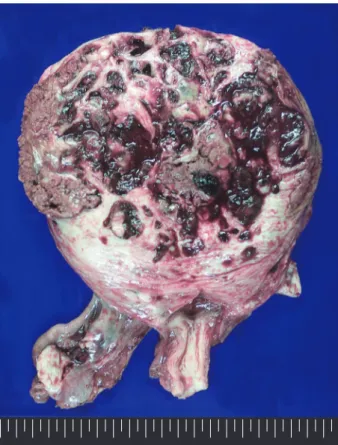
Laparotomy revealed a 5 cm sized uterine mass occupying the lower pelvic cavity, and both adnexae were small and atrophied (Fig. Uterine cervix could not be palpated definitely.
We report a case of endometriosis presenting in a postmenopausal woman with no history of endometriosis before hormone replacement therapy.. Transvaginal ultrasound showing a
Therefore, we report an extremely rare case of central sclerosing hemangioma in 58-year-old wom- an with radiological-pathological findings, which was initially misdiagnosed
This report is a very rare case of uterine prolapse in a young healthy primigravid woman, resulting in a successful vaginal delivery.. Keywords: Pregnancy;
We present a case of a cavernous hemangioma of the ilium mimicking an aggressive malignant bone tumor, which demonstrated a large osteolytic lesion with cortical
