• 검색 결과가 없습니다.

Massive Hemoptysis due to Endotracheal Hemangioma: A Case Report and Literature Review

N/A
N/A
Protected

Academic year: 2021

Share "Massive Hemoptysis due to Endotracheal Hemangioma: A Case Report and Literature Review"

Copied!
6
0
0

로드 중.... (전체 텍스트 보기)

전체 글

(1)

Massive Hemoptysis due to Endotracheal Hemangioma: A Case Report and Literature Review

Yeonsil Yu, M.D.

1

, Suhyeon Lee, M.D.

1

, Jinyoung An, M.D.

1

, Jeongmin Lee, M.D.

1

, Jihoon Kim, M.D.

2

, Youngkyung Lee, M.D, Ph.D.

3

, Eunah Jung, M.D.

4

, Sookhee Song, M.D.

1

, Hyeok Kim, M.D., Ph.D.

1

and Suhyun Kim, M.D.

1

Departments of

1

Internal Medicine,

2

Otorhinolaryngology-Head and Neck Surgery,

3

Radiology, and

4

Pathology, Seoul Medical Center, Seoul, Korea

Tracheal hemangioma is a rare benign vascular tumor in adults. We reported a case of massive hemoptysis caused by a cavernous hemangioma in a 75-year-old man. This is the first report, to our knowledge, of a tracheal cavernous hemangioma that presented with massive hemoptysis. The lesion was removed with a CO

2

laser under rigid laryngoscopy. Endovascular tumors, such as tracheobronchial hemangiomas, should be considered a diagnostic option in cases of massive hemoptysis without a significant underlying lung lesion.

Keywords: Hemangioma, Cavernous; Hemoptysis; Trachea

life-threatening manifestations of the disease. We reported a case of tracheal hemangioma in an adult with massive he- moptysis. We furthermore reviewed the literature to date.

Case Report

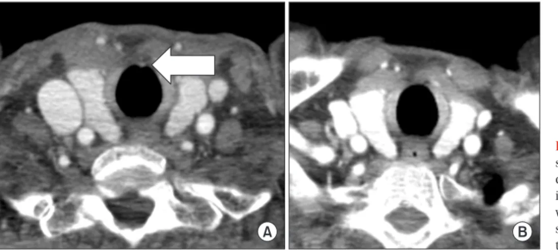
A 75-year-old man presented with hemoptysis in our outpa- tient clinic. He had recurrent episodes of blood tinged sputum and minor hemoptysis during the past year. He had a history of smoking an average of 55 pack-year of cigarettes and had bronchial asthma. Recently, he had also been diagnosed with smear-negative pulmonary tuberculosis (TB) in the public health center and was treated with anti-TB drugs. The results of hematologic and chemical laboratory tests were unremark- able. Chest radiography showed small nodules with linear opacities in the left upper lung zones. Low dose chest com- puted tomography (CT) scans without contrast enhancement showed small calcified nodules with linear opacities in the left upper lobe. The opacities in the left upper lobe were un- changed in comparison with low dose chest CT scans exam- ined 10 months ago. Occasional hemoptysis persisted despite the ongoing anti-TB therapy and oral tranexamic acid admin- istered for about a month. Flexible bronchoscopy performed under local anesthesia, revealed an approximately 6 mm- Copyright © 2015

The Korean Academy of Tuberculosis and Respiratory Diseases.

All rights reserved.

Introduction

Primary tracheal tumors are rare with an approximate in- cidence of 2.7 new cases per million per year, with vascular tumors accounting for less than 10%

1,2

. Tracheal tumors are more commonly malignant than benign in adults, whereas the reverse is true in children

3

. Hemangioma of the tracheo- bronchial tree occurs more frequently in young children and regresses steadily, while, tracheal hemangioma is exceptional in adults

1

. Hemoptysis is one of the most serious and possibly

Address for correspondence: Suhyun Kim, M.D.

Department of Internal Medicine, Seoul Medical Center, 156 Sinnae-ro, Jungnang-gu, Seoul 131-795, Korea

Phone: 82-2-2276-7808, Fax: 82-2-2276-7820 E-mail: [email protected]

Received: Oct. 7, 2014 Revised: Nov. 10, 2014 Accepted: Nov. 26, 2014

cc

It is identical to the Creative Commons Attribution Non-Commercial

License (http://creativecommons.org/licenses/by-nc/3.0/).

(2)

sized, well-circumscribed, reddish hypervascular, multilocu- lated, polypoid lesion found on the ventral wall of the tracheal midline, 5 mm below the vocal cords (Figure 1A). The other parts of the trachea and bronchi were unremarkable. Bleeding from the polypoid lesion occurred on intra-procedural cough- ing. The bleeding stopped after endobronchial instillation of 1:10,000 diluted epinephrine. However, forcep biopsy without laser coagulation equipment was not attempted due to the possibility of profuse intra-procedural bleeding.

The patient was admitted and treated with intravenous tranexamic acid. Subsequently, no further hemoptysis was found until 1-week postbronchoscopy. Chest CT scans with contrast enhancement showed a focal enhancement <5 mm in the midline of the anterior tracheal wall (Figure 2A, arrow).

The polypoid lesion was diagnosed as a presumptive tracheal hemangioma. We referred the patient to a tertiary referral hos- pital for therapeutic bronchoscopic laser ablation. The hospi- tal staff recommended a wait-and-see policy; however, 3 days later he was admitted to our hospital for recurrent episodes of massive hemoptysis. During the follow-up bronchoscopy, ap-

proximately 500 mL of blood was suctioned from the tracheal polypoid lesion (Figure 1B). A consult was arranged with the otorhinolaryngology department. The patient underwent an emergency operation for the tracheal lesion, under general anesthesia on the 10th day of hospitalization. A 4-mm-sized, hemorrhagic and polypoid lesion was found about 5 mm be- low the glottis. The lesion was removed by CO

2

laser (2 watt, super-pulse, continuous mode) under rigid laryngoscopy.

After removal, bleeding from tracheal mucosa was controlled by coagulation with a suction Bovie. The patient had no fur- ther episodes of hemoptysis during the 12 months postopera- tive follow-up period. Histopathology showed several dilated spaces generally with degenerated lining cells; the dilated spaces lined by flattened cells focally, contained few red blood cells (Figure 3). A diagnosis of tracheal cavernous heman- gioma was made. One-week follow-up flexible bronchoscopy showed healing and scarring of the lesion with no evidence of bleeding (Figure 1C). The tracheal nodular lesion had disap- peared in the 6-month follow-up chest CT scans (Figure 2B).

A B C

A B

Figure 1. Bronchoscopic findings. (A) Initial bronchoscopy showed a polypoid lesion on the ventral wall of the trachea, 5 mm below the vocal cord. (B) Follow-up bronchoscopy showed massive bleeding on the polypoid lesion. (C) Postoperative bronchoscopy showed healing and scarring of the lesion with no evidence of bleeding.

Figure 2. Chest computed tomography

scans. (A) Initial imaging showed a fo-

cal enhancement smaller than 5 mm

in the midline of the anterior tracheal

wall (arrow). (B) Postoperative imaging

shows disappearance of the previous

tracheal nodular lesion.

(3)

Discussion

Hemangioma is a common benign tumor of the head and neck in children, which is characterized by proliferative development during infancy and gradual involution later in childhood

4

. Sweetser classified hemangiomas of the air- ways into the infantile and adult varieties

4

. Hemangiomas in adults usually arise from the upper airway, including the laryngopharynx, nose, and tongue, and seldom arise from the subglottic trachea

5

. Hoarseness with slight or no dyspnea is the most common manifestation, and hemoptysis occurs frequently

5

. Hemangiomas can be divided into cavernous and capillary types, depending on the diameter of the vascular channels that comprise the tumor

6

. Capillary hemangioma is the most common variant of hemangiomas that occur in the skin, subcutaneous tissues, and mucous membranes of the oral cavities and lips, as well as in internal viscera

6

. Mi- croscopically, cavernous hemangioma is a well-demarcated lesion composed of large spaces filled with red blood cells and thrombotic material, while capillary hemangiomas do not have such large vascular spaces

6

. Cavernous hemangioma, as in this case, have a higher risk of bleeding due to large cavern- ous spaces than the capillary hemangioma

6

.

Tracheobronchial hemangioma is an extremely rare dis- ease with only few cases reported to date (Table 1)

1-14

. The most frequent symptom is minor to massive hemoptysis

4

. To the best of our knowledge, this is the first report of a tracheal cavernous hemangioma that presented with massive he- moptysis. The amount of bleeding is usually correlated with length of hospital stay and outcome in patients

15

. Our patient presented with recurrent episodes of blood tinged sputum and minor hemoptysis, followed by massive hemoptysis of

about 500 mL during bronchoscopy. Emergency laryngomi- crosurgery was performed on the tracheal hemangioma un- der general anesthesia on the 10th day of hospitalization. The patient was discharged without intensive care unit care on the 14th day. The two previously reported cases of capillary hemangiomas with massive hemoptysis were treated with Nd:YAG laser and interventional radiology, respectively

1,6

. The first patient presented with hemoptysis (coughed up vol- ume, 500 mL) and a 1 cm hemangioma 7 cm below the vocal cord on bronchoscopy, which was coagulated with Nd:YAG laser

6

. The second patient manifested massive hemoptysis of unknown volume and an irregular excrescent mass lead- ing to 30% to 40% stenosis of the proximal trachea on bron- choscopy. After catheter embolization of the pathologic right intercostal bronchial trunk, the patient was kept sedated for 24 hours with orotracheal intubation and insufflation of the orotracheal tube balloon at the hemangioma

1

.

Bronchiectasis, TB, mycetomas, necrotizing pneumonia and bronchogenic carcinomas are the main underlying con- ditions of massive hemoptysis. Several alterations in vascular anatomy may occur in these chronic inflammatory or infec- tious lung diseases

15

. Some authors suggest that CT scans should replace bronchoscopy as a first-line investigational approach in patients with large (<300 mL/24 hr) or massive hemoptysis (>300 mL/24 hr), because of its higher diagnostic yield. It is crucial, however, to perform fiber-optic bronchos- copy (FOB) for diagnosis of endoluminal lesions as a bleeding focus in patients without definite lung parenchymal lesions or vascular abnormalities in CT scans. Although the optimal tim- ing for a diagnostic FOB in hemoptysis remains controversial, early FOB might be more helpful than delayed FOB in patients without underlying lung disease. Chest radiograph and chest

A B

Figure 3. Photograph of histopathological specimen showing multiple dilated vascular spaces containing a few red blood cells (A, H&E stain,

×200; B, H&E stain, ×400).

(4)

Table 1. Review of case reports detailing tracheobronchial hemangiomas

Source Gender/

Age (yr)

Underlying lung disease

Clinical symptoms (amount of hemoptysis)

Bronchoscopic findings and locations of the lesions

Treatment

Brown and Rebeiz (1993)

7

M/47 None Hemoptysis

(5 mL)

5-mm-sized red, pedunculated mass on the anterior tracheal wall just below the subglottis

Endoscopic biopsy and Nd:YAG laser

Strausz and Soltesz (1999)

8

M/55 None Hemoptysis (NA) 2×2-mm-sized reddish lesion with capillarized surface on the ventral wall of the middle trachea

Bronchoscopy with forceps excisional biopsy

Strausz and Soltesz (1999)

8

F/70 None Chronic cough 5×4-mm-sized smooth surface lesion on the left upper lobe bronchus

Nd:YAG laser

So et al. (1999)

6

F/32 None Massive hemop- tysis (500 mL)

1-cm-sized smooth surface mass 7 cm below the vocal cords

Nd:YAG laser

Irani et al.

(2003)

9

F/72 None Cough, hemop-

tysis (NA)

2–3-mm-sized polypoid tracheal tumor with hyperemic mucosa 3 cm below the vocal cords

Bronchoscopy and forceps biopsy

Zambudio et al.

(2003)

1

F/66 ITP Massive hemop-

tysis (NA)

Irregular excrescent tracheal mass on the lumen between the first and third tracheal rings

Embolization of branch of right intercostal artery by interven tional radiology (ICU care)

Cordos et al.

(2005)

10

M/50 None Dyspnea, hemop-

tysis (NA)

Not available Interventional endoscopy and surgical resection, Nd:YAG laser

Madhumita et al.

(2004)

3

F/40 None Foreign body

sensation, hemoptysis (NA)

10×5-mm-sized cherry-red tracheal mass in the upper one-third, 20 cm from the upper incisor tooth

Bronchoscopic excision under general anesthesia

Rose and Mathur (2008)

5

M/66 Mitral stenosis Hemoptysis (NA) Three mucosal polyps lateral to the carina at the proximal left mainstem bronchus

Bronchoscopy with forceps biopsy and APC

Chawla et al.

(2010)

11

M/62 PTE with IVC filter, asthma

Hemoptysis (NA) Soft lesion at the 9 o’clock position in the distal trachea

Bronchoscopy with forceps exci sional biopsy and Nd:YAG laser Kim et al.

(2010)

12

F/28 None Cough, hemop-

tysis (25 mL)

10-mm-sized polypoid lesion in the lateral wall of proximal trachea 2 cm below the vocal cords

Removed by rigid bronchoscopy

Amy and Enrique (2012)

13

M/22 Hypogonadism with testosterone therapy

Hemoptysis (2–4 tablespoons)

15-mm-sized purple, vascular lesion 3 cm from the carina at the 5 o’clock position in the distal trachea

Bronchoscopic removal with electrocautery loose snare

Jie et al. (2012)

14

M/35 None Cough, dyspnea, hemoptysis (NA)

15×20-mm-sized hyperemic mucosal polypoid lesion in the lateral wall of the proximal left main bronchus

Tracheal endoscopic elec tro- cautery, APC, and brachy- therapy

Cho et al. (2013)

4

M/61 Asthma, COPD, old TB, HTN, DM

Hemoptysis (NA) 3-mm-sized polypoid lesion on the medial side of left lingular segmental bronchus

Bronchoscopy with forceps biopsy

Prakash et al.

(2014)

2

F/23 Misdiagnosed as refractory asthma, pregnant

Acute respiratory failure

40×20-mm-sized polypoid lesion in the posterior tracheal wall, extending from the tip of the endotracheal tube

Surgical debulking with ECMO (ICU care)

NA: not available; Nd:YAG: neodymium:yttrium-aluminum-garnet; ITP: idiopathic thrombocytopenic purpura; ICU: intensive care unit; APC:

argon plasma coagulation; PTE: pulmonary thromboembolism; IVC: inferior vena cava; COPD: chronic obstructive pulmonary disease; TB:

tuberculosis; HTN: hypertension; DM: diabetes mellitus; ECMO: extracorporeal membrane oxygenation.

(5)

CT scans are typical, and bronchoscopy is fundamental for di- agnosis and obtaining specimens for histologic confirmation of tumor like tracheal hemangioma, as in our case.

Maximal growth of hemangioma occurs within a few weeks and the definitive pathophysiologic mechanism remains unknown. Hormonal changes during pregnancy, viral or bac- terial infection, trauma, and preexisting arteriovenous malfor- mations have been considered as potential etiologic factors

11

. Prakash et al.

2

described a case who presented at 32-week gestation with life-threatening respiratory failure resembling acute severe asthma, requiring invasive ventilation that was extremely difficult. Amy and Enrique

13

reported tracheal hem- angioma in patient recently started on testosterone therapy, although the relationship between testosterone and angio- genesis has not been confirmed. However, other previously reported cases of tracheobronchial hemangiomas had no definite predisposing factors (Table 1). Hemangiomas were mostly located on the trachea, especially the upper one-third of the trachea, except four cases that were located on the left bronchus

4,5,8,14

.

The mainstay of treatment in symptomatic cases is surgical debulking or excision using various methods

2

. There are sev- eral therapeutic alternatives such as systemic steroids, local injection of sclerosing substances, excisional forceps biopsy with adequate hemostasis, Nd:YAG laser, argon plasma coagu- lation, arteriographic embolization and endotracheal brachy- therapy

4

. Although surgical excision is the curative treatment of choice, surgical treatment was only selected in two cases within the literature. One patient with minimal hemoptysis underwent partial bronchial resection combined with in- terventional bronchoscopy with Nd:YAG laser

7

. The other patient presented with respiratory failure and the tumor was subsequently debulked and the trachea stented by bi-femoral veno-venous extra-corporeal membrane oxygenation. Most of the cases listed in Table 1 were treated with therapeutic bron- choscopy using Nd:YAG laser or forcep biopsy. Although the tumor may be completely removed with a flexible broncho- scopic excisional biopsy and local instillation of adrenaline for bleeding control

4,8,9

, there are reports of massive and fatal hemorrhage from endobronchial vascular lesion injury during bronchoscopic biopsy

4,13

. Interventional bronchoscopy (e.g., rigid bronchoscopy) during the removal is highly suggested if bleeding is a potential risk, such as in case of vascular tu- mors

13

. Interventional bronchoscopy was unavailable and the lesion was located in the upper trachea, hence, we performed laryngomicrosurgery instead of bronchoscopic excisional biopsy. Embolization is currently not used as a therapeutic option in interventional radiology of tumor vascularization.

However, Zambudio et al.

1

reported a case of a tracheal capil- lary hemangioma with idiopathic thrombocytopenic purpura, which began as massive hemoptysis and was treated success- fully with embolization by interventional radiology; the lesion was large enough to be visualized on CT.

In conclusion, tracheal cavernous hemangioma that may produce massive hemoptysis, can be controlled efficiently and definitively with laser by rigid laryngoscopy, if located in the upper trachea, especially subglottis. Endovascular tumor like tracheobronchial hemangiomas should be considered as a diagnostic option in case of massive hemoptysis without significant underlying lung lesion in radiologic finding. While tracheobronchial hemangiomas can be treated with several methods according to lesion size and amount of hemoptysis, definitive treatment has not been established. The literature suggests the various available modalities for treatment and most cases have responded successfully to such treatment methods with favorable outcomes.

Conflicts of Interest

No potential conflict of interest relevant to this article was reported.

References

1. Zambudio AR, Calvo MJ, Lanzas JT, Medina JG, Paricio PP.

Massive hemoptysis caused by tracheal hemangioma treated with interventional radiology. Ann Thorac Surg 2003;75:1302- 4.

2. Prakash S, Bihari S, Wiersema U. A rare case of rapidly enlarg- ing tracheal lobular capillary hemangioma presenting as dif- ficult to ventilate acute asthma during pregnancy. BMC Pulm Med 2014;14:41.

3. Madhumita K, Sreekumar KP, Malini H, Indudharan R. Tra- cheal haemangioma: case report. J Laryngol Otol 2004;118:

655-8.

4. Cho NJ, Baek AR, Kim J, Park JS, Jang AS, Park JS, et al. A case of capillary hemangioma of lingular segmental bronchus in adult. Tuberc Respir Dis 2013;75:36-9.

5. Rose AS, Mathur PN. Endobronchial capillary hemangioma:

case report and review of the literature. Respiration 2008;76:

221-4.

6. So SC, Kwack KK, Park HK, Kim JH, Shin HM, Lyu DY, et al. A case of tracheal hemangioma manifested massive hemopty- sis. Tuberc Respir Dis 1999;47:704-8.

7. Brown JS, Rebeiz EE. Pathologic quiz case 1. Capillary hem- angioma. Arch Otolaryngol Head Neck Surg 1993;119:700-2.

8. Strausz J, Soltesz I. Bronchial capillary hemangioma in adults.

Pathol Oncol Res 1999;5:233-4.

9. Irani S, Brack T, Pfaltz M, Russi EW. Tracheal lobular capillary hemangioma: a rare cause of recurrent hemoptysis. Chest 2003;123:2148-9.

10. Cordos I, Ulmeanu R, Mihaltan FD, Orghidan M, Galbenu P,

Dragomir A, et al. Sequential approach in a case of tracheal

hemangioma: segmental tracheal resection after Nd-YAG la-

(6)

ser application. Pneumologia 2005;54:191-4.

11. Chawla M, Stone C, Simoff MJ. Lobular capillary heman- gioma of the trachea: the second case. J Bronchology Interv Pulmonol 2010;17:238-40.

12. Kim HJ, Bae SY, Sung YK, Song SY, Jeon K, Koh WJ, et al. A case of tracheal capillary hemangioma in an adult. Tuberc Respir Dis 2010;69:385-8.

13. Amy FT, Enrique DG. Lobular capillary hemangioma in the

posterior trachea: a rare cause of hemoptysis. Case Rep Pul- monol 2012;2012:592524.

14. Jie S, Hong-rui L, Fu-quan Z. Brachytherapy for tracheal lobu- lar capillary haemangioma (LCH). J Thorac Oncol 2012;7:

939-40.

15. Sakr L, Dutau H. Massive hemoptysis: an update on the role

of bronchoscopy in diagnosis and management. Respiration

2010;80:38-58.

수치

Figure 2. Chest computed tomography  scans. (A) Initial imaging showed a  fo-cal enhancement smaller than 5 mm  in the midline of the anterior tracheal  wall (arrow)
Figure 3. Photograph of histopathological specimen showing multiple dilated vascular spaces containing a few red blood cells (A, H&amp;E stain,
Table 1.  Review of case reports detailing tracheobronchial hemangiomas Source Gender/ Age (yr) Underlying lung disease Clinical  symptoms  (amount of  hemoptysis)

참조

관련 문서

Should a module be used with any of the absolute maximum ratings exceeded, the characteristics of the module may not be recovered, or in an extreme case, the module

• Time stamps that are used to stamp documents with the time, and a turret clock used in a clock tower. • &#34;program clock&#34; is a timer that can be programmed with

In case any of the information is found to be false, plagiarized or written by someone else, applicant will be disqualified and acceptance can be canceled

In conclusion, it is suggested that: (a) there be educational programs for developing personality on the part of juveniles in order to prevent and

 Polymers containing amide, ester, or urethane may be attacked by hot acids or alkalies, and hydrolyzed.. 

Therefore, if small cafés and effective spaces in the town can be re-created as cultural spaces of the village by adding humanistic values in accordance

Firstly, through the literature study, the detailed elements and operational definitions and analysis framework which can be reflected in the science class

• Pyrolysis process has been shown to be able to produce high yields of liquid products which can be easily shipped and stored and then utilised in small and medium size