Oligodeoxynucleotides on Vascular Smooth Muscle Cell Proliferation In Vitro and Neointimal Formation In Vivo
Jong Deok Ahn, Ryuichi Morishita, Yasufumi Kaneda, Sang-Jun Lee, Ki-Young Kwon, Se-Young Choi, Ki-Up Lee, Joong-Yeol Park, Ik-Jae Moon, Jong-Gu Park, Masao Yoshizumi,
Yasuyoshi Ouchi, In-Kyu Lee
Abstract—Excessive proliferation of vascular smooth muscle cells (VSMCs) and neointimal formation are critical steps in the pathogenesis of atherosclerosis and restenosis after percutaneous transluminal angioplasty. In this study, we investigated the hypothesis that the activator protein-1 (AP-1) plays an important role in neointimal formation after vascular injury. A circular dumbbell AP-1 decoy oligodeoxynucleotide (CDODN) was developed as a novel therapeutic strategy for restenosis after angioplasty. This CDODN was more stable than the conventional phosphorothioate linear decoy ODN (PSODN) and maintained structural integrity on exposure to exonuclease III or serum. Transfection with AP-1 decoy ODNs strongly inhibited VSMC proliferation and migration, as well as glucose- and serum-induced expression of PCNA and cyclin A genes. Administration of AP-1 decoy ODNs in vivo using the hemagglutinating virus of Japan (HVJ)-liposome method virtually abolished neointimal formation after balloon injury to the rat carotid artery.
Compared with PSODN, CDODN was more effective in inhibiting the proliferation of VSMCs in vitro and neointimal formation in vivo. Our results collectively indicate that AP-1 activation is crucial for the mediation of VSMC proliferation in response to vascular injury. Moreover, the use of stable CDODN specific for AP-1 activity in combination with the highly effective HVJ-liposome method provides a novel potential therapeutic strategy for the prevention of restenosis after angioplasty in humans. (Circ Res. 2002;90:1325-1332.)
Key Words: activator protein-1
䡲 dumbbell 䡲 smooth muscle cell proliferation 䡲 HVJ-liposome 䡲 neointimal formationN eointimal formation that results from the excessive proliferation and migration of vascular smooth muscle cells (VSMCs) is a critical step in the pathogenesis of restenosis after percutaneous transluminal coronary angio- plasty (PTCA).1,2A number of pharmacological agents have been tested for their ability to reduce the incidence and rate of restenosis after PTCA, but with no satisfactory results. Over the last decade, antigene therapy focusing on the inhibition of VSMC proliferation has emerged as a potentially attractive strategy for reducing restenosis after PTCA.
3–5
Previous studies showed that extracellular signal-regulated kinase (ERK) and the c-Jun NH
2-terminal kinase (JNK) (members of the mitogen-activated protein kinase [MAPK]
family) are rapidly and transiently activated after balloon injury.
6 –7ERK2 and JNK1 activities in the injured vessel wall rapidly increase and reach high levels by 5 minutes after injury. Furthermore, a sustained increase in ERK2 kinase activity is observed in the arterial wall over a 7-day period
and in neointima for 14 days after injury.
6,8JNK and ERK translocate to the nucleus and activate c-Jun and c-Fos, which dimerize to form the activator protein-1 (AP-1) complex.
AP-1 binds specific DNA sequences present in a large number of genes associated with cell proliferation and ECM production.
9 –10These results suggest that enhanced AP-1 binding is critical in VSMC proliferation in response to vascular injury. However, it is not known whether inhibition of AP-1 binding prevents neointimal formation.
Transfection of a double-stranded cis-element oligode- oxynucleotide (decoy ODN) results in the removal of all
trans-factors from the endogenous cis-element of the samesequence, with subsequent inhibition of gene expression.
11–13We hypothesized that transfection of VSMCs with a suffi- cient amount of decoy ODN containing the AP-1 binding site should effectively result in binding to AP-1, consequently preventing the trans-activation of essential genes associated with cell proliferation and inhibiting neointimal formation.
Original received January 11, 2002; revision received May 10, 2002; accepted May 10, 2002.
From the Department of Microbiology (J.D.A.), Kyungpook National University; Departments of Internal Medicine (S.-J.L., K.-Y.K., I.-K.L.), Thoracic Surgery (S.-Y.C.), and Medical Molecular Biology (I.-J.M., J.-G.P.), Keimyung University School of Medicine, Taegu, Korea; Division of Gene Therapy Science (R.M., Y.K.), Osaka University Medical School, Osaka, Japan; Department of Internal Medicine (K.-U.L., J.-Y.P.), University of Ulsan School of Medicine, Seoul, Korea; and the Department of Geriatric Medicine (M.Y., Y.O.), The University of Tokyo, Tokyo, Japan.
Correspondence to In-Kyu Lee, MD, PhD, Dept of Internal Medicine, Keimyung University School of Medicine, Taegu 700-712, Korea. E-mail [email protected]
© 2002 American Heart Association, Inc.
Circulation Research is available at http://www.circresaha.org DOI: 10.1161/01.RES.0000023200.19316.D5 1325
The use of decoy ODNs for reducing the trans-activity of transcription factors is an innovative and attractive strategy for gene therapy. However, in vivo usage of decoy ODN is hampered by nuclease digestion. Consequently, chemical modification procedures, such as phosphorothioation and methylphosphonation, were used to increase the stability of ODNs against nucleases. A number of problems were en- countered with these modified ODNs, including insensitivity to RNaseH, lack of sequence specificity, and immune acti- vation.
14 –18To overcome these limitations, covalently modi- fied ODNs were developed by the enzymatic ligation of 2 identical molecules, thereby preventing degradation by exo- nucleases. These novel ODNs possess increased nuclease resistance and are transported more efficiently into cells than their chemically modified linear counterparts.
19,20In the present investigation, novel AP-1 decoy ODNs with a circular dumbbell structure (CDODN) were developed and transfected into VSMCs using the hemagglutinating virus of Japan (HVJ)-liposome method to clarify the role of AP-1 activation in balloon injury. Moreover, we evaluated the stability and effectiveness of CDODN, both in vitro and in vivo. Our data indicate that AP-1 activation plays a critical role in VSMC proliferation in response to injury and that transfection of a novel CDODN, before the balloon injury procedure, almost completely prevents neointimal formation in the injured rat artery.
Materials and Methods Animals
Nine- to ten-week-old male Sprague-Dawley (SD) rats (Hyochang, Taegu, Korea) weighing 280 to 320 g were used in the experiments.
All procedures used were in accordance with the institutional guidelines for animal research.
Cell Culture
Human VSMCs were isolated from thoracic aortas of donors during heart transplant by the explant method.21 Tissue collection was approved by the local Ethics Committee. Rat VSMCs were harvested from the thoracic aorta of adult male SD rats, using the explant method. VSMCs were cultured in DMEM (Gibco BRL) containing 20% FBS (Gibco BRL). VSMC purity was characterized by positive staining with smooth muscle–specific␣-actin monoclonal antibodies (Sigma). Cells from the third and fifth passages were used in all the experiments.
Construction and Stability of CDODN
The sequences of dumbbell-shaped, phosphorothioated AP-1 decoy (PSODN) and mismatched AP-1 decoy ODN (MODN) are as follows:
CDODN (note: consensus sequences are underlined), 5⬘- GGATCCATGACTCAGAAGACGACACACGTCTTCTGAGTCAT- 3⬘; PSODN, 5⬘-AGCTTGTGACTCAGAAGCT-3⬘; MODN, 5⬘- GGATCCAAATCTCAGAAGACGACACACGTCTTCTGAGATTT- 3⬘. ODNs were annealed for 2 hours with a steady temperature descent from 80°C to 25°C. T4 DNA ligase (1 unit) was added to the mixture, followed by incubation for 24 hours at 16°C to generate a covalently ligated dumbbell-shaped decoy ODN molecule. CDODN comprises 2 loops and 1 stem containing two AP-1 consensus sequences in tandem (Figure 1A). To determine the stability of CDODN, 1g nonligated phosphodiester ODN, PSODN, and CDODN were incubated with human serum, FBS, fetal calf serum, exonuclease III or S1 nuclease, as described previously.14
Effects of AP-1 Decoy ODN on VSMC Growth
VSMCs were seeded onto 96-well tissue culture plates. At 30%
confluence, VSMCs were rendered quiescent by 24 hours incubation
in defined serum-free medium. Next, lipofectin containing 100 nmol/L decoy ODN was added to the wells. After 2 to 3 days, the cell proliferation index was determined with a WST cell counting kit (Wako).
Cell Migration Assays
Migration assays were performed as described previously,22using modified Boyden’s chamber method with an 8-m pore size poly- carbonate membrane separating the 2 chambers. VSMCs suspended in control medium were added to the upper chamber (2⫻105cells), and samples to be tested were placed in the bottom chamber.
Recombinant VEGF165(10 ng/mL, R&D System) was added to the lower chamber as a chemotactic stimulus. After 24 hours incubation at 37°C, cells adhering to the upper side of the membrane were scraped away, whereas the cells on the lower side were fixed and stained with hematoxylin and eosin. The average number of cells from 4 randomly chosen high-power (200⫻) fields on the lower surface of filters was recorded.
Electrophoretic Mobility Shift Assay (EMSA) and Northern Blot Analysis
Total RNA extraction, Northern analysis, and preparation of nuclear extracts from VSMCs and EMSA were performed as described previously.23
Luciferase Assay
AP-1 luciferase [pAP1(PMA)-TA-Luc (Clontech)] and cyclin A promoter luciferase constructs were used.24 To analyze luciferase Figure 1. Structure and molecular stability of AP-1 decoy ODN.
A, Structure of AP-1 decoy ODN, which comprises 2 identical stem loops covalently ligated to form the CDODN molecule.
CDODN contains 2 binding sites for AP-1 in the stem region. B, Stability of decoy ODNs in the presence of exonuclease III (left), S1 nuclease (left), or serum (right) is shown. Exo III indicates decoy ODN treated with exonuclease III; S1, decoy ODN treated with S1 nuclease; CS, calf serum; D, CDODN; P, PSODN; and L, the annealed form of CDODN before ligation.
expression, transfected cells were washed twice with PBS and lysed with 200L 1X Reporter lysis buffer (Promega). Each lysate (50
L) was examined for luciferase activity.
Balloon Injury and In Vivo Gene Transfer
HVJ-liposomes were prepared as described previously.23A 2 French Fogarty catheter was used to induce vascular injury in male SD rats.
Rats were anesthetized with pentobarbital and the left common carotid artery was surgically exposed. After the introduction of a cannula into the common carotid artery, vascular injury was induced by the passage and inflation of a balloon catheter through an arteriotomy in the external carotid artery 3 times. The injured segment was transiently isolated with temporary ligatures. Before or after balloon injury, 20L HVJ-liposome complex containing AP-1 decoy ODN or HVJ-liposome alone was incubated within the lumen for 10 minutes at room temperature. After transfection, blood flow to the common carotid artery was restored with ligature release, and the wound was subsequently closed.
Histological Analysis
After transfection, rats were euthanized, and vessels were perfusion- fixed with 4% paraformaldehyde. Intimal and medial areas were measured with a digitizing system (model INTUOS 6⫻8, Wacom).
Sections were incubated with rabbit anti–proliferating cell nuclear antigen (PCNA) antibody (1:200 dilution, Santa Cruz Biotechnol- ogy) and processed for immunohistochemistry using standard techniques.
Statistical Analyses
Results are expressed as mean⫾SEM. Variance analysis with a subsequent Duncan’s test was used to determine significant differ- ences in multiple comparisons. A value of P⬍0.05 was considered statistically significant. All experiments were performed at least 3 times.
Results
Construction of Dumbbell AP-1 Decoys With Enhanced Stability
The structural stability of various decoy ODNs was initially determined by examining the ability to resist degradation by nucleases. CDODN was resistant to exonuclease III, whereas both PSODN and annealed decoy ODN were completely degraded after 2 hours incubation with the enzyme (Figure 1B). The molecular characteristics of CDODN were further examined using S1 nuclease, which digests single-stranded regions of DNA. The stem regions of both the dumbbell decoy (72 bases) and PSODN (38 bases) were protected from the action of S1 nuclease, although annealed decoy ODN was not (Figure 1B).
Both PSODN and annealed decoy ODN were significantly hydrolyzed on 24 hours incubation with either human serum, FBS or fetal calf serum. However, CDODN remained largely intact after 24 hours incubation with these sera, suggesting significantly enhanced stability, compared with PSODN and annealed decoy ODN (Figure 1B).
Specific Binding of AP-1 to Dumbbell Decoy ODN Containing the AP-1 Target Site
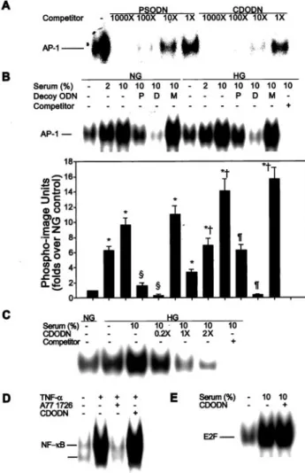
In vitro competitive binding assays were performed to exam- ine sequence-specific interactions between decoy ODN and the AP-1 protein. An increase in concentration of unlabeled AP-1 decoy ODN was accompanied by a corresponding decrease in the intensity of the retarded band. This indicates the presence of an ODN-AP-1 protein complex (Figure 2A).
A 1000-fold molar excess of unlabeled PSODN almost completely out-competed AP-1 binding to the labeled probe.
However, only a 100-fold molar excess of unlabeled CDODN was required to completely inhibit AP-1 binding to the labeled probe.
As expected, treatment with high glucose resulted in a significant increase in binding of AP-1 to DNA, compared with binding at low glucose concentrations ( P ⬍0.001; Figure 2B).
Figure 2. Effects of CDODN on the DNA binding activity of AP-1.
A, Varying amounts of AP-1 complexes were formed between the labeled probe and AP-1 protein in the presence of different con- centrations of unlabeled oligonucleotides. B, Typical gel shift assay is shown for VSMCs transfected with AP-1 decoy ODN. The experiment was repeated 5 times. C, Dose-dependent effects of CDODN on the DNA-binding activity of AP-1. After treatment of VSMCs with increasing concentrations of CDODN (20, 100, or 200 nmol/L), DNA binding activity was determined. D, Effects of CDODN on TNF-␣–induced NF-B DNA binding activity. Cells were incubated in the presence or absence of TNF-␣ (10 ng/mL) for 2 hours. Additionally, cells were treated with 10mol/L A77 1726 (TNF-␣–induced NF-B specific inhibitor) for 2 hours before the addition of TNF-␣. E, Effect of CDODN on serum-induced E2F DNA binding activity. NG indicates normal glucose (5.5 mmol/L
D-glucose); HG, high glucose (25 mmol/LD-glucose); Decoy ODN, VSMCs transfected with 100 nmol/L AP-1 decoy ODN; P, PSODN;
D, CDODN; and M, mismatched AP-1 decoy ODN. EMSA results are expressed as the mean⫾SEM of 5 independent experiments.
Statistical significance was determined as *P⬍0.001 compared with NG, †P⬍0.01 compared with HG, §P⬍0.001 compared with NG⫹10% serum, or ¶P⬍0.01 compared with HG⫹10% serum.
Similarly, the addition of serum also led to a dose-dependent increase in the DNA binding activity of AP-1 (P ⬍0.001; Figure 2B). Transfection of both PSODN and CDODN significantly attenuated AP-1 binding to DNA induced by high glucose or serum (P ⬍0.01), with CDODN exhibiting more potent inhibi- tory activity (P ⬍0.001; Figure 2B).
Next, the effects of CDODN on AP-1, NF- B, and E2F binding activity were evaluated to confirm that CDODN specifically inhibits AP-1 binding to DNA. CDODN trans- fection significantly reduced high glucose- and serum- induced AP-1 binding activity in a dose-dependent manner (Figure 2C). However, TNF- ␣ (10 ng/mL)–induced NF-B (Figure 2D) and serum-induced E2F binding activity (Figure 2E) remained unaffected by CDODN transfection.
Effect of AP-1 Decoy ODN on VSMC Gene Expression
Reporter gene constructs containing the AP-1 binding site in their promoter regions were used to investigate the effects of the AP-1 decoy ODN on promoter activity. To determine the specific role of the AP-1 binding site on cyclin A promoter activity induced by high glucose levels, we transfected a series of luciferase reporter gene plasmids containing various lengths of the human cyclin A 5 ⬘ flanking sequence into VSMCs treated with high glucose. Of these constructs, 2 plasmids (pCA ⫺266/⫹205 mt and pCA⫺133/⫺205 mt) containing mutated sequences in the activating transcription factor site that interacts with AP-1 transcription factor in cyclin A gene expression,
25exhibited significantly decreased luciferase activity (P ⬍0.001, compared with pCA⫺266/
⫹205 and pCA⫺133/⫹205; Figure 3A). Therefore, our data suggest that the AP-1 decoy ODN downregulates the pro- moter activity of cyclin A in VSMCs induced by high glucose.
Next, we examined the effects of AP-1 decoy ODN on the glucose- and serum-induced activities of luciferase reporter plasmids (ie, pAP1(PMA)-TA-Luc and the cyclin A promoter luciferase construct (pCA ⫺266/⫹205) containing the AP-1 binding site). As expected, cotransfection of luciferase report- ers with both PSODN and CDODN markedly attenuated high glucose- and serum-stimulated luciferase gene expression (P ⬍0.01; Figures 3B and 3C), with CDODN being more effective than PSODN. The effect of AP-1 decoy ODNs on the endogenous expression of cell cycle regulatory genes in vitro was also investigated. For these experiments, expression levels of the PCNA and cyclin A genes required for cell cycle progression from G1 to S phase were determined by Northern blotting. As shown in Figure 3D, high glucose and serum stimulated PCNA and cyclin A mRNA expression in human and rat VSMCs. Transfection of AP-1 decoy ODN (but not mismatched ODN) led to a reduction in glucose- and serum- induced expression of these genes. Moreover, the inhibitory effect of CDODN on gene expression was more significant than that of PSODN.
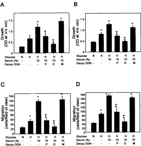
Effect of AP-1 Decoy ODN on VSMC Growth and Migration In Vitro
Treatment with high glucose and serum stimulated the growth of cultured primary human and rat VSMCs, compared with
control levels (Figures 4A and 4B). Transfection of AP-1 decoy ODN resulted in significant inhibition of the cell growth stimulated by high glucose or serum (P ⬍0.05).
Notably, CDODN almost completely inhibited VSMC growth (P ⬍0.05, compared with PSODN). Similarly, the increase in VSMC migration in the presence of high levels of both glucose and serum (P ⬍0.05; Figures 4C and 4D) was considerably suppressed by AP-1 decoy ODN (P ⬍0.01). The inhibitory effect of CDODN on VSMC migration was greater than that of PSODN (P ⬍0.01).
Effects of CDODN on Neointimal Formation in Rat Balloon-Injured Carotid Artery
To determine transfection efficiencies, PSODN and CDODN were labeled using the Label IT fluorescein nucleic acid
Figure 3. Effects of CDODN on VSMC gene expression. A, VSMCs were cotransfected with decoy ODN and serial deletion or mutant constructs of the cyclin A promoter under high glu- cose conditions. Statistical significance was determined as*P⬍0.001 compared with pCA⫺266/⫹205 or †P⬍0.001 com- pared with pCA⫺133/⫹205. Cells were cotransfected with decoy ODN and either AP1(PMA)-TA-Luc (B) or pCA⫺266/⫹205 (C). The activity of decoy ODNs is reflected in their ability to downregulate luciferase activity. Values are presented as mean⫾SEM of 6 independent experiments. Statistical signifi- cance was determined as *P⬍0.01 compared with NG, †P⬍0.01 compared with HG⫹10% serum, or ‡P⬍0.001 compared with HG⫹10% serum. D, Representative Northern blot analysis on gene expression of cyclin A (top) and PCNA (bottom) in rat (left) or human VSMCs (right). N indicates normal glucose (5.5 mmol/L
D-glucose); H, high glucose (25 mmol/LD-glucose); Decoy ODN, VSMCs transfected with 100 nmol/L AP-1 decoy ODN; P, PSODN;
D, CDODN; and M, mismatched AP-1 decoy ODN.
labeling kit (Mirus). We tested the efficiency of ODN transfection into the rat carotid artery with the HVJ-liposome method using fluorescein-labeled AP-1 decoy ODN (see online Figure found in the online data supplement available at http://www.circresaha.org). Transfection of labeled ODN re- sulted in a strong fluorescent signal (Figures 5A and 5B), which was readily detected in all layers of the artery. No significant differences were observed between the transfec- tion efficiencies of fluorescein-labeled PSODN and CDODN with the HVJ-liposome method (data not shown).
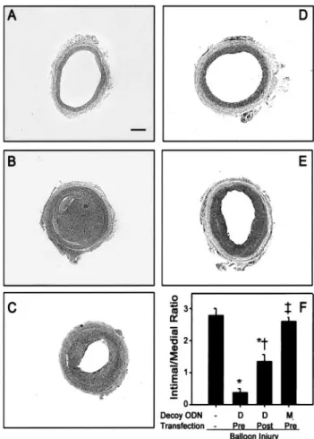
Next, we examined the effects of AP-1 decoy ODN on neointimal formation in the rat carotid balloon-injury model.
As shown in Figure 5, neointimal formation resembling that in injured vessels was detected in vessels transfected with mismatched ODN, 2 weeks after transfection. In contrast, a single administration of PSODN or CDODN resulted in significantly reduced neointimal formation (P ⬍0.01). In agreement with the above in vitro data, the inhibitory effect of CDODN on neointimal formation was more potent than that of PSODN (P ⬍0.05, compared with PSODN).
The process of neointimal formation is largely dependent on the expression of immediate early genes.
6 – 8Therefore, we compared the effects of pretreatment and posttreatment with AP-1 decoy ODN on the inhibition of neointimal formation in injured carotid arteries. As shown in Figure 6, pretreatment of the rat carotid artery with AP-1 decoy ODN before balloon injury was more effective than posttreatment (P ⬍0.0001 compared with balloon injured vessels; P ⬍0.01 compared with posttreatment with CDODN) in inhibiting neointimal formation.
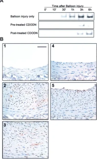
Effects of AP-1 Decoy ODN on the DNA-Binding Activity of AP-1 and Gene Expression In Vivo To determine whether AP-1 decoy ODN effectively blocks the DNA binding activity of AP-1 in vivo, we performed gel mobility shift assays, using cells from injured arteries. As shown in Figure 7A, the DNA binding activity of AP-1 increased 30 minutes after injury and peaked at 3 hours after injury. This increase in binding activity was inhibited by treatment with CDODN. Furthermore, pretreatment with decoy ODN was more effective than posttreatment in the attenuation of AP-1 activity.
PCNA staining (a widely used marker of cell proliferation in both normal and diseased states) was used to confirm the inhibitory effects of AP-1 decoy ODN on cell proliferation.
As shown in Figure 7B, no PCNA staining was detected in uninjured arteries. However, 2 weeks after injury, a marked increase in PCNA staining was observed in both neointimal and regenerated endothelial cells. Significantly, the level of cells stained with PCNA in vessels treated with AP-1 decoy ODN was much lower than that in untransfected vessels.
Discussion
Accumulating evidence indicates that activation of MAP kinase and AP-1 cascades are key events in the proliferation and growth of VSMCs in response to injury.
6 – 8However, direct evaluation of the role of AP-1 in the pathogenesis of neointimal formation is hampered by the lack of specific and potent pharmacological AP-1 inhibitors. To test our hypoth- esis that AP-1 plays a critical role in the pathogenesis of VSMC proliferation and neointimal formation, we used a
Figure 4. Inhibitory effects of CDODN on human (A) and rat (B) VSMC proliferation were examined. Results are presented as
mean⫾SEM of 6 independent measurements.
Effects of CDODN on glucose- and serum- potentiated migration of cultured human (C) and rat (D) VSMCs stimulated with 10 ng/mL VEGF for 24 hours. Average numbers of cells from 4 randomly chosen high-power (200⫻) fields on the lower surface of the filter are shown. Results are presented as mean⫾SEM of 4 separate experiments. Statistical signifi- cance was determined as *P⬍0.01 compared with NG, †P⬍0.05 compared with HG⫹10%
serum, or ‡P⬍0.01 compared with HG⫹10%
serum.
novel AP-1 ODN method. Our data demonstrate that a novel AP-1 decoy ODN prevents VSMC proliferation in vitro and neointimal formation in vivo after balloon injury.
The transcription factor AP-1 binds to AP-1 consensus sequences present in numerous genes associated with cell proliferative response and extracellular matrix production that are also important in neointimal formation,
26 –28such as
c-myc, fibroblast growth factor, transforming growthfactor- , endothelin-1, and plasminogen activator inhibitor-1.
It is reported that treatment with AP-1 decoy ODN abolishes
the expression of several genes, including plasminogen acti- vator inhibitor-1, transforming growth factor- , and endothelin-1.
12,23,29However, further studies are required to elucidate the mechanisms by which AP-1 controls gene expression during neointimal formation.
In this investigation, we used high glucose and serum as stimulants to enhance the proliferation and migration of VSMCs. These stimulants additionally induce many early genes, growth factors, and mitogens via MAPK and AP-1 cascades.
30 –34In VSMCs, AP-1 ODNs effectively abolish the expression of cyclin A and PCNA genes induced by the elevated glucose and serum levels, which are required for cell cycle progression from G1 to S phase. Additionally, cotrans- fection of AP-1 decoy ODN (but not mismatched ODN) with the luciferase reporter constructs completely eliminates lucif- erase expression of cyclin A induced by high glucose and
Figure 5. Effects of AP-1 decoy ODN on neointimal formationoccurring after balloon injury to rat carotid artery. Fluorescent microscopy image of the left common carotid artery treated only with fluorescein-labeled CDODN (A) or fluorescein-labeled CDODN with HVJ-liposomes (B). Cross-section of the left com- mon carotid artery of control rat (C) is shown 14 days after bal- loon injury (D), 14 days after balloon injury with MODN using the HVJ-liposome method (E), with the HVJ-liposome method and PSODN (F), or with the HVJ-liposome method and CDODN (G).
Average ratio of intimal/medial area of the left carotid artery in groups transfected with HVJ-liposomes containing AP-1 decoy ODN is shown (H). Bars signify the neointima/media ratio of common carotid arteries after balloon injury from each group of animals studied (n⫽10). Values are presented as mean⫾SEM, with statistical significance determined as *P⬍0.01 compared with balloon-injured arteries or †P⬍0.05 compared with PSODN treated arteries. Original magnification:⫻100 (A and B) and ⫻25 (C-G). Scale bar⫽200m.
Figure 6. Effect of CDODN treatment times on the inhibition of neointimal formation in injured carotid arteries. Cross-section of the left common carotid artery of control rat (A), 14 days after balloon injury (B), after pretreatment with MODN using HVJ- liposome method (C), after pretreatment with CDODN (D), and after posttreatment with CDODN (E). Average ratio of intimal/
medial area of the left carotid artery in groups transfected with HVJ-liposomes containing AP-1 decoy ODN (F) is shown. Bars represent the neointima/media ratio of common carotid arteries after balloon injury from each group of animals studied (n⫽10).
Values are presented as mean⫾SEM, with statistical signifi- cance determined as *P⬍0.005 compared with balloon injured arteries, †P⬍0.01 compared with pretreated arteries with CDODN, or ‡P⬍0.01 compared with posttreated arteries with CDODN. Original magnification:⫻25. Scale bar⫽200m.
serum. These observations, together with in vitro data show- ing that transfection of AP-1 decoy ODN inhibits VSMC proliferation and migration, demonstrate that AP-1 decoy ODN inhibits neointimal formation via the suppression of VSMC growth and migration.
Interestingly, we observed that pretreatment with decoy ODN was more effective than posttreatment. This finding is explained by the time-course of AP-1 activation in injured arteries. Previous studies report that levels of ERK and JNK activity in the vessel wall rapidly increase and reach a plateau within 5 minutes after balloon angioplasty, which is main- tained for 1 hour thereafter.
6,8In addition, the expression of immediate early genes (c-jun and c-fos) peaked at 30 minutes after balloon injury. As shown in our results, AP-1 activity is
detectable 30 minutes after balloon injury and reaches max- imum levels at 3 hours. These findings collectively indicate that signal transduction in response to balloon injury is rapid, and that time is an important factor in blocking the flow of signals induced as a result of the injury.
Importantly, CDODN is structurally more stable and effec- tive than chemically modified decoy ODN. CDODN contains two AP-1 binding sites in a single closed-ended decoy molecule, thus allowing the targeting of more than 1 promoter site. In concurrence with recent reports,
19,20our CDODN was more stable than PSODN in the presence of serum, exonu- clease III, and S1 nuclease. In addition, sequence specificity of CDODN, assessed by an in vitro competition assay, was nearly 10 times greater than that of PSODN. The inhibitory effect of CDODN on glucose- and serum-induced AP-1 DNA binding activity was greater than that of PSODN. These results suggest that CDODN has a higher affinity for AP-1 protein than PSODN. Consistent with these in vitro data, CDODN is more effective in preventing neointimal formation after vascular injury.
The HVJ-liposome technique was used to transfect AP-1 decoy ODN into rat carotid arteries. In this delivery system, DNA is packaged into a liposome comprising phospholipids and cholesterol. The liposome is fused to UV-inactivated HVJ to form an HVJ-liposome, which subsequently attaches to the cell surface and delivers the DNA molecule directly to the cytoplasm. This method is very effective in gene transfer, even into the medial VSMC of the intact artery not subjected to endothelial denudation.
4We recently showed that trans- fection of a fluorescein-labeled AP-1 decoy ODN using the HVJ-liposome method into cultured human VSMCs resulted in stronger (approximately 100 times) fluorescence than the conventional LipopectAMINE method.
23Consistent with this report, transfection of fluorescein-labeled ODN by the HVJ- liposome method in vivo resulted in strong fluorescence, which was readily detected in all layers of the artery.
In conclusion, our novel dumbbell decoy ODNs are mark- edly more stable than the previously characterized chemically modified ODNs. Moreover, inhibition of AP-1 activity by decoy ODN effectively decreases in vitro proliferation and migration of cells, as well as neointimal formation in vivo.
Our data clearly demonstrate that the transcription factor, AP-1, is crucial for the mediation of VSMC proliferation and neointimal formation after vascular injury. Furthermore, we present a novel potential therapeutic strategy for the treatment of restenosis, which involves the utilization of CDODN with minimal side-effects and the highly effective HVJ-liposome gene delivery technique.
Acknowledgments
This study was supported by a grant from the Korea Health 21 R&D project, Ministry of Health and Welfare, Republic of Korea (HMP-99-M-08-0004).
References
1. Liu MW, Roubin GS, King SB III. Restenosis after coronary angioplasty.
Circulation. 1989;79:1374 –1387.
2. Pauletto P, Sartore S, Pessina AC. Smooth-muscle-cell proliferation and differentiation in neointima formation and vascular restenosis. Clin Sci.
1994;87:467– 479.
Figure 7. A, Analysis of AP-1 binding activity in arterial extracts.
Gel mobility shift assays were performed using nuclear extracts from cells of carotid arteries harvested at the indicated times after injury (n⫽10). Before (pretreated with CDODN) or after (posttreated with CDODN) balloon injury, 20L HVJ-liposome complex containing CDODN was incubated within the lumen for 10 minutes at room temperature. B, PCNA expression in rat carotid artery after balloon injury. PCNA staining of control ves- sels (1), balloon-injured vessels (2), arteries pretreated with MODN (3), arteries pretreated with CDODN (4), and arteries posttreated with CDODN (5) are shown. PCNA-positive cells appear brownish-black. All figures are depicted at 200⫻ magni- fication. Scale bar⫽50m.
3. Simons M, Edelman ER, DeKeyser JL, Langer R, Rosenberg R.
Antisense c-myb oligonucleotides inhibit intimal arterial smooth muscle cell accumulation in vivo. Nature. 1992;359:67–73.
4. Morishita R, Gibbons GH, Ellison KE, Nakajima M, von der Leyen H, Zhang L, Kaneda Y, Ogihara T, Dzau VJ. Intimal hyperplasia after vascular injury is inhibiter by antisense cdk 2 kinase oligonucleotides.
J Clin Invest. 1994;93:1458 –1464.
5. Morishita R, Sugimoto T, Aoki M, Kida I, Tomita N, Moriguchi A, Maeda K, Sawa Y, Kaneda Y, Higaki J, Ogihara T. In vivo transfection of cis element “decoy” against NF-B binding site prevented myocardial infarction as gene therapy. Nat Med. 1997;3:894 – 899.
6. Hu Y, Cheng L, Hochleitner BW, Xu Q. Activation of mitogen-activated protein kinases (ERK/JNK) and AP-1 transcription factor in rat carotid arteries after balloon injury. Arterioscler Thromb Vasc Biol. 1997;17:
2808 –2816.
7. Pyles JM, March KL, Franklin M, Mehdi K, Wilensky RL, Adam LP.
Activation of MAP kinase in vivo follows balloon overstretch injury of porcine coronary and carotid arteries. Circ Res. 1997;81:904 –910.
8. Izumi Y, Kim S, Namba M, Yasumoto H, Miyazaki H, Hoshiga M, Kaneda Y, Morishita R, Zhan Y, Iwao H. Gene transfer of dominate- negative mutants of extracellular signal-regulated kinase and c-Jun NH2- terminal kinase prevents neointimal formation in balloon-injured rat artery. Circ Res. 2001;88:1120 –1126.
9. Karin M. The regulation of AP-1 activity by mitogen-activated protein kinases. J Biol Chem. 1995;270:16483–16486.
10. Whitmarch AJ, Davis RJ. Transcription factor AP-1 regulation by mitogen-activated protein kinase signal transduction pathway. J Mol Med.
1996;74:589 – 607.
11. Bielinska A, Shivdasani RA, Zhang L, Nabel GJ. Regulation of gene expression with double-stranded phosphorothioate oligonucleotides.
Science. 1990;250:997–1000.
12. Morishita R, Gibbons GH, Horiuchi M, Kaneda Y, Ogihara T, Dzau VJ.
Role of AP-1 complex in angiotensin II-mediated transforming growth factor- expression and growth of smooth muscle cells: using decoy approach against AP-1 binding site. Biochem Biophys Res Commun.
1998;243:361–367.
13. Sawa Y, Morishita R, Suzuki K. A novel strategy for myocardial pro- tection using in vivo transfection of cis element ‘decoy’ against NFB binding site. Circulation. 1997;96:II-280 –II-285.
14. Moon IJ, Choi K, Choi YK, Kim JE, Lee Y, Schreiber AD, Park JG.
Potent growth inhibition of leukemic cells by novel ribbon-type antisense oligonucleotides to c-myb1. J Biol Chem. 2000;275:4647– 4653.
15. Hosoya T, Takeuchi H, Kanesaka Y, Yamakawa H, Miyano-Kurosaki N, Takai K, Yamamoto N, Takaku H. Sequence-specific inhibition of a transcription factor by circular dumbbell DNA oligonucleotides. FEBS Lett. 1999;461:136 –140.
16. Gao WY, Han FS, Storm C, Egan W, Cheng YC. Phosphorothioate oligodeoxynucleotides are inhibitors of human DNA polymerases and Rnase H: implications for antisense technology. Mol Pharmacol. 1992;
41:223–229.
17. Brown DA, Kang SH, Gryaznov SM, DeDionisio L, Heidenreich O, Sullivan S, Xu X, Nerenberg MI. Effect of phosphorothioate modification of oligodeoxynucleotides on specific protein binding. J Biol Chem. 1994;
269:26801–26805.
18. Burgess TL, Fisher EF, Ross SL, Bready JV, Qian YX, Bayewitch LA, Cohen AM, Herrera CJ, Hu SS, Kramer TB, Lott FD, Martin FH, Pierce GF, Simonet L, Farrell CL. The antiproliferative activity of c-myb and c-myc antisense oligonucleotides in smooth muscle cells is caused by a
nonantisense mechanism. Proc Natl Acad Sci U S A. 1995;92:
4051– 4055.
19. Chu BCF, Orgal L. The stability of different forms of double-stranded decoy DNA in serum and nuclear extracts. Nucleic Acids Res. 1992;20:
5857–5858.
20. Abe T, Takai K, Nakada S, Yokota T, Takaku H. Specific inhibition of influenza virus RNA polymerase and nucleoprotein gene expression by circular dumbbell RNA/DNA chimeric oligonucleotides containing antisense phosphodiester oligonucleotides. FEBS Lett. 1998;425:91–96.
21. Dubey RK, Gillespie DG, Mi Z, Jackson EK. Adenosine inhibits growth of human aortic smooth muscle cells via A2b receptors. Hypertension.
1998;31:516 –521.
22. Wang Z, Castresana MR, Newman WH. Reactive oxygen and NF-B in VEGF-induced migration of human vascular smooth muscle cells.
Biochem Biophys Res Commun. 2001;285:669 – 674.
23. Ahn JD, Morishita R, Kaneda Y, Lee KU, Park JY, Jeon YJ, Song HS, Lee IK. Transcription factor decoy for activator protein-1 (AP-1) inhibits high glucose- and angiotensin II-induced type 1 plasminogen activator inhibitor (PAI-1) gene expression in cultured human vascular smooth muscle cells. Diabetologia. 2001;4 4:713–720.
24. Yoshizumi M, Wang H, Hsieh CM, Sibinga NE, Perrella MA, Lee ME.
Down-regulation of the cyclin A promoter by transforming growth factor-1 is associated with a reduction in phosphorylated activating transcription factor-1 and cyclin AMP-responsive element-binding protein. J Biol Chem. 1997;272:22259 –22264.
25. Sylvester AM, Chen D, Krasinski K, Andres V. Role of c-fos and E2F in the induction of cyclin A transcription and vascular smooth muscle cell proliferation. J Clin Invest. 1998;101:940 –948.
26. Shi Y, Fard A, Galeo A, Hutchinson HG, Vermani P, Dodge GR, Hall DJ, Shaheen F, Zalewski A. Transcatheter delivery of c-myc antisense oli- gomers reduces neointimal formation in a porcine model of coronary artery balloon injury. Circulation. 1994;90:944 –951.
27. Daley SJ, Gotlieb AI. Fibroblast growth factor receptor-1 expression is associated with neointimal formation in vitro. Am J Pathol. 1996;148:
1193–1202.
28. DeYoung MB, Tom C, Dichek DA. Plasminogen activator inhibitor type 1 increases neointima formation in balloon-injured rat carotid arteries.
Circulation. 2001;104:1972–1977.
29. Lauth M, Wagner AH, Cattaruzza M, Orzechowski HD, Paul M, Hecker M. Transcriptional control of deformation-induced preproendothelin-1 gene expression in endothelial cells. J Mol Med. 2000;78:441– 450.
30. Miano JM, Vlasic N, Tota RR, Stemerman MB. Smooth muscle cell immediate-early gene and growth factor activation follows vascular injury. Arterioscler Thromb. 1993;13:211–219.
31. Inaba T, Ishibashi S, Gotoda T, Kawamura M, Morino N, Nojima Y, Kawakami M, Yazaki Y, Yamada N. Enhanced expression of platelet- derived growth factor- receptor by high glucose involvement of plate- let-derived growth factor in diabetic angiopathy. Diabetes. 1996;45:
507–512.
32. Di Paolo S, Gesualdo L, Ranieri E, Grandaliano G, Schena FP. High glucose concentration induces the overexpression of transforming growth factor- through the activation of a platelet-derived growth factor loop in human mesangial cell. Am J Pathol. 1996;149:2095–2106.
33. Schwartz SM, de Blois DM, O’Brien ERM. The intima: soil for athero- sclerosis and restenosis. Circ Res. 1995;77:445– 465.
34. Lindner V, Lappi DA, Baird A, Majack RA, Reidy MA. Role of basic fibroblast growth factor in vascular lesion formation. Circ Res. 1991;68:
106 –113.