Korean Circulation Journal
Print ISSN 1738-5520 • On-line ISSN 1738-5555
Clinical Outcomes in Patients with Intermediate Coronary Stenoses:
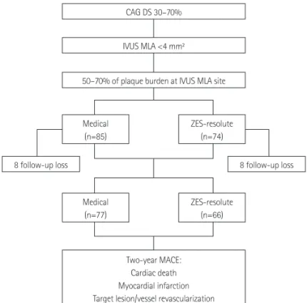
MINIATURE Investigators (Korea MultIceNter TrIal on Long-Term Clinical Outcome According to the Plaque Burden and Treatment Strategy in Lesions with MinimUm Lumen ARea lEss Than 4 mm 2 Using Intravascular Ultrasound)
Young Joon Hong, MD 1 , Yun Ha Choi, MS 1 , Soo Young Park, BS 1 , Chang Wook Nam, MD 2 , Jang Hyun Cho, MD 3 , Won Yu Kang, MD 4 , Sang Rok Lee, MD 5 , Sung Yun Lee, MD 6 , Sang Wook Kim, MD 7 , Sang Yeob Lim, MD 8 , Kyung Ho Yun, MD 9 , Jung Sun Kim, MD 10 , Jin Won Kim, MD 11 , Woong Chol Kang, MD 12 , Ki Seok Kim, MD 13 ,
Jin Ho Choi, MD 14 , Joong Wha Chung, MD 15 , Soo Joong Kim, MD 16 , Youngkeun Ahn, MD 1 , and Myung Ho Jeong, MD 1
1
Division of Cardiology, Chonnam National University School of Medicine, Gwangju,
2
Division of Cardiology, Keimyung University College of Medicine, Dongsan Medical Center, Daegu,
3
Division of Cardiology, Saint Carollo Hospital, Suncheon,
4Division of Cardiology, Gwangju Veterans Hospital, Gwangju,
5
Division of Cardiology, Chonbuk National University College of Medicine, Jeonju,
6Division of Cardiology, Inje University College of Medicine, Ilsan Paik Hospital, Goyang,
7
Division of Cardiology, Chung-Ang University College of Medicine, Seoul,
8Division of Cardiology, Korea University College of Medicine, Ansan Hospital, Ansan,
9
Division of Cardiology, Wonkwang University College of Medicine, Iksan,
10Division of Cardiology, Yonsei University College of Medicine, Severance Hospital, Seoul,
11
Division of Cardiology, Korea University Guro Hospital, Seoul,
12Division of Cardiology, Gachon University Gil Medical Center, Incheon,
13
Division of Cardiology, Jeju National University College of Medicine, Jeju,
14Division of Cardiology, Samsung Medical Center, Seoul,
15