DOI 10.17480/psk.2017.61.2.75
호흡기 상피 배상세포에서의 점액 과다분비와 관련된 뮤신 유전자 발현 및 생성에 대한 리나룰, 올레아놀산, 베타 카리오필렌의 약리작용
이 현 재*
삼육대학교 교양대학 및 보건관리학과
(Received December 13, 2016; Revised March 16, 2017; Accepted April 3, 2017)
Pharmacological action of Linalool, Oleanolic acid and β-caryophyllene on Gene Expression and Production of Mucin from Goblet cells in Respiratory Epithelium
Hyun Jae Lee*
Department of Health Management and Smith Liberal Arts College, Sahmyook University, 815 Hwarang-ro, Nowon-gu, Seoul 01795, Korea
Abstract — We examined whether linalool and other natural products affect airway MUC5AC mucin gene expression and production induced by phorbol 12-myristate 13-acetate (PMA) from NCI-H292 cells. Cells were pretreated with each agent for 30 min and then stimulated with PMA for 24h. Linalool, β-caryophyllene and oleanolic acid inhibited the gene expres- sion and production of MUC5AC mucin. This result suggests that these three compounds can inhibit the production and gene expression of mucin, through directly acting on airway epithelial cells.
Keywords Airway, mucin, linalool
호흡기는 공기의 통로인 기도와 산소-이산화탄소의 교환이 일 어나는 폐로 구성되어 있는데, 호흡기에 존재하는 점액(mucus) 은 기도를 통해 유입되는 환경성 유해입자, 유해 화학물질, 다양 한 병원성 미생물 등에 대한 인체의 방어 기능 수행 과정에서 점 액섬모성 운반작용(mucociliary transport)을 통해 중요한 역할을 한다. 이러한 기도 점액의 생체방어 작용은 점액의 생화학적 주 구성요소인 뮤신의 점탄성(viscoelasticity)에 기인한다. 그러나, 뮤 신의 양과 질의 이상은 기도 생리의 이상뿐 아니라 인체의 방어 작용에 영향을 주어 심각한 병리 현상을 유발할 수 있다. 즉, 천 식, 만성 기관지염, 폐기종, 기관지 확장증, 낭포성 섬유증 등의 기도 질환에서 관찰되는 점액(객담)의 과다 분비는 이러한 질환 의 예후를 악화시키는 주된 요인으로 알려져 있다.1-4)
과다 생성 및 분비된 점액을 기도로부터 제거하는 데는 두 가 지 방법이 있을 수 있다. 첫째, 물리적 방법에 의한 점액의 제거, 즉 점액의 점도를 낮춘 뒤 흡인해 내는 방법이고, 둘째는, 점액 생성 자체를 억제할 수 있는 약물을 투여하는 방법이다. 물리적 방법은 기도 내부의 자극을 유발하고, 반사기전에 의해 점액 분 비를 오히려 자극하게 된다. 마취 하에서 그런 방법이 시도된다 고 해도, 점액의 제거는 feedback mechanism을 통해 점액의 생 성과 분비를 더욱더 자극하게 된다. 따라서, 점액에 점성을 부여 하는 주 구성요소인 뮤신의 생성 자체를 조절하거나 혹은 분비 를 조절하기 위한 약물학적 접근은 기도질환의 치료에 있어 중 요한 방향이 될 수 있다.5)이러한 전략에 근거하여, 항염증, 항 산화, 항암 효능을 발현하는 것으로 알려진 다수의 천연물 중에 서 호흡기 염증성 질환에 동반되는 기도 뮤신의 생성 및 과다분 비를 조절할 가능성이 있는 후보물질의 탐색은 유망한 접근 방 법이라 할 수 있을 것이다.
전통의학 문헌과 다수의 연구보고에 의하면, 곽향(Agastachis Herba), 당귀(Angelicae Gigantis Radix), 천궁(Cnidii Rhizoma), 목통(Akebiae Caulis) 등은 경험적으로 호흡기 질환을 포함한 다양 한 염증성 질환의 조절을 위해 사용되어 왔으며,6)곽향에 함유된
#Corresponding Author Hyun Jae Lee
Department of Health Management and Smith Liberal Arts Col- lege, Sahmyook University, 815 Hwarang-ro, Nowon-gu, Seoul 01795, Korea
Tel.: 02-3399-1909 Fax.: 02-3399-1821 E-mail: [email protected]
Short Report종설
아카세틴(acacetin), 유지놀(eugenol), 메틸 유지놀(methyleugenol), 리나룰(linalool), 베타 카리오필렌(β-caryophyllene) 등과 당귀와 천 궁 등에 함유된 페룰산(ferulic acid), 목통에 함유된 올레아놀산 (oleanolic acid) 등의 천연물은 항염증 작용을 포함한 다양한 생 리활성을 나타내는 것으로 보고되어 있다.7-10)그러나, 현재까지 인간 호흡기 상피세포에서 뮤신 유전자의 발현 및 생성에 대해 곽향에서 유래한 아카세틴, 유지놀, 메틸 유지놀, 리나룰, 베타 카리오필렌 등과 당귀와 천궁 등에 함유된 페룰산, 목통에 함유 된 올레아놀산 등의 천연물이 어떠한 영향을 미치는 지 여부는 검증된 바가 없었다. 따라서, 본 연구에서는 이러한 천연물들이 인간 기도 상피세포에서 포볼 에스터인 PMA에 의해 증가된 뮤 신의 유전자 발현 및 생성에 대해 어떠한 약리작용을 나타내는
지를 검증함으로써, 효과적인 기도점액 과다생성(분비) 조절 신 약의 개발을 위한 기초과학적 정보를 제공하고자 하였다.
실험 방법
세포주 및 시약(Cell line and reagents)
NCI-H292 세포는 American Type Culture Collection 사 (Manassas, VA, U.S.A.)에서 구입하였다. 잘 건조된 후 세절된 Agastache rugosa(3.0 kg)의 지상부에 함유된 성분을, 70% 에탄올 로 3시간 동안 추출하였다. 이 에탄올 추출물을 증류수에 현탁시 키고 이어서 n-hexane, dichloromethane, ethylacetate, n-butanol (각 분획물, 28.2 g, 16.8 g, 14.8 g, 21.4 g) 등의 용매를 이용하여
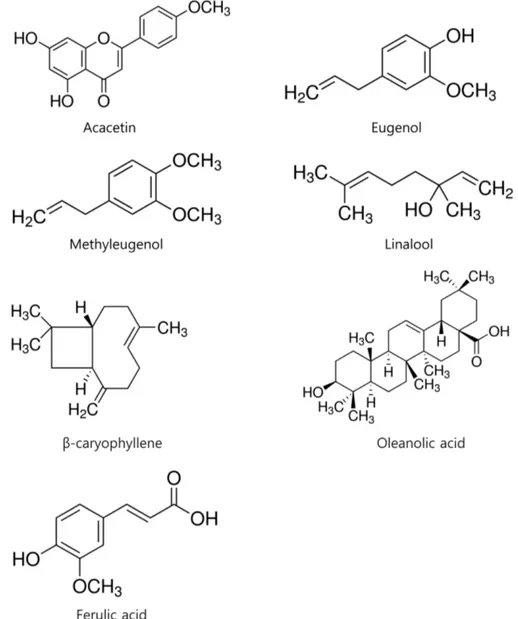
Fig. 1 − Chemical structure of acacetin, eugenol, methyleugenol, linalool, β-caryophyllen, oleanolic acid and ferulic acid.
분획하였다. 이후, 칼럼 크로마토그래피 및 고속액체 크로마토그 래피 등을 이용, 아카세틴(acacetin, 순도 95%), 유지놀(eugenol, 순도 95%), 메틸 유지놀(methyleugenol, 순도 95%), 리나룰 (linalool, 순도 95%), 베타 카리오필렌(β-caryophyllene, 순도 95%) 등의 물질을 분리, 정제하였는데, 제반 과정은 동국대학교 바이오학부 천연물화학 교실 이동웅 교수 연구실에서 수행되었 다. 페룰산(ferulic acid, 순도 95%)과 올레아놀산(oleanolic acid, 순도 98%) 은 Sigma(St. Louis, MO, U.S.A.) 사에서 구입하였다 (Fig. 1). Protease inhibitor cocktail은 Roche사(Indianapolis, IN, U.S.A.)에서, mouse anti-MUC5AC clone 45M1 및 HRP-Goat Anti-Mouse IgG Conjugate은 NeoMarkers사(Freemont, CA, U.S.A.)에서, trypsin-EDTA, phorbol 12-myristate 13-acetate (PMA), tumor necrosis factor-α(TNF-α), Tween 20, bovine serum albumin (BSA), HEPES, dimethyl sulfoxide(DMSO), 3,3',5,5'- tetramethyl- benzidine peroxide solution(TMB), NP-40, EDTA, EGTA, HEPES, ethidium bromide, diethylpyrocarbonate (DEPC), 등은 Sigma(St. Louis, MO, U.S.A.) 사에서, Easy-Blue RNA extraction kit 는 INTRON biotechnology(Kyunggi-do, Korea) 사에서, Accuprep RT premix kit는 Bioneer(Daejeon, Korea) 사에서, 그리고 PCR Master Mix 는 ABgene(Rochester, NY, U.S.A.) 사에서, penicillin-G, streptomycin, fetal bovine serum(FBS), RPMI 1640은 GIBCO-BRL사(Grand Island, New York, U.S.A.)에서, 기타 제반 시약들은 일급시약 등급 이상의 것 들을 구입하여 사용하였다.
인간 기도상피 세포(NCI-H292) 배양 및 각 약물의 처리 (Incubation of human airway epithelial cells (NCI-H292) and treatment of each drug)
세포는 습도가 충분히 유지되며 95% 공기, 5% CO2를 함유하는 37oC 배양기 내에서 HEPES(25 mM), penicillin G(100 U/ml), streptomycin(100μg/ml), FBS(10%, V/V) 등이 첨가된 RPMI 1640 배양액에서 배양하였는데, 1주에 2회 빈도로 subculture하 였고 배양액은 2일마다 1회씩 교체해 주었다. 뮤신 생성, 분비 및 그 유전자 발현에 대한 약물의 작용을 검증하기 위하여, 뮤신 생성 및 분비량 검증을 위해서는 24 well culture plate 를 기준 으로, well 당 2.0×10⁴ cells/well 의 밀도로, 뮤신 유전자 발현 정도의 검증을 위해서는 6 well culture plate 를 기준으로, well 당 5.0×10⁴ cells/well 의 밀도로 각각 세포를 도포하고 배양하였 다. 세포가 각 well의 70−80% 정도를 차지할 정도로 자라면, FBS 의 농도를 0.2%로 감소시킨 배양액을 주고 24 시간 동안 배양하고, serum을 첨가하지 않은 배양액(serum-free medium) 으로 세포를 세척한 후 약물 1−100 μM을 함유하는 배양액 200μl(24 well plate 기준)를 well마다 가하였다. 30분이 경과한 후 PMA (10 ng/ml) 혹은 TNF-α 0.2 nM을 세포에 투여한 후
37oC에서 추가로 24시간 동안 배양하였다.11-13)
인간 기도상피 세포에서의 MUC5AC 뮤신 분비량 및 생성량 측정(Measurement of secretion and production of MUC5AC mucin from human airway epithelial cells)
각 약물의 처리 기간이 종료된 후, 배양액으로 분비된 뮤신을 정량하기 위하여 배양 상층액을 전량 수거해 둔 후, 배상세포 내 생성되어 저장되어 있는 뮤신을 정량하기 위하여, 세포 용해용 완충액(20 mM Tris, 0.5% NP-40, 250 mM NaCl, 3 mM EDTA, 3 mM EGTA, protease inhibitor cocktail)을 가하여 세 포 내에 존재하는 MUC5AC 뮤신을 추출하였다. 즉시로, 효소연 계 면역흡착법(enzyme-linked immunosorbent assay, ELISA) 을 이용하여 뮤신의 분비량 및 생성량을 다음과 같이 측정하였 다. 뮤신을 함유하고 있는 배양 상층액 및 수거된 cell lysate를 각각 PBS로 1/10배 희석하고, 희석된 각 sample을 ELISA 전용 의 96-well plate에 각각 100 μl씩 분포시킨 후 42oC에서 완전히 건조시켰다. 그 후 PBS-Tween 20(0.05%, PBS-T) 용액 200 μl/
well을 이용, 각 well 당 3회씩 세척하였다. 세척 후 PBS-T에 용 해된 2% BSA 용액 200 μl를 각 well당 가하고 다시 1시간 동안 incubation하였다. 1시간 후 PBS-T 200 μl로 3회 세척하고 MUC5AC에 대한 monoclonal antibody인 mouse anti-MUC5AC clone 45M1을 2% BSA에 1 : 200의 비율로 희석한 후에, 각 well 당 100 μl씩 첨가하고 1시간 동안 incubation하였다. 1시간 후 PBS-T로 3회 세척하고 2차 항체인 Horse radish peroxidase (HRP)-Goat Anti-Mouse IgG Conjugate를 2% BSA에 1 : 3,000의 비율로 희석한 후, 각 well당 100 μl씩 첨가하고 1시 간 동안 incubation하였다. PBS-T로 다시 3회 세척 후 3,3',5,5'- tetramethyl-benzidine peroxide (TMB) 용액 100 μl를 각 well 에 첨가하고 5분 후 1N H2SO4 50μl를 첨가하여 반응을 정지시 켰다. 450 nm에서 각 well의 흡광도를 측정함으로써 대조군과 약물 처리군에 존재하는 MUC5AC 뮤신을 정량하였다.11-13)
인간 기도상피 세포 내에 존재하는 total RNA의 분리 (Extraction of total RNA exists in human airway epithelial cells)
24 시간 동안 각 약물을 처리한 세포를 냉각된 PBS 로 2회 세척하였다. 세포에 trypsin-EDTA 용액을 처리하여 배양 용기 바닥으로부터 분리하고, 세포들의 혼합물을 1.5 ml 용량의 microtube 에 옮겨 원심 분리함으로써 세포들만 수거하였다. 이 어서, total RNA를 분리하고자 INTRON biotechnology 사의 Easy-Blue RNA extraction kit(total RNA isolation reagent) 를 이용해(0.5 ml/4×105 cells) 세포를 lysis 시키고, 상온에서 5 분간 방치하였다. 5분 후 즉시, microtube 에 chloroform 을 첨
가, 15초간 vortexing 하고 상온에 2-3분간 방치한 후 4oC, 13,000 rpm(Hanil centrifuge, MICRO 17 R)에서 10분간 원심 분리하여 얻은 상층액 400 μl를 새 microtube 에 옮겼다. 상층액 에 동량의 isopropanol 을 첨가하여 잘 혼합한 후 상온에서 10분 간 방치하고 다시 4oC, 13,000 rpm에서 10분간 원심 분리하여 RNA 침전물을 얻었다. 이 침전물에 diethylpyrocarbonate DEPC) 가 함유된 75% ethanol 을 가하고 4oC, 10,000 rpm에서 10분간 원심 분리함으로써 세척하였다. 수거된 RNA 침전물을 5분간 대 기 중에서 건조시킨 후, 20 μl의 RNase-free water로 부유시키 고, spectrophotometer(Beckman, DU-650)를 사용하여 260 nm 파장에서 흡광도를 측정함으로써 RNA의 농도를 측정한 후 실 험에 사용하였다(1.0A260=single strand RNA 40 μg/ml).14)
PCR(Polymerase Chain Reaction)을 위한 primer 제조 (Preparation of primers for PCR)
PCR에 사용된 primer는 전문 제조회사인 Genotec(주) (Daejeon, Korea)에 주문, 합성하였다. NCI-H292 세포에서의 human MUC5AC 유전자 합성을 위해 사용한 sense primer 의 염기서열은 5‘-TGA TCA TCC AGC AGC AGG GCT-3', antisense primer 의 염기서열은 5’-CCG AGC TCA GAG GAC ATA TGG G-3' 이었다. 정량적 대조 유전자로 사용된 Rig/S15 유전자 primer의 염기서열은(sense primer) 5'-TTC CGC AAG TTC ACC TAC C-3' 및(antisense primer) 5'-CGG GCC GGC CAT GCT TTA CG-3'이었다.
RNA의 역전사 반응 및 중합효소 연쇄반응(RT-PCR) (Reverse transcription and polymerase chain reaction)
수거된 total RNA를 이용, 역전사 반응(Reverse Transcription) 으로 cDNA 를 만들고, 이를 중합효소 연쇄반응(PCR)으로 증폭 시켰다. 즉, 얻어진 total RNA 1 μg을 75oC에서 5분간 가열함으 로써 denaturation 시키고, 이를 얼음에 담가 급냉시킨 후 RT premix kit의 사용자 설명서에 따라 역전사 반응을 진행시켰다.
MUC5AC 유전자에 대한 PCR은, 각각의 역전사 반응에서 얻은 cDNA 산물 2 μl를 PCR premix kit 의 사용자 설명서에 따라 진 행시켰다. 증폭반응을 위하여, PCR을 40회 실시(PCR thermal cycler, Takara MP-300, Japan) 하였으며, denaturation 은 94oC 에서 30초, annealing은 60oC에서 30초, extension은 72oC에서 30초간 각각 시행하였다.
전기영동에 의한 중합효소 연쇄반응 산물의 확인(Verification of RT-PCR products by electrophoresis)
RNA의 역전사 반응 및 중합효소 연쇄반응으로 증폭된 cDNA 산물들을 전기영동으로 분리함으로써 MUC5AC 유전자 발현 변 동여부를 관찰하였다. 즉, 증폭된 PCR 산물 10 μl를 10×gel
loading buffer(0.25% bromphenol blue, 0.25% xylene cyanol FF, 50% glycerol)와 잘 혼합한 다음, Tris-acetate-EDTA buffer (40 mM Tris-acetate, 1 mM EDTA) 용액 및 1 μg/ml의 ethidium bromide 가 포함된 1.0% agarose gel에서 전기 영동하였다. Gel 상에서 이동된 각각의 DNA band 는 자외선 투사기(ultraviolet transilluminator)를 이용하여 관찰하고, 사진 촬영하였다.
통계처리(Statistics)
모든 측정 결과는 Mean±S.E.M. 으로 환산한 후, 약물 처리 군의 측정치는 대조군 측정치의 백분율로 나타냈다. 통계처리는 one-way ANOVA 및 post-hoc test 로서 Holm-Sidak test 를 이 용하였으며 p<0.05인 경우 통계적으로 유의성이 있는 것으로 판 정하였다.
실험 결과
아카세틴, 유지놀, 메틸 유지놀, 리나룰, 베타 카리오필렌, 올레 아놀산, 페룰산이 PMA 로 자극된 MUC5AC 기도뮤신 유전자 발현에 미치는 영향
그림 2에서 볼 수 있는 것처럼, 리나룰, 베타 카리오필렌, 올 레아놀산은 PMA처리로 증가된MUC5AC 유전자 발현을 억제하 는 경향을 보여주었다(Fig. 2 (D), (E), (F)). 그러나, 아카세틴, 유지놀, 메틸 유지놀, 페룰산은 기도 MUC5AC 유전자 발현에 영향을 주지 못하였다(Fig. 2 (A), (B), (C), (G)).
아카세틴, 유지놀, 메틸 유지놀, 리나룰, 베타 카리오필렌, 올레 아놀산, 페룰산이 PMA로 자극된 MUC5AC 기도뮤신 생성 현 상에 미치는 영향
그림 3에서 볼 수 있는 것처럼, 리나룰, 베타 카리오필렌, 올 레아놀산은 10 μM의 PMA처리로 증가된 MUC5AC 기도뮤신의 생성을 각각 억제하였다. 각 처리 농도별 뮤신의 양은, 대조군, PMA 10μM 단독 처리군, 리나룰 10-6 M + PMA, 리나룰 10-5 M + PMA, 리나룰 10-4 M + PMA처리군에서 각각 100±9%, 306±52%, 224±17%, 202±12%, 174±15% 이었고, 대조군, PMA 10μM 단독 처리군, 베타 카리오필렌 10-6 M + PMA, 베타 카리오필렌 10-5 M + PMA, 베타 카리오필렌 10-4 M + PMA처리 군에서 각각 100±9%, 209±10%, 136±9 %, 102±4%, 83±7%
이었으며, 대조군, PMA 10 μM 단독 처리군, 올레아놀산 10-6 M + PMA, 올레아놀산 10-5 M + PMA, 올레아놀산 10-4 M + PMA처리군에서 각각 100±8%, 240±6%, 122±12%, 116±6%, 82±14% 이었다(Fig. 3 (D), (E), (F)). 그러나, 아카세틴, 유지놀, 메틸 유지놀, 페룰산은 MUC5AC 기도뮤신의 생성에 유의한 영 향을 미치지 못하였다. 각 처리 농도별 뮤신의 양은, 대조군, PMA 10μM 단독 처리군, 아카세틴 10-6 M + PMA, 아카세틴 10-5 M
+ PMA, 아카세틴 10-4 M + PMA처리군에서 각각100±4%, 390±51%, 411±24 %, 401±60%, 351±51% 이었고, 대조군, PMA 10μM 단독 처리군, 유지놀 10-6 M + PMA, 유지놀 10-5 M + PMA, 유지놀 10-4 M + PMA처리군에서 각각 100±8%, 308±40%, 374±14%, 327±26%, 294±15% 이었으며, 대조군, PMA 10 µM 단독 처리군, 메틸 유지놀 10-6 M + PMA, 메틸 유 지놀 10-5 M + PMA, 메틸 유지놀 10-4 M + PMA처리군에서 각 각 100±13%, 376±57%, 381±46%, 383±46%, 358±18% 이 었고, 대조군, PMA 10 μM 단독 처리군, 페룰산 10-6 M + PMA,
페룰산 10-5 M + PMA, 페룰산 10-4 M + PMA처리군에서 각각 100±8%, 205±17%, 185±15%, 198±19%, 191±8% 이었다 (Fig. 3 (A), (B), (C), (G)).
고 찰
PMA는protein kinase C(PKC), diacylglycerol(DAG)의 내인 성 활성화 인자로 알려진 물질로서, 유전자의 전사와 세포분화 를 조절할 수 있는 염증성 자극인자이다. PMA는 인간의 호흡기 Fig. 2 − Effect of acacetin, eugenol, methyleugenol, linalool, β-caryophyllen, oleanolic acid or ferulic acid on PMA-induced MUC5AC gene
expression in NCI-H292 cells
NCI-H292 cells were pretreated with varying concentrations of acacetin, eugenol, methyleugenol, linalool, β-caryophyllen, oleanolic acid or ferulic acid 30 min and then stimulated with PMA (10 ng/mL) for 24 h. MUC5AC gene expression was measured by RT-PCR. As a quantitative control, primers for Rig/S15 rRNA, which encodes a small ribosomal subunit protein, a housekeeping gene that was constitutively expressed, were used. Three independent experiments were performed and the representative data were shown (A-G). (cont:
control, concentration unit is μM.)
에 존재하는 뮤신 유전자의 발현 및 뮤신 당단백질 생성 현상의 조절과 연관된 연구모델로 자주 사용되는 인간 기도 상피세포인 NCI-H292 세포에 대해서도, 뮤신 유전자 발현을 유도할 수 있
음이 보고되어 있다. 호흡기에 존재하는 뮤신은 펩티드 골격과 탄수화물 가지로 이루어진 수백만 dalton의 분자량을 가진 당단 백질(glycoprotein)로서, 뮤신의 펩티드 골격을 coding 하는 유전 Fig. 3 − Effect of acacetin, eugenol, methyleugenol, linalool, β-caryophyllen, oleanolic acid or ferulic acid on PMA-induced MUC5AC mucin
production from NCI-H292 cells
NCI-H292 cells were pretreated with varying concentrations of acacetin, eugenol, methyleugenol, linalool, β-caryophyllen, oleanolic acid or ferulic acid for 30 min and then stimulated with PMA (10 ng/mL) for 24 h. Cell lysates were collected for measurement of MUC5AC mucin production by ELISA. Each bar represents a mean±S.E.M. of 3 culture wells in comparison with that of control set at 100%. Three independent experiments were performed and the representative data were shown (A-G).
*significantly different from control (p<0.05).
†significantly different from PMA alone (p<0.05).
(cont: control, concentration unit is μM.)
자를 MUC로 약칭하는데, 현재까지 MUC 1, 2, 3A, 3B, 4, 5AC, 5B, 6, 7, 8, 9, 11, 12, 13, 16, 17, 18, 19, 20 등 20여종의 MUC 유전자가 존재하는 것으로 보고되어 있다. 이 중에서 MUC5AC와 MUC5B 유전자의 산물인 MUC5AC와 MUC5B 뮤 신이 인간의 호흡기에서 발견되는 겔 형성 뮤신(gel-forming mucin) 을 구성하고 있다.15-19,21)
임상적으로 호흡기 점액의 과다 생성 및 분비를 유의하게 조 절할 수 있는 유망한 약물은 당질 코르티코이드계 약물로 알려 져 있으나 동반되는 광범위한 부작용이 치료 약물로서의 효용성 을 제한하고 있는 실정이다.5)호흡기 질환의 임상에서 과다 분 비된 점액의 효율적 제거를 목적으로 다수의 점액용해제 및 거 담제 등이 사용되고 있으나 그 작용 및 작용 기전이 불명확하며 약물 투여 및 점액의 물리적 제거에 따르는 반사적 과다분비 현 상 등으로 인하여 점액 과다분비 질환의 효율적 조절은 쉽지 않 은 것으로 알려져 있다.1,5,15,22)
상기와 같은 선행 연구보고에 근거하여 수행된 본 연구의 결 과에서 볼 수 있는 바, 약리작용이 조사된 총 7개의 천연물 중 곽향에서 유래한 성분인 리나룰, 베타 카리오필렌과 목통에서 유 래한 올레아놀산은 PMA로 증가(자극) 된 MUC5AC 뮤신의 유 전자 발현을 억제하는 경향을 보였다(Fig. 2). 유전자 발현의 결 과로 야기되는 점액성 당단백질인 MUC5AC 뮤신의 생성에 대 한 리나룰, 베타 카리오필렌, 올레아놀산 등 세 약물의 작용 역 시 유전자 발현에 대한 영향과 동일한 경향, 즉 뮤신 생성을 억 제하는 작용이 있음을 보여주었다(Fig. 3).
이러한 약리학적 작용을 보이는 세 약물의 분자 수준에서의 작용기전은 아직 규명된 바 없으나, PMA는 특정 유형의 PKC isoform 을 활성화시키고, 그 PKC는 matrix metalloproteinases (MMPs) 를 활성화시키며, 이어서 MMP는 pro-EGFR ligands를 세포 표면에서 분리시켜 성숙한 EGFR ligands로 전환시킨다. 이 EGFR ligands는 EGF 수용체에 결합하고 intracellular tyrosine kinase를 인산화시킨다. 이어서 MEK, ERK의 활성화가 유발되 고 전사인자인 Sp1 등이 활성화되어 MUC5AC gene promoter 상의 특이 결합 부위에 결합함으로써, MUC5AC 유전자의 전사 와 mucin protein의 생성이 촉발되는 것으로 알려져 있다.18)동 시에, MUC5AC 유전자의 주요 전사조절 인자로 알려진 NF-κB 는 세포질 내에서 IκB(inhibitory kappa B)와 결합되어 불활성 상태로 있지만, PMA 등에 의한 자극에 의해 비활성 상태의 IKK (inhibitory kappa B kinase)가 활성화 됨으로써 이어 IκB-α 가 인산화되면, p65로 대표되는 단백질들이 세포핵 내로 이동함으 로써 MUC5AC 유전자의 전사를 활성화시키는 것으로 알려져 있다.23,24)
그러므로, 향후의 연구를 통하여 리나룰, 베타 카리오필렌, 올 레아놀산 등 세 약물이NCI-H292 세포에서, 상술한 PKC-EGFR- MEK-ERK-Sp1 신호전달 경로 혹은 PMA-유도성 NF-kB p65의
핵 내로의 이동 (translocation) 에 대하여 어떠한 작용을 나타내 는지, 즉 호흡기 상피 배상세포 내 Sp1 혹은NF-kB p65 단백질 의 활성화 과정에 미치는 세 약물의 영향을 검증함으로써 분자 수준에서의 약리작용 기전을 일부라도 규명할 수 있을 것으로 판 단된다.
결 론
본 연구를 종합하여 보면, 리나룰, 베타 카리오필렌, 올레아놀 산 등 세 약물은 호흡기 상피세포층 배상세포에서의 뮤신 유전 자의 발현 및 생성 과정에 걸쳐 일관된 억제작용을 나타냄으로 써, 유망한 뮤신 생성 및 분비 조절약물로의 개발 가능성을 제시 하고 있다. 비록 한계가 있겠으나 본 연구에서 얻어진 이러한 지 견들은 호흡기 점액의 과다생성 및 분비를 보이는 만성 폐쇄성 폐질환, 천식 등 다양한 호흡기 염증성 질환의 진행 과정에서 기 도 뮤신의 과다한 생성 및 분비 억제에 초점을 둔, 점액 조절용 신약후보물질 개발에 대한 기초과학적 정보를 제공함에 있어서 일부 기여할 수 있을 것으로 생각된다.
References
1) Voynow, J. A. and Rubin, B. K. : Mucins, mucus, and sputum.
Chest 135, 505 (2009).
2) Kim, K. C., Rearick, J. I., Nettesheim, P., and Jetten, A. M. : Biochemical characterization of mucous glycoproteins synthesized and secreted by hamster tracheal epithelial cells in primary culture. J. Biol. Chem. 260, 4021 (1985).
3) Ko, K. H., Lee, C. J., Shin, C. Y., Jo, M.-J. and Kim, K. C. : Inhibition of mucin release from airway goblet cells by polycationic peptides. Am. J. Physiol. 277, L811 (1999).
4) Kim, K. C., Opaskar-Hincman, H. and Bhaskar, K. R. : Secretions from primary hamster tracheal surface epithelial cells in culture: Mucin-like glycoproteins, proteoglycans, and lipids. Exp. Lung Res. 15, 299 (1989).
5) Mutschler, E. and Derendorf, H. : Drug actions. CRC press, Inc., Boca Raton, Florida, p. 410 (1995).
6) Jang, I. M. : Treatise on asian herbal medicines. Haksul-pyunsu- kwan in Research institute of natural products of Seoul National University, Seoul (2003).
7) Guzmán-Gutiérrez, S. L., Bonilla-Jaim,e H., Gómez-Cansino, R., Reyes-Chilpa, R. : Linalool and β-pinene exert their antidepressant-like activity through the monoaminergic pathway.
Life Sci. 128, 24 (2015).
8) Yang, C. H., Huang, Y. C., Tsai, M. L., Cheng, C. Y., Liu, L. L., Yen, Y. W., Chen, W. L. : Inhibition of melanogenesis by β- caryophyllene from lime mint essential oil in mouse B16
melanoma cells. Int. J. Cosmet. Sci. 37, 550 (2015).
9) Jeon, S. J., Lee, H. J., Lee, H. E., Park, S. J., Gwon, Y., Kim, H., Zhang, J., Shin, C. Y., Kim, D. H., Ryu, J. H. : Oleanolic acid ameliorates cognitive dysfunction caused by cholinergic blockade via TrkB-dependent BDNF signaling. Neuropharmacology. 113, 100 (2017).
10) Yu, C. L., Zhao, X. M., Niu, Y. C. : Ferulic Acid Protects Against Lead Acetate-Induced Inhibition of Neurite Outgrowth by Upregulating HO-1 in PC12 Cells: Involvement of ERK1/2- Nrf2 Pathway. Mol. Neurobiol. 53, 6489 (2016).
11) Shao, M. X., Ueki, I. F. and Nadel, J. A. : TNF-alpha converting enzyme mediated MUC5AC mucin expression in cultured human airway epithelial cells. Proc. Natl. Acad. Sci. USA. 100, 11618 (2003).
12) Heo, H. J., Lee, H. J., Kim, Y. S., Kang, S. S., Son, K. H., Seok, J. H., Seo, U. K. and Lee, C. J. : Effects of baicalin and wogonin on mucin release from cultured airway epithelial cells.
Phytother. Res. 21, 1130 (2007).
13) Song, K. S., Lee, W. J., Chung, K. C., Koo, J. S., Yang, E. J., Choi, J. Y. and Yoon, J. H. : IL-1beta and TNF-alpha induced MUC5AC overexpression through a mechanism involving ERK/p38 mitogen-activated protein kinase-MSK1-CREB activation in human airway epithelial cells. J. Biol. Chem. 278, 23243 (2003).
14) Karlinsey, J., Stamatoyannopoulos, G. and Enver, T. : Simultaneous purification of DNA and RNA from small numbers of eukaryotic cells. Anal. Biochem. 180, 303 (1989).
15) Rogers, D. F. and Barnes, P. J. : Treatment of airway mucus hypersecretion. Ann. Med. 38, 116 (2006).
16) Park, S. J., Kang, S. Y., Kim, N. S. and Kim, H. M. : Phosphatidylinositol 3-kinase regulates PMA-induced differentiation and superoxide production in HL-60 cells.
Immunopharmacol. Immunotoxicol. 24, 211 (2002).
17) Hong, D. H., Petrovics, G., Anderson, W. B., Forstner, J. and Forstner, G. : Induction of mucin gene expression in human colonic cell lines by PMA is dependent on PKC-epsilon. Am. J.
Physiol. 277, G1041 (1999).
18) Hewson, C. A., Edbrooke, M. R. and Johnston, S. L. : PMA induces the MUC5AC respiratory mucin in human bronchial epithelial cells, via PKC, EGF/TGF-alpha, Ras/Raf, MEK, ERK and Sp1-dependent mechanisms. J. Mol. Biol. 344, 683 (2004).
19) Takeyama, K., Dabbagh, K., Lee, H., Agusti, C., Lausier, J. A., Ueki, I. F., Grattan, K. M. and Nadel, J. A. : Epidermal growth factor system regulates mucin production in airways. Proc.
Natl. Acad. Sci. USA. 6, 3081 (1999).
20) Cohn, L., Whittaker, L., Niu, N. and Homer, R. J. : Cytokine regulation of mucus production in a model of allergic asthma.
Novartis. Found. Symp. 248, 201 (2002).
21) Fischer, B. M., Rochelle, L. G., Voynow, J. A., Akley, N. J. and Adler, K. B. : TNF-alpha stimulates mucin secretion and cyclic GMP production by guinea pig tracheal epithelial cells in vitro.
Am. J. Respir. Cell. Mol. Biol. 20, 413 (1999).
22) Kim, K. D., Lee, H. J., Lim, S. P., Sikder, A., Lee, S. Y. and Lee, C. J. : Silibinin regulates gene expression, production and secretion of mucin from cultured airway epithelial cells.
Phytother. Res. 26, 1301 (2012).
23) Ishinaga, H., Takeuchi, K., Kishioka, C., Suzuki, S. and Basbaum, C. : Pranlukast inhibits NF-kappaB activation and MUC2 gene expression in cultured human epithelial cells.
Pharmacol. 73, 89 (2005).
24) Li, J. D., Dohrman, A. F., Gallup, M., Miyata, S., Gum, J. R., Kim, Y. S., Nadel, J. A., Prince, A. and Basbaum, C. B. : Transcriptional activation of mucin by Pseudomonas aeruginosa lipopolysaccharide in the pathogenesis of cystic fibrosis lung disease. Proc. Natl.
Acad. Sci. USA. 94, 967 (1997).