고려대학교 의과대학 내과학교실, 1고려대학교 의료원 안산병원 내과계 중환자실, 2고려대학교 의료원 구로병원 중환자실 김세중, 서정수1, 손명희1, 김수연2, 정기환, 강은해, 이승룡, 이상엽, 김제형, 신 철, 심재정, 인광호, 유세화, 강경호
The Effects of Intra-Abdominal Hypertension on the Prognosis of Critically Ill Patients in the Intensive Care Unit (ICU)
Se Joong Kim, M.D., Jeong-Su Seo, R.N.1, Myeung-Hee Son, R.N.1, Soo-Youn Kim, R.N.2, Ki Hwan Jung, M.D., Eun-Hae Kang, M.D., Sung Yong Lee, M.D., Sang Yeub Lee, M.D., Je-Hyeong Kim, M.D., Chol Shin, M.D., Jae Jeong Shim, M.D., Kwang Ho In, M.D., Se Hwa Yoo, M.D., Kyung Ho Kang, M.D.
Department of Internal Medicine, College of Medicine, Korea University, Seoul, Korea,
1Intensive Care Unit, Ansan Hospital, Korea University Medical Center,
2Intensive Care Unit, Guro Hospital, Korea University Medical Center
Background: Intra-abdominal hypertension (IAH) is defined as the presence of either an intra-abdominal pressure (IAP) ≥ 12 mmHg or an abdominal perfusion pressure (APP = mean arterial pressure – IAP) ≤ 60 mmHg. Abdominal compartment syndrome (ACS) is defined as the presence of an IAP ≥ 20 mmHg together with organ failure. The purpose of this study was to investigate the prevalence of IAH and ACS on the day of admission and the effects of these maladies on the prognosis of critically ill patients in the ICU.
Methods: At the day of admission to the ICU, the IAP was recorded by measuring the intravesicular pressure via a Foley catheter. The APACHE II and III scores were checked and SAPS II was also scored during the days the patients were in the ICU. The primary end point was the prevalence of IAH and ACS at the day of admission and the correlation between them with the 28-days mortality rate. The measurement of IAP continued until the 7th day or the day when the patient was transferred to the general ward before 7th day, unless the patient died or a Foley catheter was removed before 7th day. Patients were observed until death or the 28th day.
Results: A total of 111 patients were enrolled. At the day of admission, the prevalence of IAH and ACS were 47.7%
and 15.3%, respectively and the mean IAP was 15.1 ± 8.5 mmHg. The rates of IAH for the survivor and the non-survivor groups were 56.5% and 71.4%, respectively, and these were not significantly different (p=0.593). Yet the rates of ACS between these two groups were significantly different (4/62, 6.5% vs. 13/49, 26.5%; Odds Ratio = 5.24, 95% CI = 1.58–
17.30, p=0.004).
Conclusion: In the present study, the prevalence of IAH was 47.7% and the prevalence of ACS was 15.3% on the day of admission. ACS was associated with a poor outcome for the critically ill patients in the ICU.
(Tuberc Respir Dis 2006; 61: 46-53)
Keywords: Abdomen, Pressure, Perfusion, Hypertension, Compartment syndrome, Critical care
Address of correspondence : Kyung Ho Kang, M.D.
Division of Pulmonary and Critical Care Medicine, Department of Internal Medicine, Korea University Guro Hospital 80, Guro-dong, Guro-gu, Seoul, 152-703, Korea
Phone : +82-2-818-6638 Fax: +82-2-865-9670
E-mail : [email protected] Received : May. 11. 2006 Accepted : Jul. 10. 2006
서 론
최근 들어, 중환자에게서 발생하는 병태생리적 변 화를 설명하는 기전으로 복강 내압 항진증 (intra-
abdominal hypertension, IAH) 및 복강 구획 증후군 (abdominal compartment syndrome, ACS)에 대한 관심이 증가하고 있다. 이는 복강 내 압력 (intra- abdominal pressure, IAP)의 증가가, 복강 내 뿐만 아 니라 복강 외 여러 장기의 기능에 영향을 미친다는 연구 결과에 의한 것이다1.
정상적인 IAP는 약 5 mmHg 이다. IAP는 체중1, 몸의 위치2, 복부 근육의 강도3, 복수, 복막염, 복강 내 출혈 등의 복강 내 질환4에 의해 영향을 받는다. IAP 의 지속적인 증가는 신경계5, 순환기계6, 호흡기계7, 소화기계8, 간9, 신장10으로 공급되는 혈류량을 감소시 켜, 장기의 기능 저하를 유발한다. IAP가 증가하는 상
태인, IAH는 외과계 환자의 사망률 증가11, 중환자실 치료 기간 중에 발생한 중증의 장기 손상12 등과 관 련된 것으로 보고된 바 있다. 또한, Malbrain 등은 복 강 관류압 {abdominal perfusion pressure, APP, 평균 동맥 혈압 (mean arterial pressure)–IAP)}이 환자 생존의 중요한 예측 인자임을 보고하였다13.
IAP를 측정하는 방법은, 요도관을 이용한 방광 내 압력 측정이 가장 많이 사용되며, 그 밖에 위비관을 통한 위장 내 압력 측정, 침습적인 압력 측정기를 직 접 복막에 삽입 후 측정하는 방법 등이 있다14,15.
IAH의 유병률은 연구에 따라 사용한 진단 기준이 각각 달라, 연구마다 차이를 보이고 있다12,16. 세계 복 강 구획 증후군 학회 (www.wsacs.org)는 2004년에 열린 세계 복강 구획 증후군 회의에서 IAH를, 4-6 시 간 간격으로 하루 3회 이상 측정한 IAP가 항상 12 mmHg 이상이거나, 1-6 시간 간격으로 하루 2회 이 상 측정한 APP가 60 mmHg 이하인 경우로 정의하였 고, ACS은 IAP가 20 mmHg 이상이며, 동시에 하나 이상의 장기 부전 (organ failure)을 동반한 경우로 정 의하였다17.
국내에서는 현재까지 중환자에서 IAH 및 ACS 등 IAP 상승과 관련된 연구는 없었다. 본 연구는 내과계 중환자에서 입원 시 IAP를 측정하여 IAH와 ACS의 유병률과, 예후에 미치는 영향을 살펴 보고자 하였다.
대상 및 방법 1) 연구 대상자
2005년 7월부터 10월까지 고려대학교 의료원 구로 병원, 안산 병원 내과계 중환자실에 입원하는 내과 또 는 신경과 환자들을 대상으로 전향적으로 연구하였 다. IAP에 영향을 미칠 수 있는 골반 골절, 요로 파열, 방광암, 신경인성 방광, 복강 내 수술력이 있는 경우 는 제외하였다.
IAP의 측정은 중환자실 입원 1일부터 환자 상태가 악화되어 중환자실에서 사망하거나, 호전되어 일반 병실로 전실하는 경우, 또는 요도관을 제거할 때까지 8시간 간격으로, 매일 3회씩 측정하였다. 매 측정 시
마다 혈압을 동시에 측정하여 APP를 계산하였고, 검 사실 소견과 함께 ACS의 유무를 평가하였다. 입원 1 일에 Acute Physiology and Chronic Health Evaluation (APACHE) II18 및 APACHE III19 점수를 평가하였고, 중환자실 입원 기간 중 매일 Simplified Acute Physiology Score (SAPS) II20 점수를 측정하 였다. 중환자실에서 7일 이상 치료하는 환자는 첫 7일 동안만 IAP와 SAPS II를 측정하였고 환자의 생존 여 부는 28일째에 평가하였다. 기계 환기, 승압제 사용 여부 및 중환자실 입원 기간을 기록하였으며, 모든 연 구 과정은 고려대학교 의료원 윤리 위원회의 승인 하 에 진행되었다.
2) IAP의 측정
IAP는 삼중관 요도관 (triple-lumen urinary catheter)을 사용하여, Cheatham과 Safcsak에 의해 수정된 방법21을 변형하여 측정하였다. 세길주입허브 (three-way infusion hub)의 주입관 (infusion cannula)에 연결된 생리 식염수와 50 ml 주사기 및 압력 변환기 (pressure transducer)를 연결한 후, 요 도관의 세척관 (irrigation lumen)에 연결하고, 생리 식염수를 관류시킨 후에 치골 결합 (symphysis pubis) 위치에서 영점을 조절하였다. 이후 누운 자세 에서 소변 배액 통로를 잠그고 50 ml 생리 식염수를 방광 내로 주입한 후에, 소변 배액 통로를 열면서 호 기 말에 압력을 측정하였다. IAP는 하루 3회, 혈압과 동시에 측정하였고, 가장 낮은 값을 선택하였다. IAH 와 ACS의 진단은 세계 복강 구획 증후군 회의의 정 의에 따랐다17. 즉, IAH는 IAP가 12 mmHg 이상 또는 APP가 60 mmHg 이하일 때로 정의하였고, IAP에 따 라 1도 (12-15 mmHg), 2도 (16-20 mmHg), 3도 (21-25 mmHg), 4도 (25 mmHg 이상)로 구분하였다.
ACS는 IAP가 20 mmHg 이상이면서 동시에 하나 이 상의 장기 부전을 동반한 경우로 정의하였다. 만약 환 자의 IAP가 20 mmHg 이상이지만 장기 부전이 없다 면 위의 정의에 따라 ACS가 아니라 IAH로 분류하였 다.
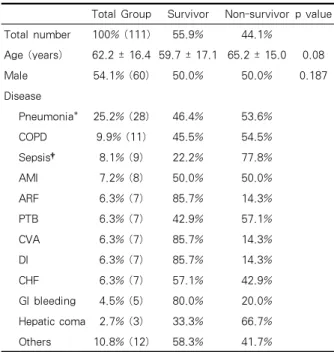
Total Group Survivor Non-survivor p value Total number 100% (111) 55.9% 44.1%
Age (years) 62.2 ± 16.4 59.7 ± 17.1 65.2 ± 15.0 0.08
Male 54.1% (60) 50.0% 50.0% 0.187
Disease
Pneumonia* 25.2% (28) 46.4% 53.6%
COPD 9.9% (11) 45.5% 54.5%
Sepsis† 8.1% (9) 22.2% 77.8%
AMI 7.2% (8) 50.0% 50.0%
ARF 6.3% (7) 85.7% 14.3%
PTB 6.3% (7) 42.9% 57.1%
CVA 6.3% (7) 85.7% 14.3%
DI 6.3% (7) 85.7% 14.3%
CHF 6.3% (7) 57.1% 42.9%
GI bleeding 4.5% (5) 80.0% 20.0%
Hepatic coma 2.7% (3) 33.3% 66.7%
Others 10.8% (12) 58.3% 41.7%
*: including pneumonia sepsis
†: extrapulmonary sepsis
COPD: Chronic Obstructive Pulmonary Disease AMI: Acute Myocardial Infarction
ARF: Acute Renal Failure PTB: Pulmonary Tuberculosis CVA: Cerebrovascular Accident DI: Drug Intoxication
GI bleeding: Gastrointestinal bleeding
Table 1. The baseline characteristics of the patients
Total patients 111 100%
Normal IAP, APP 41 36.9%
IAH 53 47.8%
Grade I (IAP 12-15 mmHg) 27 50.9%
Grade II (IAP 16-20 mmHg) 17 32.1%
Grade III (IAP 21-25 mmHg) 3 5.7%
Grade IV (IAP > 25 mmHg) 6 11.3%
ACS 17 15.3%
IAP: Intra-abdominal pressure APP: Abdominal perfusion pressure IAH: Intra-abdominal hypertension ACS: Abdominal compartment syndrome Table 2. The prevalence of IAH and ACS 3) 통계
모든 통계는 SPSS 11.0 for Windows (SPSS Inc., Chicago, IL, USA)를 사용하였다. 연속 변수는 평균
± 표준 편차로 표현하였다. IAH 또는 ACS가 있는 환 자와 없는 환자의 비교는 카이제곱 (chi-square)을 사용하였다. IAH와 APP가 예후에 미치는 영향 및 기 계 환기나 승압제를 사용한 환자에서 IAP와 APP의 비교는 t-검정 (t-test)으로 분석하였다. 중환자의 예 후와 IAP, APP, IAH, ACS, 기계 환기, 승압제 사용 과의 관계는 다중 회귀 분석을 사용하였다. 모든 p값은 0.05 이하일 때 통계적으로 유의한 것으로 판단하였다.
결 과 1) 환자 특성
총 122명의 환자 중 요로 파열, 신경인성 방광, 복 강 내 수술력이 있는 11명은 제외하고, 111명이 연구 에 포함되었다 (Table 1). 고대 구로 병원에서 70명, 고대 안산 병원에서 41명이 포함되었으며 두 군 간에 APACHE II 점수 (13.9 vs 14.2, p=0.79), APACHE III 점수 (49.5 vs 54.3, p=0.21), SAPS II 점수 (38.7 vs 43.9, p=0.07), 사망률 (41.4% vs 48.7%, p=0.45)에 통 계적으로 유의한 차이는 없었다. 생존군은 62명 (55.9%), 사망군은 49명 (44.1%)이었다. 대상 환자의 평균 연령은 62.2 ± 16.4세이며, 사망군은 65.2 ± 15.0 세로 생존군의 59.7 ± 17.1세 보다 더 높았으나 통계 적 의미는 없었다 (p=0.08). 남성은 60명 (54.1%)으로 여성 51명 (45.9%) 보다 많았으나 예후와는 상관 관 계가 없었다 (남성 사망률 50.0%, 여성 사망률 37.3%, p=0.178). 대상 환자의 진단은 폐렴 (28명, 25.2%), 만 성 폐쇄성 폐질환 (11명, 9.9%), 폐외 패혈증 (9명, 8.1%), 급성 심근 경색증 (8명, 7.2%) 순이었다. 평균 APACHE II, III 점수는 각각 14.0 ± 6.4, 52.5 ± 19.7 이었고, 입원 첫날 SAPS II 점수는 42.0 ± 16.3 이었 다. 입원 1일부터 기계 환기기 치료를 시행 받은 환자 는 60명 (54.0%) 이었고, 승압제 치료를 시작한 환자 는 42명 (37.8%) 이었다.
2) IAH 및 ACS의 유병률
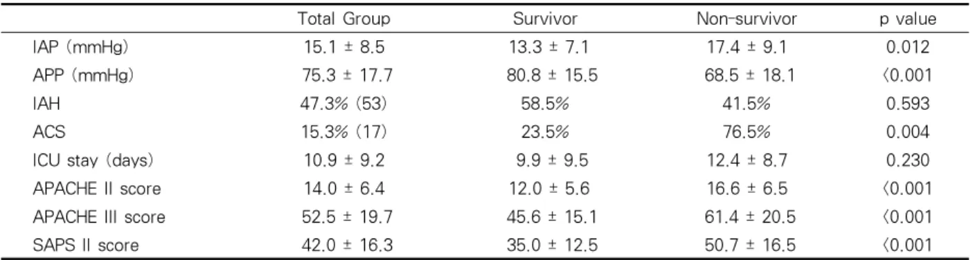
입원 1일 평균 IAP는 15.1 ± 8.5 mmHg 이었고 평 균 APP는 75.3 ± 17.7 mmHg 이었다. IAH는 53명 (47.7%)에서 진단되었고, 대부분 12-15 mmHg로 1도
Total Group Survivor Non-survivor p value
IAP (mmHg) 15.1 ± 8.5 13.3 ± 7.1 17.4 ± 9.1 0.012
APP (mmHg) 75.3 ± 17.7 80.8 ± 15.5 68.5 ± 18.1 <0.001
IAH 47.3% (53) 58.5% 41.5% 0.593
ACS 15.3% (17) 23.5% 76.5% 0.004
ICU stay (days) 10.9 ± 9.2 9.9 ± 9.5 12.4 ± 8.7 0.230
APACHE II score 14.0 ± 6.4 12.0 ± 5.6 16.6 ± 6.5 <0.001
APACHE III score 52.5 ± 19.7 45.6 ± 15.1 61.4 ± 20.5 <0.001
SAPS II score 42.0 ± 16.3 35.0 ± 12.5 50.7 ± 16.5 <0.001
IAP: Intra-abdominal pressure, APP: Abdominal perfusion pressure
IAH: Intra-abdominal hypertension, ACS: Abdominal compartment syndrome
APACHE: acute physiology and chronic health evaluation, SAPS: Simplified acute physiology score Table 3. The data of 111 patients at the day of admission to ICU
7 14 21
1 2 3 4 5 6 7
S urvivo r g ro up
N o n-s urvivo r g ro up
*
*
n=62
n=49 n=55
n=47
n=39
n=47
n=40 n=35 n=32
n=32 n=25
n=28 n=19
n=22
Day
IAP (m mHg)
Figure 1. The IAP values of the survivor and non-survivor groups.
The IAP was higher in the non-survivor group than in the survivor group at 1st and 2nd day (p<0.05), but there were no differences from 3rd to 7th days.
*: Survivor group vs. Non-survivor group, p< 0.05 n: number of patients
IAP: Intra-abdominal pressure
였다 (50.1%). ACS는 17명 (15.3%)에서 진단되었다 (Table 2).
3) IAP와 사망과의 관계
입원 1일 IAH는 사망과의 연관성은 없었다
(p=0.593). 반면, ACS는 사망과 유의한 연관성이 있 었다 (비교 위험도 5.24, 95% 신뢰 구간 1.58-17.30, p=0.004, Table 3).
하지만 IAH의 두 요소인, IAP와 APP의 영향을 각 각 따로 분석한 결과, 입원 1일 IAP는 사망군에서 생 존군보다 유의하게 높았고 (17.4 ±9.1 mmHg vs. 13.3
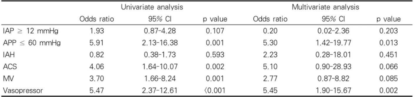
Univariate analysis Multivariate analysis
Odds ratio 95% CI p value Odds ratio 95% CI p value
IAP ≥ 12 mmHg 1.93 0.87-4.28 0.107 0.20 0.02-2.36 0.203
APP ≤ 60 mmHg 5.91 2.13-16.38 0.001 5.30 1.42-19.77 0.013
IAH 0.82 0.38-1.73 0.593 2.23 0.28-18.01 0.451
ACS 4.06 1.64-10.07 0.002 5.10 0.90-28.93 0.066
MV 3.70 1.66-8.24 0.001 2.77 0.87-8.82 0.085
Vasopressor 5.47 2.37-12.61 <0.001 5.45 1.90-15.67 0.002
IAP: Intra-abdominal pressure, APP: Abdominal perfusion pressure
IAH: Intra-abdominal hypertension, ACS: Abdominal compartment syndrome MV: Mechanical Ventilator, 95% CI: 95% Confidence Interval
Table 4. The analysis of mortality risk by multiple variables
n=62
n=49 n=55
n=47
n=47 n=39
n=35 n=32
D a y
n=25
n=28 n=19
n=22
60 75 90
1 2 3 4 5 6 7
Survivor group
Non-survivor group
* * * *
n=62 n=55
n=47 n=39
n=32 n=25 n=19
n=49 n=47 n=40
n=35 n=32
n=28 n=22
APP ( m m H g )
Day
Figure 2. The APP values of the survivor and non-survivor groups.
The APP was lower in the non-survivor group than in the survivor group from 1st to 4th day (p<0.001). The APP was more significantly related with mortality than the IAP, especially at 3rd to 4th day.
*: Survivor group vs. Non-survivor group, p< 0.001 n: number of patients
APP: Abdominal perfusion pressure
± 7.7 mmHg, p=0.012, Figure 1), 입원 2일에도 역시 사망군에서 더 높은 IAP를 보였으나 (p=0.04), 내원 3 일부터 7일까지는 유의한 차이가 없었다. APP는 입 원 1일부터 4일까지 사망군에서 생존군에 비해 유의 하게 낮았다 (Figure 2). APP는 IAP에 비해 입원 3, 4일에 사망과 더 유의한 연관성이 있었다.
단순 회귀 분석을 통하여 사망과 관련한 위험요인 을 분석한 결과, 입원 1일 ACS 발생, APP 60 mmHg 이하, 기계 환기 및 승압제의 사용, APACHE II, III, SAPS II 점수의 증가는 사망과 유의한 상관 관계가 있었다. 다중 회귀 분석을 통하여 혼란 변수에 대하여 상호 보정한 이후에는, 입원 1일 APP 60 mmHg 이하
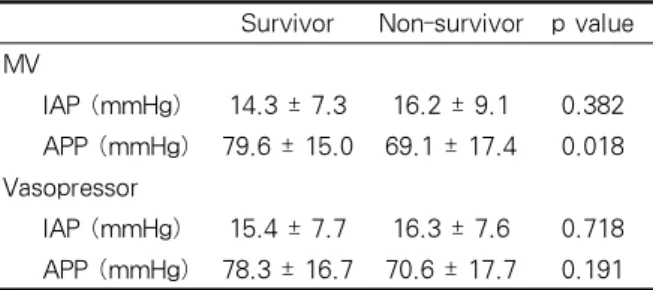
Survivor Non-survivor p value MV
IAP (mmHg) 14.3 ± 7.3 16.2 ± 9.1 0.382 APP (mmHg) 79.6 ± 15.0 69.1 ± 17.4 0.018 Vasopressor
IAP (mmHg) 15.4 ± 7.7 16.3 ± 7.6 0.718 APP (mmHg) 78.3 ± 16.7 70.6 ± 17.7 0.191 MV: Mechanical Ventilator
IAP: Intra-abdominal pressure APP: Abdominal perfusion pressure
Table 5. The differences of IAP and APP between the survivor and non-survivor groups treated with mechanical ventilation or vasopressor
(비교 위험도 5.30, 95% 신뢰 구간 1.42-19.77, p=0.013), 승압제의 사용 (비교 위험도 5.45, 95% 신뢰 구간 1.90-15.67, p=0.002)이 사망과 유의한 연관성이 있었다 (Table 4). 기계 환기를 시행한 환자군에서, APP는 사망과 연관성이 있었으나 IAP는 사망과 유 의한 연관성이 없었다. 승압제 사용 환자군에서, APP 와 IAP 모두 사망과 연관성이 없었다 (Table 5). 7일 동안 IAP 및 APP의 변동과 SAPS II 점수는 유의한 상관 관계가 없었다. 또한, IAH의 발생, 중환자실 입 원 기간 역시 사망과 연관성이 없었다.
고 찰
본 연구에서 대상 환자의 입원 1일 평균 IAP는 15.1 ± 8.5 mmHg, IAH 및 ACS의 유병률은 각각 47.7%, 15.3% 이었다. 입원 1일 IAH는 사망과 상관 관계가 없었으나, ACS는 사망과 유의한 상관 관계가 있었다. 하지만 IAH의 두 가지 요소인 IAP와 APP를 독립적으로 분석한 결과에서는, IAP의 증가와 APP 의 감소가 사망과 유의한 연관성이 있었다. 또한 기계 환기를 시행한 환자에서 APP가 사망과 유의한 상관 관계가 있었다.
최근 IAP는 중환자에서의 생리적 변화를 설명하는 요인으로 관심을 받고 있다. IAP가 증가하면 여러 생 체 장기 기능이 악화된다22. 호흡기계에서는 IAP의 증가가 횡격막을 상승시켜 일호흡량 (tidal volume) 의 감소를 가져와 폐의 환기 장애를 유발하고17, 순환
기계에서는 일회 박출량 (stroke volume)과 전부하 (preload)를 감소시키고, 후부하 (afterload)는 증가시 켜 심박출량의 저하를 가져온다23.
Malbrain 등12은 APP를 고려하지 않은 상태에서, IAH를 IAP 12 mmHg 이상으로만 정의한 바 있다.
하지만 이후 세계 복강 구획 증후군 회의에서는17, IAH를 IAP 12 mmHg 이상 또는 APP 60 mmHg 이 하로 정의하였다. 그러나, 본 연구에서는 세계 복강 구획 증후군 회의에서 정의한 IAH는 사망과 유의한 연관성이 관찰되지 않았다.
2004년 Malbrain 등16은 중환자에서 평균 IAP는 9.8 ± 4.7 mmHg, IAH 및 ACS의 유병률은 각각 50.5%, 8.2%로 보고한 바 있다. 같은 그룹에서 시행 한 2005년 추적 연구에서 평균 IAP는 10.0 ± 4.8 mmHg, IAH 및 ACS의 유병률은 각각 32.1%, 4.2%
이었다12. 본 연구에서는 기존의 연구 결과들보다 더 높은 평균 IAP를 보였고, IAH 및 ACS의 유병률 역 시 높았다. 이러한 차이는 대상 환자군의 차이에서도 기인할 수 있으나, IAP를 측정하는 기술의 정확성 및 재현성의 문제일 가능성을 배제할 수 없다22. 연구 기 간 동안 2개 병원에서 각각 3명의 인원이 IAP를 측정 하였기 때문에, 검사자들 사이의 측정 오차가 있을 수 있다. 이러한 이유로 이전에 시행한 연구 결과보다 더 높은 평균 IAP 결과를 가져왔을 가능성이 있다.
한편, 본 연구에서는 8명의 문맥압 항진증을 동반 한 간경변 환자가 포함되었다. 간경변 환자는 만성적 으로 복압이 상승하며, 이런 경우 복벽의 순응도가 좋 기 때문에 IAP를 상승시키지 않는것으로 알려져 있 다17. 본 연구에서도 간경변 환자의 IAP는 다른 질환 의 환자 IAP와 비교하였을 때 유의한 차이를 보이지 않았다 (14.6 mmHg vs 15.2 mmHg, p=0.85).
기존의 연구에서는 중환자에 있어 IAH가 사망률 을 유의하게 증가시키고12, APP는 환자의 생존을 예 측하는데 있어 IAP 보다 우월한 것으로 보고된 바 있 다13. 본 연구에서도 혼란 변수에 대해 상호 보정한 다 중 회귀 분석에서, IAP는 환자의 예후와 상관 관계가 없었으나 APP는 유의한 상관 관계를 보였다.
특히, 기계 환기를 시행한 환자군에서는 APP의 감 소가 사망과 유의한 연관성이 있었다. 이는 기계 환기
로 인한 기도 압력의 증가에 의해 심박출량이 줄어들 고22, 이런 상황에서 APP가 감소하면 위장관24, 신장25, 간22 등 여러 복강 내 장기의 관류 장애를 초래하기 때문인 것으로 판단된다.
결론적으로, 본 연구에서는 내과계 중환자에서 입 원 1일에 측정한 IAP의 증가, APP의 감소 및 ACS 발생이 환자의 예후에 영향을 미치는 것으로 관찰되 었다.
요 약
연구배경: IAP의 증가는 혈류량을 감소시켜 여러 장기에 영향을 준다. IAH는 IAP가 12 mmHg 이상 또는 APP가 60 mmHg 이하로, ACS는 IAP가 20 mmHg 이상이면서 동시에 하나 이상의 장기 손상이 있는 경우로 각각 정의한다. 저자들은 중환자에서 IAH 및 ACS의 유병률과 이들이 환자의 예후에 미치 는 영향을 고찰하고자 하였다.
방 법: 고려대학교 의료원 중환자실에 입원하는 내과계 환자를 대상으로 하여, APACHE II 및 III 점 수, SAPS II 점수를 기록하였다. IAP는 삼중관 요도 관을 통하여, 입원 7일째까지 매일 3회 측정하였고, 환자가 사망하거나 일반 병실로 전실 또는 요도관을 제거할 때까지 계속하였다. 환자의 예후는 28일을 기 준으로 판단하였다.
결 과: 총 111명 대상 환자들의 입원 1일 IAH 및 ACS의 유병률은 각각 47.7%, 15.3%였다. 사망군과 생존군 사이에 IAH의 유병률은 유의한 차이가 없었 으나, ACS는 사망군에서 더 높았다 (p=0.004). 사망 군에서 IAP는 더 높았고 (p=0.012), APP는 유의하게 낮았다 (p<0.001).
결 론: 중환자에서 입원 1일에 측정한 IAP의 증 가, APP의 감소 및 ACS 발생은 환자의 사망과 연관 이 있을 것으로 판단된다.
참 고 문 헌
1. Sugerman HJ. Increased intra-abdominal pressure in obesity. Int J Obes Relat Metab Disord 1998;22:1138.
2. Hering R, Wrigge H, Vorwerk R, Brensing KA, Schroder S, Zinserling J, et al. The effects of prone positioning on intraabdominal pressure and cardio- vascular and renal function in patients with acute lung injury. Anesth Analg 2001;92:1226-31.
3. Duggan JE, Drummond GB. Abdominal muscle activity and intraabdominal pressure after upper abdominal surgery. Anesth Analg 1989;69:598-603.
4. Navarro-Rodriguez T, Hashimoto CL, Carrilho FJ, Strauss E, Laudanna AA, Moraes-Filho JP. Reduction of abdominal pressure in patients with ascites redu- ces gastroesophageal reflux. Dis Esophagus 2003;16:
77-82.
5. Rosenthal RJ, Friedman RL, Kahn AM, Martz J, Thiagarajah S, Cohen D, et al. Reasons for intra- cranial hypertension and hemodynamic instability during acute elevations of intra-abdominal pressure:
observations in a large animal model. J Gastrointest Surg 1998;2:415-25.
6. Ishizaki Y, Itoh T, Shimomura K, Noie T, Abe H, Izezuki Y. Cardiovascular effects of increasedintra- abdominal pressure during pneumoperitoneum: pre- liminary report. Nippon Geka Gakkai Zasshi 1991;
92:614.
7. Rouby JJ, Puybasset L, Nieszkowska A, Lu Q. Acute respiratory distress syndrome: lessons from computed tomography of the whole lung. Crit Care Med 2003;
31:S285-95.
8. Polat C, Aktepe OC, Akbulut G, Yilmaz S, Arikan Y, Dilek ON, et al. The effects of increased intra- abdominal pressure on bacterial translocation. Yonsei Med J 2003;44:259-64.
9. Markou N, Grigorakos L, Myrianthefs P, Boutzouka E, RizosM, Evagelopoulou P, et al. Venous pressure measurements in the superior and inferior vena cava:
the influence of intra-abdominal pressure. Hepatoga- stroenterology 2004;51:51-5.
10. Reddy VG. Prevention of postoperative acute renal failure. J Postgrad Med 2002;48:64-70.
11. Sugrue M, Jones F, Deane SA, Bishop G, Bauman A, Hillman K. Intra-abdominal hypertension is an inde- pendent cause of postoperative renal impairment.
Arch Surg 1999;134:1082-5.
12. Malbrain ML, Chiumello D, Pelosi P, Bihari D, Innes R, RanieriVM, et al. Incidence and prognosis of intraabdominal hypertension in a mixed population of critically ill patients: a multiple-center epidemiolo- gical study. Crit Care Med 2005;33:315-22.
13. Cheatham ML, White MW, Sagraves SG, Johnson JL, Block EF. Abdominal perfusion pressure: a superior parameter in the assessment of intra-abdominal hy- pertension. J Trauma 2000;49:621-6.
14. Cheatham ML, Safcsak K. Intraabdominal pressure:
a revised method for measurement. J Am Coll Surg 1998;186:594-5.
15. Kron IL, Harman PK, Nolan SP. The measurement of intra-abdominal pressure as a criterion for abdominal re-exploration. Ann Surg 1984;199:28-30.
16. Malbrain ML, Chiumello D, Pelosi P, Wilmer A, Brienza N, Malcangi V, et al. Prevalence of intra- abdominal hypertension in critically ill patients: a multicentre epidemiological study. Intensive Care Med 2004;30:822-9.
17. Sugrue M. Abdominal compartment syndrome. Curr Opin Crit Care 2005;11:333-8.
18. Knaus WA, Draper EA, Wagner DP, Zimmerman JE.
APACHE II: a severity of disease classification system. Crit Care Med 1985;13:818-29.
19. Knaus WA, Wagner DP, Draper EA, Zimmerman JE, Bergner M, Bastos PG, et al. The APACHE III prognostic system: risk prediction of hospital mor- tality for critically ill hospitalized adults. Chest 1991;
100:1619-36.
20. le Gall JR, Lemeshow S, Saulnier F. A new Simpli-
fied Acute Physiology Score (SAPS II) based on a European/North American multicenter study. JAMA 1993;270:2957-63.
21. Malbrain ML. Different techniques to measure intra- abdominal pressure (IAP): time for a critical re- appraisal. Intensive Care Med 2004;30:357-71.
22. Malbrain ML. Is it wise not to think about intra- abdominal hypertension in the ICU? Curr Opin Crit Care 2004;10:132-45.
23. Malbrain ML, Deeren D, de Potter TJ. Intra- abdominal hypertension in the critically ill: it is time to pay attention. Curr Opin Crit Care 2005;11:156-71.
24. Doty JM, Oda J, Ivatury RR, Blocher CR, Christie GE, Yelon JA, et al. The effects of hemodynamic shock and increased intra-abdominal pressure on bacterial translocation. J Trauma 2002;52:13-7.
25. Kotzampassi K, Metaxas G, Paramythiotis D, Pidonia I, Rekka H, Karamouzis M, et al. The influence of continuous seven-day elevated intra-abdominal pres- sure in the renal perfusion in cirrhotic rats. J Surg Res 2003;115:133-8.