단 보
집속이온빔으로 제작한 황사 입자 시편의 투과전자현미경 분석
Transmission Electron Microscopy of Asian Dust Particle Specimens Prepared by Focused Ion Beam
정 기 영(Gi Young Jeong)*
안동대학교 지구환경과학과
(Department of Earth and Environmental Sciences, Andong National University, Andong 760-749, Korea)
*Corresponding author: +82-54-820-5619, E-mail: [email protected]
서 언
광물먼지(mineral dust)는 태양광 및 지구 복사 를 반사 및 흡수하거나 구름 응결핵이 되어 지구 대기의 에너지 수지에 직간접적인 영향을 미친다 (Tegen and Lacis, 1996; Formenti et al., 2011).
태양광이나 지구 복사의 흡수와 산란은 입사하는 전자기파의 파장과 먼지 입자의 크기, 형태, 복소 굴절률(complex refractive indices) 등에 따라서 결 정된다(Seinfeld and Pandis, 2006; Durant et al., 2009). 따라서 광물먼지와 지구기후의 관계, 그리 고 광물먼지의 원격탐사를 다루는 대기환경분야에 서 광물먼지의 물성에 대한 많은 연구들이 있었다 (Okada et al., 1990; Anderson et al., 1996; Reid
et al., 2003; Ro et al., 2005; Kandler et al., 2007;
Chou et al., 2008; Jeong, 2008).
광물먼지의 물성은 리다(lidar), 태양광도계(sun photometer)나 인공위성 등의 원격탐사를 이용하 거나, 에어로졸 필터에 수집된 시료를 분석하여 얻 어진다. 필터 시료의 전반적 화학조성이나 광물조 성을 알아보기 위한 X선형광분석이나 X선회절분 석을 적용하기도 하지만, 흔히 필터에 채집된 시료 량이 수 mg 정도로 매우 작기 때문에 신뢰도가 높 지 않다. 반면에 주사전자현미경(SEM)이나 투과전 자현미경(TEM) 등을 이용한 단일입자 분석은 시
료량이 작아도 먼지 입자의 크기나 형태학적 특성 을 정량적으로 측정할 수 있고, X선 신호를 이용 한 화학 분석도 가능하므로 물성과 광물조성에 관 한 다양한 정보를 제공한다.
그러나 지금까지의 전자현미경 단일입자 분석은 입자의 외형을 관찰하거나 입자의 대략적 광물 특 성을 분석하는 것이며, 입자 내부에 대한 정보는 아직 없다(Nousiainen, 2009). 먼지 입자가 단일 광물이거나 단일 입자인 경우는 거의 없으며, 대부 분 여러 광물이나 입자의 집합체임이 이전 연구들 에서 밝혀진 바 있다(Jeong, 2008). 광물먼지의 대 기에너지수지 기여도 규명 및 원격탐사자료 처리 알고리듬의 개선을 위해서는 광물먼지 입자와 전 자기파의 상호작용 광학모델의 수립이 가장 중요 하다. 지금까지는 이러한 광학모델에 광물먼지의 외부 형태나 광물 조성만이 고려되었으나, 입자 내 부의 광물배열상태, 공극의 크기 및 분포, 광물종 분포 등의 입자 내부적 특징이 함께 고려되어야 한 다(Nousiainen, 2009). 먼지 입자의 내부적 특징은 사실 이전까지는 기술적 한계 때문에 연구가 거의 불가능하였으나, 최근의 집속이온빔(FIB) 장비의 빠른 보급으로 가능하게 되었다. 이 방법은 SEM 으로 시편을 관찰하면서 특정 부위에 Ga 이온빔을 집중 조사하여 절제하는 것으로, 시편의 단순 단면 을 SEM으로 관찰하거나 수십 nm 정도의 얇은 박
편을 떼어내어 TEM으로 분석한다. 저자가 집속이 온빔을 지질시료에 적용한 예로서 제주도 화산유 리의 변질과정(Jeong and Sohn, 2009; 2011) 및 중국 황사 퇴적물인 뢰스(loess) 입자에 대한 연구 (Jeong and Lee, 2010)가 있다. 이 단보에서는 FIB 를 이용하여 제작한 우리나라 황사 입자 단면 시편 의 조직과 광물구성에 대한 TEM 분석결과를 소개 하고자 한다.
시료 및 분석방법
황사 시료는 2012년 3월 31일에서 4월 1일 사이 에 인천특별시 옹진군 덕적도 정상에 설치된 황사 측정소에서 총분진측정기(TSP sampler)로 테플론 코팅 유리섬유 필터에 채집하였다. 이 시기 황사는 중국 북부와 몽골 사이의 고비사막에서 발생하여 하루 만에 경기도 서해안에 도달하였는데, 연구에 의하면 예년에 비하여 입자 직경이 큰 특성을 갖고 있다(Jeong et al., 2013). 황사 입자의 SEM 관찰 을 위하여 황동판에 전도성 탄소 양면테이프를 붙 힌 후, 필터에 가볍게 접촉하여 황사 입자들을 테 이프에 옮기고 백금 코팅을 하였다. 황사 입자들의 대략적인 광물 특성, 형태, 집합상태 분석을 위하 여 JEOL JSM 6300 SEM 및 JEOL JSM 6700F 전계방출형 SEM으로 황사 입자들을 관찰하는 동 시에 에너지분산X선분광기(EDS)로 정성적 화학조 성을 분석하였다(Jeong, 2008).
SEM 관찰을 바탕으로 내부구조 관찰을 위한 입 자를 선정하였으며, SMI 3050TB FIB 장비로 TEM 관찰을 위한 시편을 제작하였다. 시편 제작은 우선 탄소를 입자에 증착하여 고정시킨 후, Ga 이온빔 으로 표면에 수직 방향으로 두께 ∼50 nm, 면적 20 µm2 정도인 박편을 제작하였다. TEM 관찰은 JEOL JEM 2100F 장비를 이용하여 시행하였으며, EDS로 관심부위의 화학조성을 분석하였다.
결과 및 토의
입자 내부조직
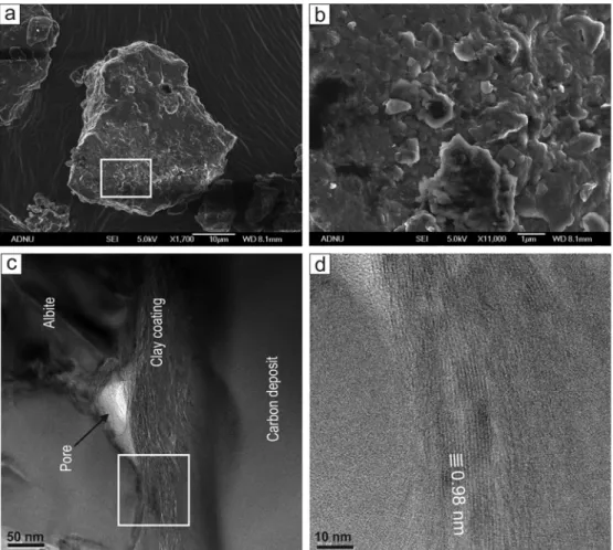
그림 1a는 SEM-EDS 분석에서 앨바이트(albite) 로 확인된 황사 입자의 SEM 사진인데, 원마도가 매우 낮은 불규칙한 형태의 입자이다. 표면의 확대 사진에서 앨바이트의 표면이 판상 점토 광물 입자 들로 얇게 피복되어 있다(그림 1b). FIB 시편의
TEM 관찰 결과, 0.5∼1 µm 정도 두께의 점토 광 물층이 요철 있는 앨바이트 표면을 피복하고 있으 며, 앨바이트와 점토 광물 피복 사이에 미세한 공 극이 형성되어 있다. 앨바이트 표면에 대체로 평행 하게 배열된 점토 광물 입자들이 피복 점토층을 구 성되어 있다. 그러나 이 피복 점토층을 구성하는 점토 광물 입자들의 두께는 매우 얇고, 상당히 헝 클어진 상태의 집합체를 형성하고 있다(그림 1c).
격자(lattice fringe) 상 관찰에 의하면, 점토 입자들 은 0.98 nm 정도의 (001) 격자 간격을 보인다(그 림 1d). 이 격자 간격은 점토 광물종이 일라이트 또 는 진공에서 탈수된 스멕타이트, 혹은 이들의 혼합 층임을 의미한다. 입자가 너무 작아서 현재로서는 화학조성에 기반하여 이 세 가지 상을 구분하기는 불가능하다.
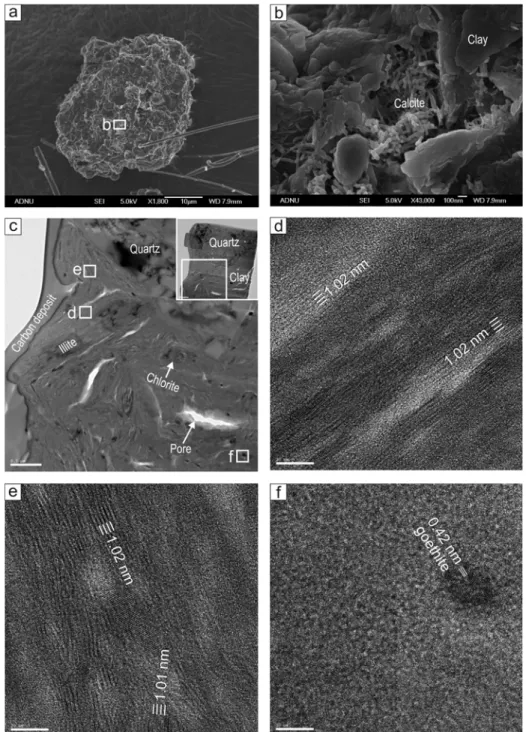
그림 2a는 SEM-EDS 분석에서 석영이 주성분이 고 점토 광물이 혼재한 입자로 감정되었으나, 표면 확대사진에서는 점토 광물과 나노 섬유상 방해석 (Jeong and Chun, 2006; Jeong, 2007)만 관찰된다 (그림 2b). FIB 시편의 TEM 관찰결과, 큰 석영 입 자가 점토 광물 입자들과 집합체를 형성하고 있음 을 알 수 있다(그림 2c). 점토 광물 입자들은 일라 이트나 녹니석처럼 좀더 크고 치밀한 입자들도 있 으나, 대부분 단 몇 개의 단위층들로 구성된 매우 얇은 나노판들의 혼돈된 집합체를 형성한다. 격자 상 관찰 결과, 이들 나노판들은 대부분 일라이트 또는 탈수 스멕타이트에 해당하는 1.0 nm 정도의 (001) 저면 격자간격을 보인다(그림 2d, 2e). 점토 집합체에는 극미립의 나노 크기 산화철 입자들이 드물게 산포되어 있는데, 격자상 관찰에서 침철석 입자들로 확인되었다(그림 2f). 그림 2c에서 여러 개의 렌즈상 미세공극들이 관찰되는데, 일부는 시 료준비 과정에서 점토 광물의 건조 수축으로 형성 되었을 가능성도 있으나, 일부는 원래 먼지 입자 내에 존재하던 것으로 보인다.
대기과학적 의미
에어로졸의 지구기후영향을 평가하거나 원격탐 사법으로 에어로졸의 분포나 이동을 연구하기 위 해서는 전자기파와 에어로졸 입자의 상호작용에 대한 물리적 모델이 수립되어야 한다(Seinfeld and Pandis, 2006). 공기분자나 극미세오염물질처럼 극 히 작은 입자들에 의한 전자기파의 산란은 레일리 산란(Rayleigh scattering)이 적용되나, 가시광선-적
Fig. 1. Internal fabric of albite-rich dust particle. (a) Irregular albite particle (SEM image). (b) Particle surface magnified from the box in a, showing coatings of submicron plates of clay minerals (SEM image). (c) TEM image of the FIB slice prepared from the particle in a. (d) TEM lattice fringe of illitic clay minerals magnified from the box in c.
외선 파장 범위 크기에 속하는 광물먼지 입자는 전 자기파와 입자의 상호작용에 대한 해(solution)인 Mie 산란이 적용된다. 입자의 Mie 산란 결과를 얻 기 위해서는 입자 형태, 크기, 광물조성 등이 입력 변수로 필요한 것으로 알려져 있다(Moosmüller et
al., 2009; Nousiainen, 2009). 따라서 광물먼지 입
자의 산란에 대한 정보를 얻기 위해서는 광물먼지 입자의 물성이 먼저 규명되어야 한다. 이러한 관점 에서 아직 불분명한 광물먼지 물성과 광물학적 특 성은 광물입자-빛 상호작용 모델의 불완전성의 원 인 중 하나이다. 대부분의 광물먼지-빛 상호작용 모델은 광물먼지의 개별 입자들이 하나의 광물로 구성되어 있다고 간주하지만, 사실 대부분의 광물 먼지 입자는 그림 1 및 2에서 보는 바와 같이 크고작은 여러 입자들의 집합체이다. 단일 입자의 산란 과 달리, 다양한 광물로 구성된 입자의 다중 산란 에는 Zubko et al. (2008) 등이 시도한 것처럼 다 른 상호작용 모델이 적용되어야 한다. 또한 그림 1 및 2에서 보는 바와 같은 먼지 입자 내 구성광물들 의 다양한 배열상태, 비등방성, 크기 분포, 공극도 빛의 산란과정에 영향을 미칠 것이다(Nousiainen, 2009).
빛의 산란 외에도 먼지 입자를 구성하는 광물들 의 다양한 굴절률은 빛의 흡수에도 영향을 미친다 (Moosmüller et al., 2009). 광물 중에서 특히 산화 철 광물(그림 2f)들은 불과 수 % 정도의 적은 양으 로도 광물먼지의 전체의 흡수도에 결정적 기여를 하는 것으로 알려져 있다(Lafon et al., 2006). 따
Fig. 2. Internal fabric of a quartz-rich dust particle. (a) Quartz-rich particle (SEM image). (b) Particle surface magnified from the box in a, showing the thick coatings of clay minerals and nanofiber calcite (SEM image).
(c) TEM image of the slice prepared by FIB, showing the large quartz grain with the aggregate of
illite-smectite series clay minerals with discrete illite and chlorite grains. (d), (e) TEM lattice fringes of
illite-smectite series clay minerals with ~1.0 nm spacings magnified from the boxes in c. (f) Nano-sized
goethite grain in the clay aggregate magnified from the box in c.
라서 빛의 흡수도를 모델링하기 위해서는 산화철 의 광물종, 크기, 먼지 입자 내 분포 등에 대한 광 물학적 자료가 필요하다.
이와 같은 광물먼지 개별 입자의 광물학적 특성 은 이전에는 SEM을 이용한 입자의 EDS 패턴으로 부터 추론할 수밖에 없었으나(Jeong, 2008), 최근 의 FIB 장비의 보급으로 본격적인 연구가 가능하 게 되었다. 이와 같은 종류의 분석은 SEM 분석과 비교하여 아직 시료준비와 분석 비용이 많이 들고, 절차가 훨씬 복잡하지만, 광물먼지-빛 상호작용의 보다 실제적인 광학모델 수립에 기여할 것으로 보 인다.
결 론
광물먼지에 의한 태양광의 산란 및 흡수 상호작 용에 관한 광학 모델은 지구 대기의 지역적 에너지 균형 평가나 원격탐사 자료처리에서 매우 중요하 다. 이와 같은 모델의 적용성을 향상시키기 위해서 는 광물먼지 입자들의 크기나 형태 등의 물성 외에 도, 개별 입자의 조성이나 구조에 대한 광물학적 자료가 필요하다. 이 연구에서 FIB 시편준비와 TEM 분석법을 황사 개별 입자의 광물학적 특성 분석에 복합적으로 적용한 결과, 황사 입자들의 복잡한 내 부구조와 다양한 광물분포를 확인할 수 있었다.
FIB를 이용한 먼지 입자 단면 시료 제작과 TEM 내부 구조 및 광물분석은 개별 광물먼지 입자의 내 부 특성 연구에 매우 효과적임을 알 수 있었다. 향 후 대표적인 광물먼지 입자들에 대한 체계적인 분 석을 통하여 그 내부 구조와 광물조성을 유형화하 는 후속연구가 필요하다.
사 사
이 연구는 한국연구재단의 지원(NRF-2011-0028597) 을 받아 수행되었다. 원고의 미비점을 지적하시고 개선 방향을 제시해주신 황진연 교수님과 익명의 심사위원께 감사드린다.
REFERENCES
Anderson, J.R., Buseck, P.R., and Patterson, T.L. (1996) Characterization of the Bermuda tropospheric aero- sols by combined individual-particle and bulk-aero- sol analysis. Atmospheric Environment, 30, 319-338.
Chou, C., Formenti, P., Maille, M., Ausset, P., Helas,
G., Harrison, M., and Osborne, S. (2008) Size dis- tribution, shape, and composition of mineral dust aerosols collected during the African Monsoon Multidisciplinary Analysis Special Observation Period 0: Dust and Biomass-Burning Experiment field cam- paign in Niger, January 2006. Journal of Geophysi- cal Research, 113, D00C10, doi:10.1029/2008JD009897.
Durant, A.J., Harrison, S.P., Watson, M., and Balkanski, Y. (2009) Sensitivity of direct radiative forcing by mineral dust to particle characteristics. Progress in Physical Geography, 33, 80-102.
Formenti, P., Schütz, L., Balkanski, Y., Desboeufs, K., Ebert, M., Kandler, K., Petzold, A., Scheuvens, D., Weinbruch, S., and Zhang, D. (2011) Recent prog- ress in understanding physical and chemical proper- ties of African and Asian mineral dust. Atmospheric Chemistry and Physics, 11, 8231-8256.
Jeong, G.Y. (2007) Nanosized calcite in the Chinese loess. Journal of the Mineralogical Society of Korea, 20, 255-260 (in Korean with English abstract).
Jeong, G.Y. (2008) Bulk and single-particle mineralogy of Asian dust and a comparison with its source soils. Journal of Geophysical Research, 113, D02208, doi:10.1029/2007jd008606.
Jeong, G.Y. and Chun, Y. (2006) Nanofiber calcite in Asian dust and its atmospheric roles. Geophysical Research Letters, 33, L24802, doi:10.1029/2006 GL028280.
Jeong, G.Y. and Lee, K.-S. (2010) A mineral tracer to- ward high-resolution dust provenance on the Chinese Loess Plateau: SEM, TEM, and sulfur isotopes of sulfate inclusions in biotite. American Mineralogist, 95, 64-72.
Jeong, G.Y. and Sohn, Y.K. (2009) Basaltic glass al- teration and lithification of the Holocene Hamori Formation, Jeju Island. Journal of the Geological Society of Korea, 45, 331-344 (in Korean with English abstract).
Jeong, G.Y. and Sohn, Y.K. (2011) Microtextures, mi- crochemistry, and mineralogy of basaltic glass alter- ation, Jeju Island, Korea, with implications for ele- mental behavior. American Mineralogist, 96, 1129- 1147.
Jeong, G.Y., Kim, J.Y., Seo, J., Kim, G.M., and Jin, H.C., and Chun, Y. (2013) Long-range transport of giant particles in Asian dust identified by physical, mineralogical, and meteorological analysis. Atmos- pheric Chemistry and Physics (in press).
Kandler, K., Benker, N., Bundke, U., Cuevas, E., Ebert, M., Knippertz, P., Rodríguez, S., Schütz, L.,
and Weinbruch, S. (2007) Chemical composition and complex refractive index of Saharan mineral dust at Izaña, Tenerife (Spain) derived by electron microscopy. Atmospheric Environment, 41, 8058-8074.
Lafon, S., Sokolik, I.N., Rajot, J.L., Caquineau, S., and Gaudichet, A (2006) Characterization of iron oxides in mineral dust aerosols: Implications for light ab- sorption, 111, D21207, doi:10.1029/2005JD007016.
Moosmüller, H., Chakrabarty, R.K., and Arnott, W.P.
(2009) Aerosol light absorption and its measure- ment: A review, Journal of Quantitative Spectrosco- py and Radiation Transfer, 110, 844-878, doi:10.1016/
j.jqsrt.2009.02.035.
Nousiainen T. (2009) Optical modeling of mineral dust particles: a review. Journal of Quantitative Spectro- scopy and Radiation Transfer, 110, 1261-1279.
Okada, K., Naruse, H., Tanaka, T., Nemoto, O., Iwasaka, Y., Wu, P.-M., Ono, A., Duce, R.A., Uematsu, M., Merrill, J.T., and Arao, K. (1990) X-ray spectrome- try of individual Asian dust-strom particles over the Japanese islands and the north Pacific Ocean, Atmo- spheric Environment, Part A, 24, 1369-1378.
Reid, J.S., Jonsson, H.H., Maring, H.B., Smirnov, A., Savoie, D.L., Cliff, S.S., Reid, E.A., Livingston, J.M., Meier, M.M., Dubovik, O., and Tsay, S. C.
(2003) Comparison of size and morphological meas- urements of coarse mode dust particles from Africa.
Journal of Geophysical Research, 108, doi:10.1029/
2002JD002485.
Ro, C.-U., Hwang, H., Kim, H., Chun, Y., and Van Grieken, R. (2005) Single-particle characterization of four “Asian dust” samples collected in Korea, us- ing low-Z particle electron probe X-ray micro- analysis, Environmental Science & Technology, 39, 1409-1419.
Seinfeld, J.H. and Pandis, S.N. (2006) Atmospheric Chemistry and Physics. John Wiley, Hoboken, N. J.
Tegen, I. and Lacis, A.A. (1996) Modeling of particle size distribution and its influence on the radiative properties of mineral dust aerosol. Journal Geophy- sical Research, 101, 19237-19244.
Zubko, E., Shkuratov, Y., Mishchenko, M., and Videen, G. (2008) Light scattering in a finite multi-particle system. Journal of Quantitative Spectroscopy &
Radiative Transfer, 109, 2195-2206.
접수일(2013년 12월 9일), 수정일(1차 : 2013년 12월 19일), 게재확정일(2013년 12월 19일), 책임편집위원 : 유재영