흉추 도수교정이 둥근어깨자세를 가진 30대 성인남녀의 통증, 관절가동범위, 근활성도에 미치는 영향
이재남, 3) 양성화, 공원태
1)대한적십자사 경인의료재활센터병원, 나사렛대학교 물리치료학과
1)Orig
inal Artic le
The Effects of Thoracic Spine Thrust Manipulation on Shoulder Pain, Range of Motion and Muscle Activity in 30′s Adults with Rounded Shoulder Posture
Jae-nam Lee, Seong-hwa Yang, Won-tae Gong
1)Dept. of Physical Therapy, Gyeong-in Medical Rehabilitation Center Hospital Redcross Dept. of Physical Therapy, Korea Nazarene University
1)Key Words:
Rounded shoulder posture, Thoracic spine manipulation, Electromyograp hy, Pain, Active range of motion
ABSTRACT
Purpose: This study was aimed to determine the effects of thoracic spine thrust manipulation on muscle activities of the scapular upward rotators and middle deltoid, active range of motion (AROM), shoulder pain, and rounded shoulder posture in young adults with rounded shoulder.
Methods: The subjects were 30 young adults (14 males, 16 females) with rounded shoulder.
Thirty subjects were randomly assigned to an experimental (manipulation) and control (placebo) groups of fifteen subjects respectively. The manipulation group received the manipulation (high velocity, low amplitude), which was performed by a physical therapist with the subject in the supine position and with the arms crossed over the chest and hands passed over the shoulders. For the sham group, the same procedure was performed, with the exception that the high-velocity thrust was not applied. Measurements were taken before and after the intervention. Muscle activity of upper and lower trapezius, serratus anterior, middle deltoid was measured using surface electromyography. Visual analog scale (VAS) was used for shoulder pain.
Goniometry was used for shoulder abduction active range of motion (AROM). Straight edge was used for supine rounded shoulder posture (RSP) distance. Results: The muscle activity of the upper trapezius, lower trapezius and middle deltoid muscle increased significantly after the intervention (p<.05). However, no significant difference was observed in serratus anterior muscle (p>.05). The VAS was significantly decreased and AROM significantly increased after the intervention (p<.05). The distance of RSP were not significant (p>.05). The control group showed no differences before and after the intervention (p>.05). Conclusion: The results of this study suggest that thoracic spine thrust manipulation can be an effective component of treatment plan to improve pain and function.
Ⅰ. 서 론
최근 스마트폰의 사용 실태를 보면, 30대의 스마트폰 보급률은 97% 이상에 이르며 하루 평균 사용시간은 4 시간이상으로 나타났다(통계청, 2015). 또한 직장과 가
교신저자: 이재남(경인의료재활센터병원, [email protected]) 논문접수일: 2016.04.06, 논문수정일: 2016.06.01,
개재확정일: 2016.06.09.
정에서 컴퓨터의 사용시간은 주당 평균 12시간이다(통 계청, 2014). 산업이 발달함에 따라 현대인들의 컴퓨터 와 휴대폰 사용 시간은 급격하게 증가하고 있으며, 장 시간의 사용으로 인해 자세변형을 유발하고 목과 어깨 통증이 나타나게 된다(Szeto 등, 2002).
일상생활이나 직장업무 중에서 습관적으로 구부정한
자세는 둥근어깨자세(rounded shoulder posture; RSP)
를 유발한다(Chansirinukor 등, 2001). RSP는 신체의 중
력선에 대해 견봉이 앞쪽으로 돌출되어 있는 자세로 정 의되며, 전방머리자세(forward head posture)와 함께 경 추의 전만과 흉추의 후만을 증가시키고, 견갑골위치의 변형, 그리고 어깨전방자세를 증가시킨다. 즉, 견갑골의 전인, 하방회전, 그리고 전방경사를 유발하고 목과 어깨 의 근육긴장과 스트레스를 증가시켜 어깨의 통증과 기 능저하, 견갑골 상방회전감소와 같은 골격근이상을 유 발한다(Sahrmann, 2002; Greenfield, 2001; Lukasiewicz 등, 1999; Wang 등, 1999). 이처럼 RSP는 정상적인 어 깨움직임을 어렵게 만든다(Lau 등, 2010).
어깨의 정상적인 외전은 상완골의 거상과 견갑골의 상방회전이 요구되며, 견갑골의 상방회전은 상승모근 (upper trapezius), 하승모근(lower trapezius), 그리고 전 거근(serratus anterior)의 활동에 의해 이뤄진다(Bagg와 Forrest, 1986; Inman 등, 1944). 또한 어깨의 정상적인 기능을 위해서는 견갑골의 위치와 움직임에 영향을 주 는 척추의 올바른 정렬이 필요하며, 그중에서도 흉추의 상태는 어깨의 움직임을 위해서는 고려되어야 한다 (Theodoridis와 Ruston, 2002, Kebaetse 등, 1999).
흉추 정렬을 바로잡기 위한 방법 중 흉추 도수교정 (thoracic spine thrust manipulation)은 빠른 속도로 짧 은 진폭의 밀어넣기(thrust)를 가하는 것으로서 흉추부 위 외에도 목, 어깨, 그리고 허리의 근골격계 질환에 적 용하는 치료법이다(Cross 등, 2011; Ho 등, 2009;
Walser 등, 2009). 이러한 흉추 도수교정은 어깨충돌증 후군(shoulder impingement syndrome)과 회전근개 건 병증(rotator cuff tendinopathy)과 같은 어깨질환의 통 증 완화와 관절가동범위 증진을 위해 적용되어 왔다 (Muth 등, 2012; Ludewig와 Cook, 2000).
이전 연구들에서는 RSP와 관련된 어깨의 문제를 치 료하기 위해 소흉근의 스트레칭과 연부조직 가동술(soft tissue mobilization)이 적용되었고(Cantu와 Grodin, 2001), 감소된 견갑골상방회전의 움직임을 회복시키기 위하여 하승모근과 전거근의 근력운동에 대한 효과가 연구되었다(Ekstrom 등, 2003). 대부분의 연구에서 근육 의 길이회복과 근력향상에 대한 중재를 통해 어깨기능 의 향상을 기대하였지만, 근육이외에 뼈의 정렬과 같은 구조적인 문제에 대한 접근은 매우 부족한 실정이다.
따라서 본 연구는 어깨움직임 중 RSP로 인해 가장 제 한되는 어깨 외전동작을 할 때 흉추 도수교정이 견관절 외전근육의 근활성도, 통증, 관절가동범위에 미치는 효 과를 알아보고자 하였다. 더불어 RSP에 어떠한 변화가 있는지를 알아보고자 한다.
Ⅱ. 연구방법
1. 연구대상자
본 연구는 인천광역시 소재 K병원에 근무 중인 30대 의 건강한 성인 남녀 30명(남 14명, 여 16명)을 대상으 로 실시하였으며, 무작위 선택방법으로 실험군 15명과 대조군 15명으로 나누어 진행하였다(Table 1). 대상자의 선정기준은 바로누운자세에서 견봉의 후면이 테이블과 2.5 ㎝이상 거리가 있는 둥근어깨자세(Sahrmann, 2002) 를 가진 대상자를 선정하였다. 연구 대상자의 제외기준 은 척추부나 어깨의 골절이나 심각한 질병이나 손상의 병력이 없으며, 실험 시 영향을 줄 수 있는 다른 질환 이 없는 자로 선정하였다. 대상자는 실험 전 연구의 목 적과 방법에 대한 충분한 설명을 듣고 자발적으로 실험 참여에 동의하였다.
2. 평가도구 및 측정방법 1) 근활성도 측정
어깨 외전 근육의 근활성도를 측정하기 위해 무선 표면 근전도인 Myotrace 400 MR-XP(Noraxon Inc.
Scottsdale, AZ, USA)를 사용하였다. 본 장비의 측정 시 검사자내 신뢰도는 r=.93으로 높다(Ludewig, 2000).
전극은 중삼각근, 상승모근, 하승모근, 전거근에 부착 하였다. 측정방법은 대상자가 벽에 등을 붙이고 선 자 세에서, 체간과 어깨의 회전을 통한 보상작용을 제한시 킨 후, 시작 신호에 따라 어깨 외전을 가능한 끝범위까 지 3초간 실시하였다. 흉추 도수교정 적용 전과 후 각 각 측정시마다 5회씩 측정하여 그 중 최소값과 최대값을 제외한 3회 평균값을 사용하였다(Reed 등, 2015)(Figure 1).
근전도 자료 처리는 주파수 대역폭을 10~250 ㎐로 하였고, 표본 추출률은 1000 ㎐로 설정하였다. 근전도 신호는 자료값을 RMS(root mean square)로 처리를 한 후 평균 근전도 신호량을 %MVIC(% maximal voluntary isometric contraction)로 변환해 사용하였다.
2) 통증수준 측정
대상자의 지각된 통증을 측정하기 위하여 시각적 상
사 척도(visual analogue scale; VAS)를 이용하여 평가
하였다. 검사자내 신뢰도는 r=.97(Bijur, 2001)이며 대상
자가 직접 자신의 통증정도를 1-10까지 표시되어 있는
10 ㎝ 일직선상에 V 표로 표시하도록 하였다. 점수는 0
점에서 10점까지 이며, 통증이 없는 상태를 0, 참을 수
없는 통증의 정도를 10점으로 정의하였다.
Figure 1. Measurement of muscle activity
3) 관절가동범위 측정
관절가동범위를 측정하기 위해 중재 전과 후에 각도 계(goniometer, Sammons Preston, USA)를 이용하여 능동관절가동범위를 측정하였다. 선행연구에서 검사자 내 신뢰도는 r=.84이다(Riddle, 1987). 측정방향은 어깨 외전의 관절가동범위를 측정하였으며, 서있는 자세에서 주관절을 신전한 상태에서 견봉의 전방부에서 상완골두 의 중심을 축으로 척추 극돌기에 평행한 흉부 전면의 외측면에서 상완골의 중심선에 이루는 각을 측정하였 다. 측정은 통증이 없는 범위 내에서 각각 3회 반복 측 정하여 평균값으로 하였다(Palmer, 2001).
4) 둥근어깨자세 측정
RSP를 측정하는 도구로서 곧은 자를 이용하여 길이 를 측정하였다. 측정방법으로는, 바로누운자세에서 테이 블바닥과 견봉의 후외측면과의 거리(㎝)를 측정하였다 (Sahrmann, 2002)(Figure 2).
3. 중재방법 1) 실험군
실험군의 흉추 도수교정 적용은 대상자의 양손을 반 대쪽 어깨에 올려놓은 자세를 취해 견갑골이 벌어질 수 있도록 하여 바로누운자세로 눕힌다. 치료사의 우측 손 은 기능장애 척추 운동단위의 아래 분절을 축으로 해서 놓는다. 치료사는 환자의 상부흉추, 목, 머리를 조절하 면서 축 바로 다음지점까지 윗 몸통을 굽힌다. 치료사
는 환자의 머리, 목, 상부흉추를 축 바로 위에서 폄 방 향으로 떨어트린다. 지레팔에 접해있는 치료사의 몸통 을 통해 빠른 속도, 짧은 진폭의 순간밀기를 축 위에서 폄 방향으로 위분절에 가한다. T3-T12을 촉지하여 흉추 의 횡돌기(transverse process)의 움직임이 적은 분절을 선택하여 한 번씩 적용하였다(Destefano, 2011)(Figure 3).
Figure 2. Measurement of Supine RSP distance
2) 대조군
대조군의 플라시보 중재는 조직의 긴장을 유발하지 않 은 상태에서 흉추도수치료와 유사한 동작을 적용하지만 밀어넣기 동작은 이뤄지지 않는다(Fernández 등, 2008).
Figure 3. Therapist and participant position for
the thoracic spine thrust manipulation procedure
4. 분석방법
두 집단의 일반적 특성에서 성별은 카이제곱 검정 (Chi-squared test)을, 나이와 몸무게는 독립표본 t-검정 을 통해 동질성을 검정하였다. 군내의 중재 전과 후 차 이를 검증하기 위해 대응표본 t-검정을 실시하였고, 군 간의 차이를 알아보기 위해 독립표본 t-검정을 실시하 였다. 모든 통계적 분석은 윈도우용 PASW Statics ver.
18.0을 사용하였으며, 유의수준은 α=.05로 하였다.
Ⅲ. 결 과
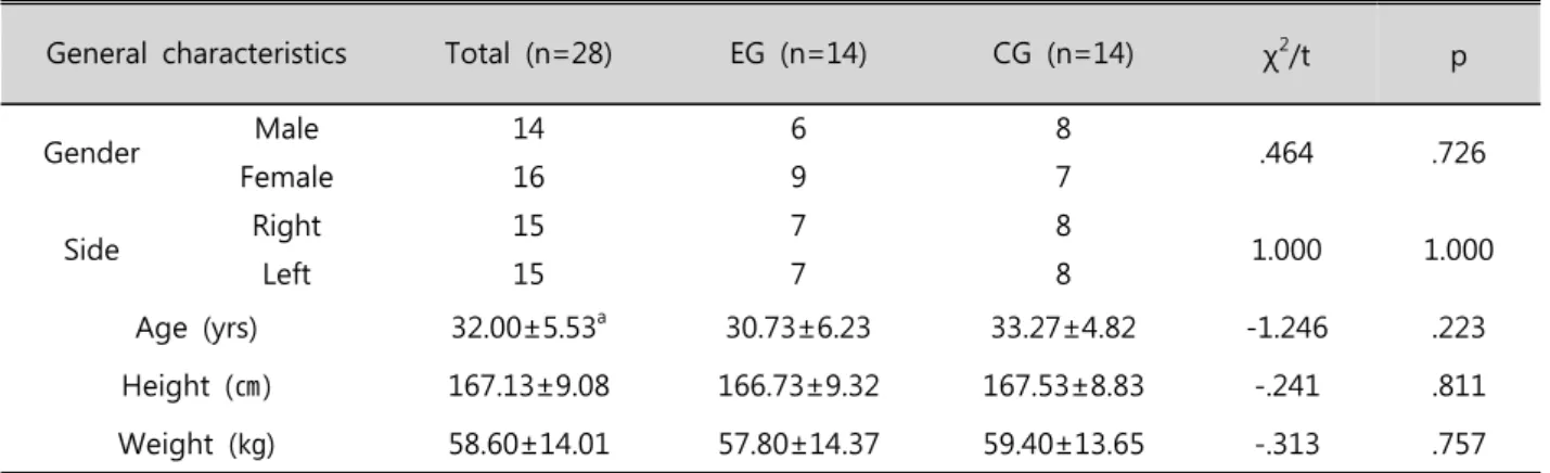
1. 연구대상자의 일반적인 특성
대상자는 실험군 15명(남 6명, 여 9명), 대조군 15명 (남 8명, 여 7명)으로 총 30명이었으며 두군 모두 동질 한 것으로 나타났다. 평균연령은 32.00±5.53세로 군 간 차이는 없었다. 신장은 167.13±9.08 ㎝이었고, 몸무게는 58.60±14.01 ㎏으로 나타나 두 군 간에 유의한 차이가 없었다(Table 1).
2. 중재 전과 후 근활성도의 변화 비교
흉추 도수교정 적용 전과 후의 근활성도의 변화는 다음과 같다. 중삼각근의 경우 흉추 도수교정 적용 전 20.49±5.30에서 적용 후 26.51±7.62로 증가하였고, 상 승모근의 경우 적용 전 16.89±8.08에서 적용 후 22.55±11.97로 증가하여 유의한 차이를 보였다. 전거근 의 경우 18.36±13.76에서 적용 후 23.85±15.32로 증가 하였으나 유의한 차이를 보이지 않았다(Table 2).
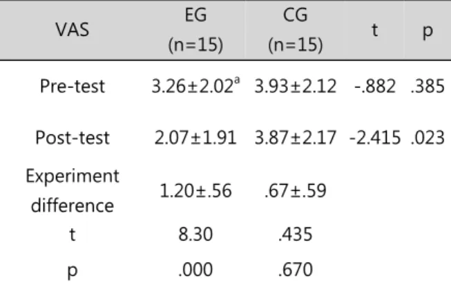
3. 중재 전과 후 통증의 변화 비교
대상자의 통증을 평가하는 VAS는 실험군이 흉추 도 수교정 적용 전 3.26±2.02점에서 적용 후 2.07±1.91점 으로 감소하여 실험 전과 후에 유의한 차이가 없었다.
두 군간의 실험 전 후를 비교한 결과 유의한 차이가 없 었다(Table 3).
4. 중재 전과 후 능동관절가동범위의 변화 비교 대상자의 관절가동범위를 평가하는 방법으로 통증범 위 내에서의 능동적 관절가동범위를 측정하였다.
실험군에서 관절가동범위는 105.07±13.6도에서 114.13±
12.92도로 9.07±8.42도의 증가를 보였으며 유의한 차이 가 있었다. 대조군은 101.87±15.72도에서 103.53±
15.24도로 1.67±5.69도의 증가를 보였으나 통계학적으 로 유의한 차이가 없었다(Table 4).
5. 중재 전과 후 둥근어깨자세의 변화 비교
대상자의 RSP를 평가하는 테이블과 견봉 후외측면 사이의 거리는 실험군에서 5.43±1.22 ㎝에서 5.33±1.25
㎝로 0.10±0.19 ㎝ 감소하였으며, 대조군에서는 5.30±
1.07 ㎝에서 5.17±0.81 ㎝로 0.13±0.39 ㎝ 감소하였으 나, 두 군 모두 통계적으로 유의한 차이가 없었다(Table 5).
Ⅳ. 고 찰
사람들은 문화적 습관, 직업, 행동양식, 나이와 관계 된 몸의 변화, 그리고 근골격계 구조를 포함하여 여러 가지 복잡한 요소에 따라 다양한 방식으로 자세를 취한 다(Shin와 Yoo, 2014; Caneiro 등, 2010; Szeto 등, 2005). 앉은 자세가 좋지 못하다면 흉추 후만의 증가와 어깨를 전방으로 이동시키는 자세가 되고 이것은 전방 머리자세를 포함하는 RSP가 된다(Yoo, 2013; Borstad와 Ludewig, 2005; Finley와 Lee, 2003). 이러한 자세는 견 갑골의 위치와 움직임, 그리고 근육활성의 변화를 가져 온다. 그리고 시간이 지나면 목과 어깨근육의 긴장과 스트레스는 증가하고, 결과적으로 통증과 감각이상, 기 능저하와 다양한 신경근증상이 나타나게 된다(Lee 등, 2013; Silva와 Johnson, 2013; Kang 등, 2012).
많은 선행연구에서 적절한 어깨자세는 근 골격계의 균형을 가져오고 상체의 스트레스와 긴장은 최소화시킨 다고 보고하였다. 척추정렬을 통한 중립자세유지는 잘 못된 척추, 흉추, 그리고 견갑골자세에 의한 부담을 줄 여줄 수 있고, 기능적인 자세를 유지시켜주는 깊은 자 세성 근육을 활성화 시킬 수 있다(Thigpen 등, 2010;
Falla 등, 2007). 이에 본 연구는 RSP로 인해 어깨움직 임의 기능저하가 있는 건강한 성인의 어깨기능을 개선 하기 위하여 흉추에 도수교정을 적용한 후 효과에 대하 여 알아보고자 하였다.
근활성도 측정 결과 실험군에서 전거근을 제외한 모
든 근육에서 유의한 차이를 보였으나, 대조군은 유의한
차이를 보이지 않았다. 척추의 정렬상태는 견갑골의 자
세와 견갑대의 기능에 영향을 미친다. 전방머리자세
(forward head posture)처럼 좋지 않은 척추의 정렬은
어깨와 목 주위근육의 긴장을 증가시키고 견갑골의 위
치와 움직임에 영향을 주며(Kebaetse 등, 1999), 견갑골
과 상완의 변화된 움직임은 이차적으로 근육의 불균형
을 유발한다. 또한 견갑골 상방회전의 길항근 역할을 하
는 견갑거근의 긴장은 견갑골의 움직임을 제한시킨다
(Weon 등, 2010).
Table 2. Comparison of muscle activity
EG (n=14) CG (n=14) t p
Middle deltiod
Pre-test 20.49±5.30
a19.61±7.13 .380 .707
Post-test 26.51±7.62 18.40±6.55 3.128 .004
Experiment difference -6.03±5.47 1.21±4.62
t -4.264 1.018
p .001 .326
Upper trapezius
Pre-test 14.46±3.93 15.03±6.40 -.294 .771
Post-test 19.30±7.38 13.46±4.63 2.597 .015
Experiment difference -4.84±4.61 1.57±4.00
t -4.074 1.517
p .001 .152
Lower trapezius
pre-test 16.89±8.08 15.16±7.41 .614 .544
post-test 22.55±11.97 13.75±6.64 2.489 .019
Experiment difference -5.65±9.31 1.41±4.03
t -2.352 1.352
p .034 .198
Serratus anterior
Pre-test 18.36±13.76 17.30±9.97 .241 .812
Post-test 23.85±15.32 15.39±9.64 1.810 .081
Experiment difference -5.49±10.96 1.91±4.64
t -1.938 1.597
p .073 .132
aMean(%MVIC)±SD, EG: experimental group, CG: control group
Table 1. General characteristics of the subjects
General characteristics Total (n=28) EG (n=14) CG (n=14) χ
2/t p
Gender Male 14 6 8
.464 .726
Female 16 9 7
Side Right 15 7 8
1.000 1.000
Left 15 7 8
Age (yrs) 32.00±5.53
a30.73±6.23 33.27±4.82 -1.246 .223 Height (㎝) 167.13±9.08 166.73±9.32 167.53±8.83 -.241 .811 Weight (㎏) 58.60±14.01 57.80±14.37 59.40±13.65 -.313 .757
aMean±SD, EG: experimental group, CG: control group
Table 3. Comparison of visual analogue scale
VAS EG
(n=15)
CG
(n=15) t p
Pre-test 3.26±2.02
a3.93±2.12 -.882 .385 Post-test 2.07±1.91 3.87±2.17 -2.415 .023 Experiment
difference 1.20±.56 .67±.59
t 8.30 .435
p .000 .670
aMean(point)±SD, EG: experimental group, CG: control group, VAS: visual analog scale
Table 5. Comparison of Supine RSP distance Distance EG
(n=15)
CG
(n=15) t p
Pre-test 5.43±1.22
a5.30±1.07 .316 .754
Post-test 5.33±1.25 5.17±.81 .415 .682 Experiment
difference .10±.19 .13±.39
t 2.010 1.246
p .064 .233
aMean(㎝)±SD, EG: experimental group, CG: control group, RSP: rounded shoulder posture
Table 4. Comparison of active range of motion
AROM EG
(n=15)
CG
(n=15) t p
Pre-test 105.07±13.60
a101.87±15.72 .596 .556
Post-test 114.13±12.92 103.53±15.24 2.055 .049 Experiment
difference -9.07±8.42 -1.67±5.69
t -4.170 -1.134
p .001 .276
aMean(°)±SD, EG: experimental group, CG: control group AROM: active range of motion