Relationships Between Rounded Shoulder Posture and Biceps Brachii Muscle Length, Elbow Joint Angle, Pectoralis Muscle Length, Humeral
Head Anterior Translation, and Glenohumeral Range of Motion
Sil-ah Choi1, MSc, PT, Heon-seock Cynn1,2, PhD, PT, Ji-hyun Lee1, PhD, PT, Da-eun Kim1, BPT, PT, A-reum Shin1, BPT, PT
1Applied Kinesiology and Ergonomic Technology Laboratory, Dept. of Physical Therapy, The Graduate School, Yonsei University
2Dept. of Physical Therapy, College of Health Science, Yonsei University
Abstract
1)Background: Rounded shoulder posture (RSP), a postural abnormality, might cause shoulder pain and pathologic conditions. Although most previous research has investigated RSP focusing on the proximal structures of the shoulder, such as the scapula and pectoralis muscles, the relationship between RSP and anterior distal structures of the upper extremity, such as the biceps brachii muscle and elbow joint, is not clearly understood.
Objects: This study aimed to investigate the correlations between RSP and the biceps brachii length, elbow joint angle (EJA), pectoralis minor length, general pectoralis major length, humeral head anterior translation (HHAT), glenohumeral internal rotation (IR), external rotation (ER), and horizontal adduction (HAD).
Methods: Twelve subjects with RSP (6 male, 6 female) were recruited. All subjects fulfilled the RSP criteria indicated by a distance ≥2.5 ㎝ from the posterior aspect of the acromion to the table in the supine position. The examiner measured each of the following parameters twice: RSP, biceps brachii length, EJA, pectoralis minor length, pectoralis major length, HHAT, glenohumeral IR, ER, and HAD.
Pearson’s correlation coefficient(r) was used to assess the correlation between RSP and all the variables.
Results: There was a significant moderate positive correlation between RSP and biceps brachii length (r=.55, p=.032), moderate negative correlation between RSP and pectoralis minor length (r=-.62, p=.015), and moderate positive correlation between RSP and HHAT (r=.53, p=.038).
Conclusion: The biceps brachii length, pectoralis minor length, and HHAT could be used to evaluate patients with RSP. Better understanding of the correlation between these factors and RSP could help in the development of effective methods to treat patients with this condition in clinical management.
Key Words: Biceps brachii muscle; Elbow joint angle; Rounded shoulder posture.
Introduction
Rounded shoulder posture (RSP) is a specific pos- tural abnormality that might cause shoulder pain and pathologic conditions such as subacromial impinge- ment syndrome, adhesive capsulitis, and rotator cuff disease (Lewis et al, 2005; Ludewig and cook, 2000;
Michener et al, 2005). In RSP cases, excessive hum- eral head anterior translation (HHAT) relative to the line of gravity of the body with glenohumeral in-
ternal rotation (IR) is common when the scapula is abducted and tilted anteriorly (Massimini et al, 2012;
Myers et al, 2006). Also, the elbow is excessively flexed, and the forearm is pronated and placed ante- rior to the hips, although the ideal alignment of the elbow is slight flexion, which indicates that the proximal and distal humerus is placed almost in the same vertical line, and the forearm is in neutral ro- tation so that the thumb is oriented anteriorly and the palm of the hand is oriented towards the body Corresponding author: Heon-seock Cynn [email protected]
(Page et al, 2009; Sahrmann, 2002).
Most previous research has investigated RSP fo- cusing on the proximal structures of the shoulder girdle, such as the scapular or humeral head position, glenohumeral range of motion (ROM), and pectoralis muscle length. To correct RSP, pectoralis muscle stretching or manual release (Borstad and Ludewig, 2006; Wong et al, 2010) and scapular repositioning or stabilization exercises such as exercises with a scap- ular brace, scapular posterior tilt exercise, and wall push-up plus are frequently used by physical thera- pists (Cole et al, 2013; Lee et al, 2015). However, the relationship between RSP and anterior distal struc- tures of the upper extremity, such as the biceps bra- chii muscle length and elbow joint angle (EJA), is not clearly understood. The biceps brachii muscle and elbow joint could be important components to consider when dealing with shoulder problems.
The biceps brachii muscle affects the glenohumeral joint function because of the two different attach- ments of the biceps brachii: the long and short heads. The long head of the biceps (LHB) is at- tached from the supraglenoid tubercle of the scapula and passes through the bicipital groove, and the short head of the biceps (SHB) is attached from the coracoid process of the scapula (Buck et al, 2011;
Drake et al, 2009; McGarry et al, 2016). Especially, the LHB has long been considered as a causative factor of anterior shoulder pain (Wilk and Hooks, 2016). Inflammation of the LHB diagnosed as biceps tendonitis has been associated with rotator cuff le- sions and subacromial impingement syndrome (Chen et al, 2005; Gill et al, 2007; Habermeyer et al, 2004).
Also, subcoracoid impingement syndrome related with the common origin of the coracoid process (the SHB and coracobrachialis) is characterized by anterior shoulder pain, cervical myofascial pain syndrome, and radiating pain to the upper arm and forearm (Karim et al, 2005; Paulson et al, 2001).
The myofascial pain syndrome may occur when the shoulder pain or injury originating from muscle imbalance, malalignment, or abnormal posture changes
the shoulder mechanics (Karim et al, 2005; Simon et al, 1999). Simon et al. (1999) reported that in pa- tients with trigger points in the pectoralis minor, bi- ceps brachii, or coracobrachialis muscles, pain can radiate from the anterior shoulder region and supra- scapular region to the arm. Clinically, upper ex- tremity pain or symptoms from sustained and re- peated postural compensation patterns, such as for- ward neck posture, RSP, or excessive kyphosis would be observed along with the myofascial meridian, for example, biceps tendinopathy, elbow epicondylitis, or carpal tunnel syndrome (Myers, 2009; Sahrmann, 2002). In this way, myofascial referral pain and the myofascial meridian may explain why the con- tributing factors to shoulder or scapular issues would involve not only the pectoralis major and minor but also the biceps brachii and elbow joint.
Therefore, the purpose of this study was to inves- tigate the correlations between RSP and the biceps brachii length, EJA, pectoralis minor length, general pectoralis major length, humeral head anterior trans- lation (HHAT), glenohumeral internal rotation (IR), ex- ternal rotation (ER), and horizontal adduction (HAD).
It was hypothesized that there would be a moderate to strong correlation between RSP and the biceps brachii muscle length, EJA, pectoralis minor muscle length, pectoralis major muscle length, HHAT, gle- nohumeral IR, ER, and HAD.
Methods
Subjects
A power analysis was performed with G*power software ver. 3.1.2 (Franz Faul, University of Kiel, Kiel, Germany) using the results of a pilot study in- volving 5 subjects. The sample size calculation was carried out with a power of .80, alpha level of .05, and effect size of .96. This provided a necessary sample size of 4 subjects for this study. Twelve subjects with RSP (6 male, 6 female) were recruited from a university population (age=21.7±1.7 years;
A
Figure 1. Measurement of biceps brachii muscle length.
B
Figure 2. Measurement of elbow joint angle.
height=167.3±8.6 ㎝; weight=59.7±11.5 ㎏; and body mass index=21.1±2.3 ㎏/㎡). All subjects fulfilled the specific RSP criteria in the preferred arm when eat- ing and writing (Yoshizaki et al, 2009). RSP was in- dicated by a distance ≥2.5 ㎝ from the posterior as- pect of the acromion to the table in the supine posi- tion (Sahrmann, 2002). Subjects were excluded if they had a history of surgery or existing pathologies of the shoulder and elbow, dysfunction that sub- stantially limited shoulder and elbow motion, current complaint of numbness or tingling in the upper ex- tremity limiting their activities, and any congenital postural abnormalities (Cole et al, 2013; Lee et al, 2015).
Prior to collecting data, the examiner informed the subjects of the study procedures and each subject completed an informed consent form. The study pro- tocol was approved by the Yonsei University Wonju Institutional Review Boar (approval number: 1041849- 201704-BM-034-01).
Procedures
The examiner measured each of the following twice: RSP, biceps brachii length, EJA, pectoralis mi- nor length, general pectoralis major length, HHAT, glenohumeral IR, ER, and HAD. The mean value from the two measurements was used for analyzing data.
1. Biceps brachii muscle length
The subject was positioned in the supine position, with the shoulder at the edge of a table with the el- bow extended and forearm pronated. After instruct- ing subjects on the motion desired, the examiner hy- perextended the shoulder completely through the available ROM while maintaining the elbow in full extension (Reese and Bandy, 2010). This passive movement allows an estimate of the available ROM. In this position, the digital inclinometer (GemRed DBB, Gain Express Holdings, Ltd., Hong Kong, China) was placed on the humerus aligned with the lateral aspect of the acromion process and lateral epicondyle of the humerus (Figure 1.).
2. Elbow joint angle
To achieve a natural resting posture, subjects were instructed to march in place 5 times, moving both the upper and lower extremities. Next, subjects stood in a relaxed position looking straight ahead with their feet shoulder width apart. Three reflective markers were placed over the acromion, humerus lat- eral epicondyle, and middle point of the radial styloid process and the ulnar head. The EJA was measured as the angle between the line connecting the acro- mion and the humerus lateral epicondyle and the line connecting humerus lateral epicondyle and the middle point of the radial styloid process and the ulnar head.
With Image J software (National Institutes of Health, Bethesda, MD, USA), the EJA was calculated auto- matically (Figure 2.).
3. Pectoralis minor muscle length
The subjects stood up straight with the forearm in neutral position and hand in resting position. To measure the muscle length of the pectoralis minor, the examiner marked the origin and insertion of the pectoralis minor on the skin (the medio-inferior as- pect of the coracoid process and just lateral to the sternocostal junction of the inferior aspect of the fourth rib). The examiner measured the distance be- tween the two bony landmarks with a digital caliper (Borstad and Ludewig, 2005).
4. General pectoralis major muscle length The subject was placed in the supine position, with the hands clasped together behind the head.
The examiner ensured that the subject maintained the hands clasped and did not flex the cervical spine any more than necessary to place the clasped hands behind the head. The subject relaxed the shoulder muscles, allowing the elbows to move toward the support surface; the lumbar spine was to remain flat against the support surface. Using a digital caliper, the examiner measured the distance between the ole- cranon process of the humerus and the support sur- face (Reese and Bandy, 2010)
5. Humeral head anterior translation
HHAT was measured with the subject lying in a relaxed position on a table, with both arms placed beside the trunk in the neutral position. The exam- iner then calculated the difference between the dis- tance from the table and the anterior peak of the humeral head, and the distance from the table and the acromion (Gong et al. 2013).
6. The range of glenohumeral internal rotation and glenohumeral external rotation
The subject was positioned in the supine position with the shoulder abducted 90° and the elbow flexed 90° to ensure a neutral horizontal position. The hu- merus was passively rotated internally and externally with the examiner stabilizing the scapula with his
other hand (Lunden et al, 2010). In this position, the digital inclinometer was placed on the dorsal surface of the forearm aligned with the long axis of the ulna for reference. A second examiner measured the angle between the forearm and a plane perpendicular to the table.
7. The range of glenohumeral horizontal adduction The examiner stood beside the table and posi- tioned the test shoulder with the elbow in 90° flexion and the shoulder abducted 90°. The examiner stabi- lized the lateral border of the scapula by providing a dorsally directed force to control scapular protraction, rotation, and abduction. The examiner held the fore- arm, and then moved the humerus into horizontal adduction passively until the limit of movement (Moore et al, 2011). An examiner recorded the ROM for horizontal adduction using a digital inclinometer aligned with the ventral midline of the humerus.
Statistical Analysis
The Kolmogorov-Smirnov Z-test was used to as- sess normal data distribution. Pearson’s correlation co- efficient (r) was used to assess the correlation be- tween RSP and the biceps brachii length, EJA, pec- toralis minor length, general pectoralis major length, humeral head anterior translation (HHAT), gleno- humeral internal rotation (IR), external rotation (ER), and horizontal adduction (HAD). As suggested by Portney and Watkins (2008), correlation coefficient values under .25, between .25 and .5, between .5 and .75, and above .75 indicated little or no relationship, fair relationship, moderate to good relationship, and good to excellent relationship, respectively. The level of significance was set at .05. All statistical analyses were performed using SPSS Statistics ver. 21.0 (SPSS, Inc., Chicago, IL, USA).
Results
All variables had a normal distribution. The meas-
Variable Mean±standard deviation
Rounded shoulder posture (㎜) 66.53±6.32
Biceps brachii length (°) 40.13±6.85
Elbow joint angle (°) 152.78±2.31
Pectoralis minor length (㎜) 136.58±22.73
General pectoralis major length (㎜) 69.55±36.93
Humeral head anterior translation (㎜) 34.00±2.71
Glenohumeral internal rotation (°) 56.60±12.12
Glenohumeral external rotation (°) 98.46±15.30
Glenohumeral horizontal adduction (°) 14.85±7.06
Table 1. Measurement values of all variables (N=12)
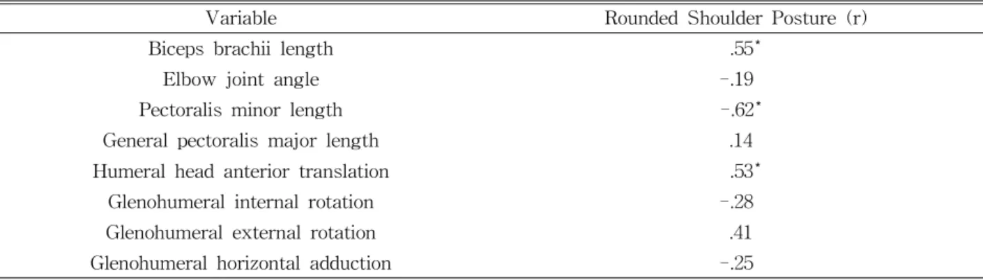
Variable Rounded Shoulder Posture (r)
Biceps brachii length .55*
Elbow joint angle -.19
Pectoralis minor length -.62*
General pectoralis major length .14
Humeral head anterior translation .53*
Glenohumeral internal rotation -.28
Glenohumeral external rotation .41
Glenohumeral horizontal adduction -.25
*p<.05.
Table 2. Pearson correlation coefficients (r) between round shoulder posture and all variables (N=12) ured values of RSP, biceps brachii length, EJA, pec-
toralis minor length, pectoralis major length, HHAT, glenohumeral IR, ER, and HAD are presented in Table 1. There was a significant moderate positive correlation between RSP and biceps brachii length (r=.55, p=.032), moderate negative correlation between RSP and pectoralis minor length (r=-.62, p=.015), and moderate positive correlation between RSP and HHAT (r=.53, p=.038) (Table 2).
Discussion
To our knowledge, this study is the first to ana- lyze the correlation between RSP and not only prox- imal structures of the shoulder girdle such as the humeral head position, the glenohumeral ROM, and pectoralis major, and minor muscles length, but also
distal structures of the upper extremity such as the biceps brachii muscle length and EJA. The results partially supported the research hypothesis.
This study found a significant moderate positive correlation between RSP and biceps brachii muscle length. RSP is related to the dominant or tight pec- toralis major muscle, which pulls the humeral head anteriorly (Konrad et al, 2006; Page et al, 2009;
Labriola et al, 2005), i.e., the glenohumeral position is regarded as extension. In the glenohumeral extension position, the biceps brachii muscle length increased, which supported our research. Many previous re- searchers found that the LHB functions as a stabil- izer for the glenohumeral joint (Itoi et al, 1993;
Pagnani et al, 1996; Rodosky et al, 1994). In a simu- lated cadaveric study, the LHP contraction prevented anterior, superior, and inferior translation of the humeral head (Pagnani et al, 1996). Especially, the
LHB develops the anterior stability of the gleno- humeral joint and resistance to torsional force, in- creasing the anterior instability when the shoulder is abducted and externally rotated (Itoi et al, 1993;
Rodosky et al, 1994). Also, the scapular position was associated with the biceps function (Kibler and McMullen, 2003; Lukasiewicz et al, 1999; Solem-Bertoft et al, 1993). Kibler and McMullen (2003) reported that secondary scapular dyskinesis from the abnor- mal scapular position or motion could affect the bi- ceps function because of coupled scapulohumeral movements. Therefore, the positive correlation be- tween RSP and biceps brachii muscle length in the present study was consistent with previous studies, which supported that the proper position of the scap- ula is needed to improve upper extremity function.
This study found a significant moderate negative correlation between RSP and pectoralis minor muscle length, supporting the research hypothesis. This finding is consistent with many previous studies (Burkhart et al, 2000; Escamilla et al, 2009; Hebert et al, 2002; Ludewig and Cook, 2000). Because the pectoralis minor is attached to the coracoid process, Escamilla et al (2009) indicated that scapular malalignment and dysfunction, especially scapular anterior tilting or winging, are associated with tightness of the pector- alis minor muscles. Hebert et al (2002) and Ludewig and Cook (2000) reported that increased tension of the pectoralis minor changes the subacromial space narrowly causing impingement syndrome. Accordingly, the importance of pectoralis minor stretching exercise is emphasized to correct RSP, thoracic kyphosis, or forward head posture, which are characterized by scapular malalignment (Borstad and Ludewig, 2005;
Finley and Lee, 2003). A previous report by Lee et al (2015) revealed that pectoralis minor stretching helped correct RSP, decreasing by 30.72%, and re- store the length of the pectoralis minor, increasing by 4.54%. These studies supported the fact that de- creased flexibility or shortness of the pectoralis mi- nor is a main factor to aggravate excessive forward scapular posture (Borstad and Ludewig, 2005).
This study found a significant moderate positive correlation between the RSP and HHAT, supporting the research hypothesis. RSP is associated with up- per crossed syndrome (USC) (Page et al, 2009). In UCS, tightness of the pectoralis major and minor on the anterior part crosses with tightness of the upper trapezius and levator scapulae on the posterior part.
The dominant or tight pectoralis major muscle pulls the humeral head anteriorly because the pectoralis major is attached to the lateral lip of the inter- tubercular groove (anteromedial area) of the humerus, so the humeral head is displaced more anterior than the distal humerus not in the same vertical line (Konrad et al, 2006; Page et al, 2009; Labriola et al, 2005). Konrad et al (2006) and Labriola et al (2005) found that the posture with HHAT reduced the gle- nohumeral stability and centration on the glenoid fossa, and pain appeared in the anteriomedial should- er during the glenohumeral motion. Similarly, Matsen et al (1991) and Bahk et al (2007) described the gle- nohumeral instability as a clinical sign of unwanted translation of the humeral head. Additionally, subjects with RSP or forward scapular posture frequently ex- perience posterior shoulder muscle tightness (Laudner et al, 2006; Tyler et al, 2000), and tight posterior shoulder muscles may cause anterior and superior translation of the humeral head on the glenoid fossa (McClure et al, 2007; Tyler et al, 2000).
Our study had anticipated originally that RSP would have significant relationship with the EJA for the following reasons. One reason is that RSP asso- ciated with the HHAT would induce the gleno- humeral extension position and deliver passive ten- sion of the long head of the biceps brachii and might cause reduction in a relaxed EJA (i.e., increased el- bow flexed position) (Chleboun et al, 1997; Dartnall et al, 2008). An altered length-tension relationship dis- turbs maintaining optimal muscle lengths for peak force generation, increases whole-muscle passive ten- sion, and reduces maximal force, which would cause difficulty in achieving an ideal joint angle (Newmann, 2002; Proske and Allen, 2005). Another possible ex-
planation is the presence of the myofascial meridian around the arm line, especially the deep front arm line, including the pectoralis minor and biceps brachii. According to Myers (2009), the fascia of the pectoralis minor connects to the short head of the biceps and coracobrachialis at the coracoid process.
As mentioned above, RSP was related to the pector- alis minor, and so we expected that the dominant pectoralis minor would tighten the biceps brachii and coracobrachialis, inducing excessive elbow flexion.
Despite these possible reasons, our study did not confirm a significant correlation between RSP and the EJA; therefore, future study with a greater num- ber of patients with RSP, chronic pain, or pathologic symptoms from RSP is needed clarify the relation- ship between RSP and the EJA.
In this study, there was no significant correlation between RSP and the glenohumeral ROM such as IR, ER, and HAD. The reason for this unexpected result is that this study did not focus on tight or stiff posterior shoulder muscles or capsule regardless of recruiting subjects with RSP. Many previous studies reported that posterior shoulder tightness leads to approximation of the humeral head to the acromion causing shoulder impingement (Choi et al, 2012; Tyler et al, 2000; Yang et al, 2012), related to altered acromioclavicular and sternoclavicular joint motion (Wong et al, 2010), and change in gleno- humeral flexion, IR, and HAD (Kibler and McMullen, 2003; Laudner et al, 2006; Tyler et al, 2000).
Additionally, posterior shoulder tightness contributed 5% more to the prediction of forward scapular pos- ture (Lee et al, 2015). Therefore, the glenohumeral ROM should be considered as a significant contrib- utor to clinical RSP.
This study has several limitations. First, our find- ings could not be generalized to the general patient population because healthy young subjects with only specific RSP criteria participated in this study.
Therefore, young as well as older subjects with var- ious clinical assessments for RSP should be inves- tigated in future studies. Second, because this study
only examined the relationship between RSP and in- dependents variables, it could not explain the cause and effect for RSP by other independents variables.
Further research about the long-term effect of ther- apeutic intervention or a randomized controlled trial is warranted to validate the results of our study.
Conclusion
This study examined the relationship between RSP and the biceps brachii length, EJA, pectoralis minor length, pectoralis major length, HHAT, glenohumeral IR, ER, and HAD. There was a significant positive correlation between RSP and biceps brachii muscle length, negative correlation between RSP and pector- alis minor muscle length, and positive correlation be- tween RSP and HHAT. While these findings do not establish a cause and effect relationship, the results suggest that the assessment of HHAT, pectoralis minor muscle length, and especially biceps brachii muscle length should be taken into consideration when evaluating RSP. Better understanding of the correlation between these factors and RSP could help in the development of effective methods to treat pa- tients with this condition in clinical management.
References
Bahk M, Keyurapan E, Tasaki A, et al. Laxity test- ing of the shoulder: A review. Am J Sports Med. 2007;35(1):131-144.
Borstad JD, Ludewig PM. Comparison of three stretches for the pectoralis minor muscle. J Shoulder Elbow Surg. 2006;15(3):324-330.
Borstad JD, Ludewig PM. The effect of long versus short pectoralis minor resting length on scapular kinematics in healthy individuals. J Orthop Sports Phys Ther. 2005;35(4):227-238.
Buck FM, Dietrich TJ, Resnick D, et al. Long biceps tendon: normal position, shape, and orientation
in its groove in neutral position and external and internal rotation. Radiology. 2011;261(3):
827-881.
Burkhart SS, Morgan CD, Kibler WB. Shoulder in- juries in overhead athletes. The “dead arm”
revisited. Clin Sports Med. 2000;19(1):125-158.
Chen CH, Hsu KY, Chen WJ, et al. Incidence and severity of biceps long head tendon lesion in patients with complete rotator cuff tears. J Trauma. 2005;58(6):1189-1193.
Chleboun GS, Howell JN, Conatser RR, et al. The relationship between elbow flexor volume and angular stiffness at the elbow. Clin Biomech (Bristol, Avon). 1997;12(6): 383-392.
Choi SA, Lee JH, Yoon TL, et al. Immediate effects of soft tissue massage on posterior shoulder muscle tightness: A preliminary study. Phys Ther Korea. 2012;19(4):8-15.
Cole AK, McGrath ML, Harrington SE, et al. Scapular bracing and alteration of posture and muscle ac- tivity in overhead athletes with poor posture. J Athl Train. 2013;48(1):12-24. https://doi.org/10.4085/
1062-6050-48.1.13
Dartnall TJ, Nordstrom MA, Semmler JG. Motor unit synchronization is increased in biceps brachii after exercise-induced damage to elbow flexor muscles. J Neurophysiol. 2008;99(2):1008-1019.
https://doi.org/10.1152/jn.00686.2007
Drake RL, Vogl W, Mitchell AWM. Gray’s Anatomy for Students. 2nd ed. Philadelphia, Churchill Livingstone, 2009:680.
Escamilla RF, Yamashiro K, Paulos L, et al. Shoulder muscle activity and function in common shoulder rehabilitation exercises. Sports Med. 2009;39(8):
663-685. https://doi.org/10.2165/00007256-200939080- 00004
Finley MA, Lee RY. Effect of sitting posture on 3-dimensional scapular kinematics measured by skin-mounted electromagnetic tracking sensors.
Arch Phys Med Rehabil. 2003;84(4):563-568.
Gill HS, El Rassi G, Bahk MS, et al. Physical ex- amination for partial tears of the biceps tendon.
Am J Sports Med. 2007;35(8):1334-1340.
Gong WT, Jun IS, Choi YR. An analysis of the cor- relation between humeral head anterior glide posture and elbow joint angle, forward head posture and glenohumeral joint range of motion.
J Phys Ther Sci. 2013;25(4):489-491.
Habermeyer P, Magosch P, Pritsch M, et al.
Anterosuperior impingement of the shoulder as a result of pulley lesions: A prospective arthroscopic study. J Shoulder Elbow Surg. 2004;13(1):5-12.
Hebert LJ, Moffet H, McFadyen BJ, et al. Scapular behavior in shoulder impingement syndrome.
Arch. Phys. Med. Rehabil. 2002;83(1):60-69.
Itoi E, Kuechle DK, Morrey BK, et al. Stabilizing function of the biceps in stable and unstable shoulders. J Bone Joint Surg Br. 1993;75(4):
546-550.
Karim MR, Fann AV, Gray RP, et al. Enthesitis of biceps brachii short head and coracobrachialis at the coracoid process: A generator of shoulder and neck pain. Am J Phys Med Rehabil.
2005;84(5):376-380.
Kibler WB, McMullen J. Scapular dyskinesis and its relation to shoulder pain. J Am Acad Orthop Surg. 2003;11(2):142-151.
Konrad GG, Jolly JT, Labriola JE, et al.
Thoracohumeral muscle activity alters gleno- humeral joint biomechanics during active abduction. J Orthop Res. 2006;24(4):748-756.
Labriola JE, Lee TQ, Debski RE, et al. Stability and instability of the glenohumeral joint: the role of shoulder muscles. J Shoulder Elbow Surg.
2005;14(1 suppl S): 32S-38S.
Laudner KG, Stanek JM, Meister K. Assessing pos- terior shoulder contracture: the reliability and validity of measuring glenohumeral joint hori- zontal adduction. J Athl Train. 2006;41(4):
375-380.
Lee JH, Cynn HS, Yoon TL, et al. The effect of scapular posterior tilt exercise, pectoralis minor stretching, and shoulder brace on scapular alignment and muscles activity in subjects with
round-shoulder posture. J Electromyogr Kinesiol.
2015;25(1):107-114. https://doi.org/10.1016/j.jelekin.
2014.10.010
Lewis JS, Wright C, Green A. Subacromial impinge- ment syndrome: The effect of changing posture on shoulder range of movement. J Orthop Sports Phys Ther. 2005;35(2):72-87.
Ludewig PM, Cook TM. Alterations in shoulder kin- ematics and associated muscle activity in people with symptoms of shoulder impingement. Phys Ther. 2000;80(3):276-291.
Lukasiewicz AC, McClure P, Michener L, et al.
Comparison of 3-dimensional scapular position and orientation between subjects with and with- out shoulder impingement. J Orthop Sports Phys Ther. 1999;29(10):574-583.
Lunden JB, Muffenbier M, Giveans MR, et al.
Reliability of shoulder internal rotation passive range of motion measurements in the supine versus sidelying position. J Orthop Sports Phys Ther. 2010;40(9):589-594.
Massimini DF, Boyer PJ, Papannagari R, et al.
In-vivo glenohumeral translation and ligament elongation during abduction and abduction with internal and external rotation. J Orthop Surg Res. 2012;7(1):29-37.
Matsen FA, Harryman DT, Sidles JA. Mechanics of glenohumeral instability. Clin Sports Med. 1991;
10(4):783-788.
McClure P, Balaicuis J, Heiland D, et al. A random- ized controlled comparison of stretching proce- dures for posterior shoulder tightness. J Orthop Sports Phys Ther. 2007;37(3):108-114.
McGarry MH, Nguyen ML, Quigley RJ, et al.
The effect of long and short head biceps loading on glenohumeral joint rotational range of motion and humeral head position. Knee Surg Sports Traumatol Arthrosc. 2016;24(6):1979-1987.
https://doi.org/10.1007/s00167-014-3318-5
Michener LA, Pidcoe PE, Frith AM. Reliability and validity of scapular muscle strength testing in patients with shoulder pain and functional loss.
Phys Ther. 2005;85(5):1128-1138.
Moore SD, Laudner KG, McLoda TA, et al. The immediate effects of muscle energy technique on posterior shoulder tightness: A randomized con- trolled trial. J Orthop Sports Phys Ther. 2011;
41(6):400-407.
Myers JB, Laudner KG, Pasquale MR, et al.
Glenohumeral range of motion deficits and poste- rior shoulder tightness in throwers with patho- logic internal impingement. Am J Sports Med.
2006;34(4):385-391.
Myers TW. Anatomy Trains: Myofascial meridians for manual and movement therapists. 2nd ed.
Churchill Livingstone, Elsevier Health Sciences, 2009:149-158.
Newmann DA. Kinesiology of the Musculoskeletal System: Foundations for physical rehabilitation.
1st ed. St Louis, Mosby, 2002:129-130.
Page P, Frank CC, Lardner R. Assessment and Treatment of Muscle Imbalance: The janda approach. 1st ed. Champaign, Human Kinetics, 2009:52-55.
Pagnani MJ, Deng XH, Warren FR, et al. Role of the long head of the biceps brachii in glenohumeral stability: A biomechanical study in cadaver. J Shoulder Elbow Surg. 1996;5(4):255-262.
Paulson MM, Watnik NF, Dines DM. Coracoid im- pingement syndrome, rotator interval re- construction, and biceps tenodesis in the over- head athlete. Orthop Clin North Am. 2001;32(3):
485-493.
Portney LG, Watkins MP. Foundations of Clinical Research: Applications to practice. 3rd ed. Upper Saddle River, NJ, Prentice Hall. 2008:525.
Proske U, Allen TJ. Damage to skeletal muscle from eccentric exercise. Exerc Sport Sci Rev. 2005;
33(2):98-104.
Rodosky MW, Harner CD, Fu FH. The role of the long head of the biceps muscle and superior glenoid labrum in anterior stability of the shoulder. Am J Sports Med. 1994;22(1):121-130.
Sahrmann SA. Diagnosis and Treatment of
This article was received April 5, 2017, was re- viewed April 5, 2017, and was accepted May 10, 2017.
Movement Impairment Syndrome. 1st ed. St.
Louis, Mosby, 2001: 231-234.
Reese NB and Bandy WD. Joint Range of Motion and Muscle Length Testing. 2nd ed. Churchill Livingstone, Elsevier Health Sciences, 2010:
146-147,156-157.
Simon DG, Travell JG, Simons LS, et al. Myofascial Pain and Dysfunction: The trigger point manual.
2nd ed. Baltimore, Williams and Wilkins, 1999:
648-651.
Solem-Bertoft E, Thuomas KA, Westerberg CE. The influence of scapular retraction and protraction on the width of the subacromial space. An MRI study. Clin Orthop Relat Res. 1993;296:99-103.
Tyler TF, Nicholas SJ, Roy T, et al. Quantification of posterior capsule tightness and motion loss in patients with shoulder impingement. Am J Sports Med. 2000;28(5):668-673.
Wilk KE, Hooks TR. The painful long head of the biceps brachii: Nonoperative treatment approaches.
Clin Sports Med. 2016;35(1):75-92. https://doi.org/
10.1016/j.csm.2015.08.012
Wong CK, Coleman D, Song J, et al. The effects of manual treatment on rounded-shoulder posture, and associated muscle strength. J Bodyw Mov Ther. 2010;14(4):326-333. https://doi.org/10.1016/
j.jbmt.2009.05.001
Yang JL, Chen SY, Hsieh CL, et al. Effects and predictors of shoulder muscle massage for pa- tients with posterior shoulder tightness. BMC Musculoskelet Disord. 2012;13:46-53. https://doi.org/
10.1186/1471-2474-13-46
Yoshizaki K, Hamada J, Tamai K, et al. Analysis of the scapulohumeral rhythm and electromyography of the shoulder muscles during elevation and lowering: Comparison of dominant and non- dominant shoulders. J Shoulder Elbow Surg.
2009;18(5):756-763. https://doi.org/10.1016/j.jse.2009.
02.021