INTRODUCTION
A major consideration in designing implants has been focused to produce surfaces that promote desirable responses in cells and tissues contacting with implants.1,2The tissue response to a dental implant may involve (1) physical factors such as size, shape, surface topography, (2) relative interfacial movement, as well as (3) chemical factors associated with composition and structure of implants.3The first factor should be considered as morphological compatibility. The second and third factors are well documented as mechanical compatibility and biological compatibility respectively. These three compatibilities are required for successful biofunctional implant system.4
Osseointegration, structural and functional bonding between living bones and surfaces of implants, is believed to be an important factor for implant success5. After implant installation, a chamber was first filled with a blood clot6. In spaces between the implant surfaces and the pristine bone, blood clot formation results in the organization of an early granulation tissue after 4 days. This granulation tissue will give rise to an osteocoating of the implant surfaces after 1 week. From 2 to 4 weeks, this primary bone spongiosa or
primary spongework was replaced with lamellar and/or parallel fibered bone7.
In the overall processes of osseointegration, the initial stage is the most important step. The initial interaction between living bone and implant surface occurs as the surface of the implant biomaterial is exposed to tissue fluids5. When blood comes in contact with foreign solid surfaces, two well-recognized processes may occur: the adsorption of proteins leading to coagulation and the adhesion of platelets, at first to the solid and then to each other8,9. The developing platelets form a thrombus and preosteoblasts adhere to the solid surface. After the preosteoblasts in blood were attached to the surface of implants, they were differentiated into osteoblasts and progressed into the procedure of osseointegration.
This initial stage was mainly affected by the surface topography, wettability and bioactive molecules. The surface topography determines the degree of cell adhesion.
There are various types of surface treatment with titanium implants. They may be subdivided into implants with roughened surfaces by coating [e.g. resorbable blast media (RBM), hydroxyapatite coated (HA)], implants with the
WETTABILITY AND DRUG DELIVERY OF FUNCTIONALLY GRADED NANO-MICRO POROUS TITANIUM SURFACE
Kwi-Dug Yun1, DDS, Mong-Sook Vang2, DDS, PhD, Hong-So Yang2, DDS, PhD, Sang-Won Park3*, DDS, PhD, Ha-Ok Park4, DDS, PhD, Hyun-Pil Lim5, DDS
1Graduate student, Department of Prosthodontics, College of Dentistry, Chonnam University
2Professor, Department of Prosthodontics, College of Dentistry, Chonnam University
3Associate professor, Department of Prosthodontics, College of Dentistry, Chonnam University
4Assistant professor, Department of Prosthodontics, College of Dentistry, Chonnam University
5Clinical instruction, Department of Prosthodontics, College of Dentistry, Chonnam University
Corresponding Author: Sang-Won Park
Department of Prosthodontics, School of Dentistry, Chonnam National University Graduate School Hak 1 dong, Dong gu, Gwangju, 504-190, Korea +82-62-220-4420: e-mail, [email protected] Received June 9, 2008 Last Revison June 17, 2008 Accepted June 20, 2008.
※ This work was supported by the Korea Research Foundation Grant funded by the Korean Government (KRF-2007-313-D00964).
roughened surfaces with electrochemical modifications (anodic oxidation) of the commercially pure titanium implants and implants with the roughened surfaces without coating [e.g. sand-blasted or acid-etched]. Anodic oxidation technique increases the thickness of the titanium oxide layer from several nanometer to several micrometer and creates pores on titanium surface10. Anodic oxidation reduces the releasement of metal ion and increases the resistance of corrosion. This kind of surface remarkably improves wettability, and accelerates adherence and activity of osteoblasts, therefore enhances osteointegration11.
Wettability can be expressed by the contact angle of liquid on solid substrate4. Wettability is determined by the degree of blood adhesion associated with osteogenic cells.
As it is more hydrophilic, microvilla and filopodia of cells can be more easily permeable to the energy barrier between the cells and materials12. It improves the migration and attachment of osteogenic cells in the blood. Accordingly, wettability is a critical factor in immobilization of biological cells13.
The boactive molecules like bone morphogenetic proteins (BMPs) belonging to the transforming growth factor-β superfamily and play an important role in osteogenesis and bone metabolism14. Among them, BMP-2 has very strong osteoinductive activity by stimulating differentiation of mesenchymal stem cells toward an osteoblastic lineage, thereby increasing the number of differentiated osteoblasts able to form new bone14. Despite its strong osteoinductive activity, clinical use of BMP-2 has been hampered by the lack of suitable delivery systems and half life of rhBMP-215. Currently clinical usage of rhBMP-2 is limited to be inserted into scaffold such as collagen or gelatin. Or it may be used as a coating material to bone. However there is little study about using for a implant as a coating. Due to its short half life, it is critical to have a capacity for delayed release.
Thus an efficacious delivery system is needed to localize BMP-2 at the defect site for prolonged periods of time with appropriate dose15.
The aim of this study is to analyze the wettability of nano tubular structures formed by anodic oxidation methods, to assess the bone healing around this new surface in vivo for clinical application and to evaluate whether this type of tubular surface structure is able to serve as the drug delivery system.
MATERIALS AND METHODS
1. Preparation of Ti samples and surface roughness test
Commercially pure titaniums (ASTM Grade Ⅱ, Kobe Steel Co., Japan) discs having 15 mm in diameter and 2 mm in thickness were used. Control and experimental groups are classified into 4 groups as follow.
Group 1 (polished group, machined surface); mechanically polished surface grinded by #1000 grit silicon carbide (SiC) metallographic papers
Group 2 (micro group, blasting surface); sandblasting on one side of the plate with 50 ㎛ alumina particles (Al2O3) at 20 psi for 1 minute with a fixed distance (1 ㎝) between the surface of the sample and a blasting tip
Group 3 (nano group, anodized-machined surface); uniform nano-size tube formation by anodic oxidation technique on the machined surface
Group 4 (nano-micro group, anodized-blasting surface);
uniform nano-size tube formation by anodic oxidation technique on Al2O3 blasting surface
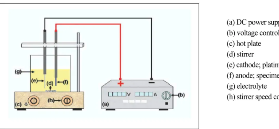
All experimental samples, prior to various treatments, were mechanically polished under the same condition as for control group. The appliance system for anodic oxidation is shown in Fig. 1.
For the removal of cutting oil and sterilization, all specimens were ultrasonically cleaned with acetone for 30 minutes and then additional ultrasonic cleansing was done with isopropyl alcohol for 30 minutes followed by drying at 200 �C for 1 hours.
Surface roughness was measured using a roughness tester (Diavite DH-7, Asmeto AG, Switzerland). The measured surface roughness was represented as Ra (arithmetical mean roughness) and ten samples per group were used.
2. Wettability test
Four kinds of experimental groups were prepared and 10 specimens per each group were used for wettability, which was evaluated from dynamic contact angle analysis.
Three different media were chosen for measuring the surface contact angles; distilled water, blood plasma and
rhBMP-2 (recombinant human bone morphogenetic protein-2) solution. Distilled (or deionized) water was normally employed for contact angle measurements. The present study also included plasma, because it played a critical role for the formation of early osseointegration stage. For the plasma, whole blood was collected from healthy individuals offered with the citrate phosphate dextrose solution, and centrifuged (2000 × g) for 4 minutes in 4 �C. Supernatant was transferred carefully to new tubes with pipettes. Because rhBMP-2 serves as a medium to evaluate the nano-size tubes for a drug delivery system, further rhBMP-2 solution is employed for contact angle measurement. rhBMP-2 was resolved in phosphate buffered saline (PBS) in the concentration of 250 ng/㎖.
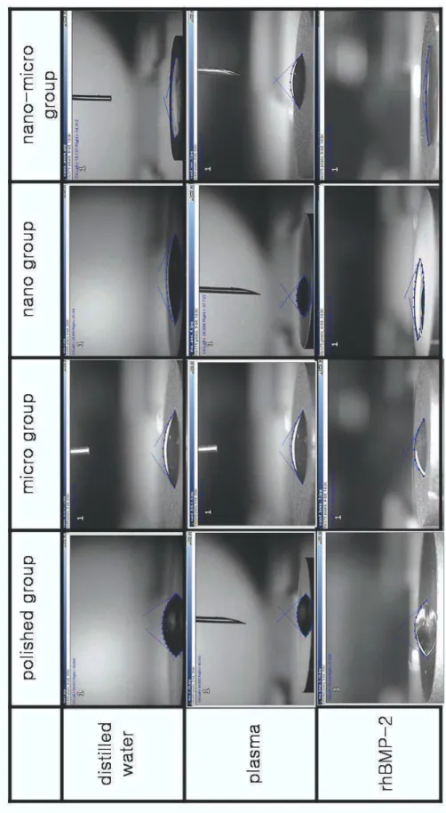
Contact angles were measured using the sessile drop method. After dropping each medium (0.025 ㎖) such as distilled water, plasma and rhBMP-2 solution on the specimen, a photo of each specimens was taken by video camera (Camscope�, Sometech, Korea). The drop image was captured and the contact angle (Θ) was calculated by an image analysis system (Dropsnake�, National Institute of Health, USA) as in Fig. 2. The values of contact angle were obtained by averaging right and left sides of drop. All specimens were read in the same environment by one trained investigator.
3. In vivo study for osseointegration
Intermezzo� (Megagen, Gwangju, Korea) implants were used in this study. The implants of 2.0 ㎜ in diameter were cut from top into 5.0 ㎜ in length to fit the thickness of the rat tibia. Two kind of implant surface were used. One was anodized-machined surface (nano group), which has uniform nano-size tube (200 nm in diameter, 500 nm in length) formed by anodic oxidation technique on the machined implant surface. The other was anodized-blasting surface (nano-micro group), which had uniform nano-size tube formed by anodic oxidation technique on resorbable Fig. 1. Schematic diagram of anodic oxidation apparatus. Titanium disc was linked on anode and platinum plate (3 ㎜ × 4 ㎜
× 0.1 ㎜) was linked on cathode. The distance between the anode and cathode was maintained in about 10 mm and main power device was used as DC power supply (Fine Power F-3005, SG EMD, Korea). Under 20 V, anodic oxidation was oper- ated in room temperature for 10 minutes. Electrolyte solution was contained 1 M phosphoric acid (H3PO4) and 1.5 wt%
hydrogen fluoride (HF).
(a) DC power supply (b) voltage controller (c) hot plate (d) stirrer
(e) cathode; platinum plate (f) anode; specimen (g) electrolyte
(h) stirrer speed controller
Fig. 2. Contact angle measurement by image analysis sys- tem.
blasting media (RBM) surface. The implant after surface treatment and cutting were sterilized by autoclaving.
Four male Wistar rats (average 400 g body weight) were used for the animal experiments. Animal care and experimental procedures were performed in accordance with the Guidelines for Animal Experimentation of Chonnam National University with the approval of the Ethics Committee for Animal Research. Four rats were anesthetized by injection of a mixture of ketamine (80 ㎍/g, Yuhan, Seoul, Korea) and xylazine (7 ㎍/g, Bayer Korea, Ansan, Korea) intraperitoneally. After shaving the animal’s right and left legs area and disinfecting with potadin, a sagittal incision around the proximal tibia was prepared manually under sterile conditions. After dissection of the proximal tibial muscle, the bone was exposed. A hole, 1.8
㎜ in diameter and depth of 5.0 ㎜, was formed 5.0 ㎜ below the knee joint with an dropping of sterile physiological saline solution. The implant sites were drilled sequentially using lance drill, 1.4 and 1.8 ㎜ in diameter at a speed of 1200 rpm. Nano group implant was inserted into right tibia, and nano-micro group implant was inserted into left tibia. The soft tissues were closed in two layers with absorbable sutures (4-0 vicryl, johnson and johnson medi- cal�, USA) and the wound region was disinfected again.
The animals were allowed to move freely without any restriction. The animals of each group were euthenized with carbon dioxide at 21 days after surgery and the tibia were prepared for further histological analysis as described below.
Directly after sacrifice, retrieved implants with the surrounding tissues were fixed in 4 % phosphate buffered formaldehyde solution. Subsequently, the specimens were dehydrated and embedded in methyl methacrylate resin.
After polymerization, sections containing tissue/implant interface were obtained using a slow speed diamond saw (Buehler Isomet saw, Buehler, Germany). The sections were then grounded using 400 and 600 grit sand paper on a wheel grinder (Alpha™and Beta™ Grinder/Polishers, Buehler, Germany) and stained with haematoxylin and eosin.
4. rhBMP-2 delivery
Four kinds of experimental group were immersed in 200
㎕ of rhBMP-2 solution (250 ng/㎖) and then shaked with
shaker (Neuronfit, Seoul, Korea) for 48 hours at 37 �C.
After 48 hours, the excess solution of rhBMP-2 on the titanium surface was removed. Then, the specimens were lyophilized for 24 hours, and immersed in 200 ㎕ of PBS solution for 72 hours. The amount of rhBMP-2 solution eluted from the solution was measured by ELISA according to manufacturer’s recommendation.
Briefly, all reagents and samples were prepared at room temperature before use. 100 ㎕ of assay diluent RD1-19 was added to each well of 96 well plate, and 50 ㎕ of standard or sample was added into well. It was incubated for 2 hours at room temperature on horizontal orbital microplate shaker set at 500 rpm. After that, each well was aspirated and washed four times. 200 ㎕ of BMP-2 conjugate was added to each well and incubated for 2 hours at room temperature on the shaker. After aspiration and washing four times, 200 ㎕ of substrate solution was added to each well, and incubated for 30 minutes at room temperature. After 30 minutes, 50 ㎕ of stop solution was added to each well. The optical density of each well were determined within 30 minutes, using microplate reader set to 450 ㎚ with standard solution.
5. Statistical analysis
One-way analysis of variance (ANOVA) was performed on the data of surface treatment and contact angle. I was also used to analyze the relationship of surface treatment and amount of rhBMP-2. Individual comparisons among group mean values were accompanied through the application of Tukey’s HSD. This conducted with the SPSS for windows software (SPSS 12.0; SPSS Inc., Chicago, IL, U.S.A) at a significance level of P<0.05.
RESULTS
1. Preparation of Ti samples and roughness test
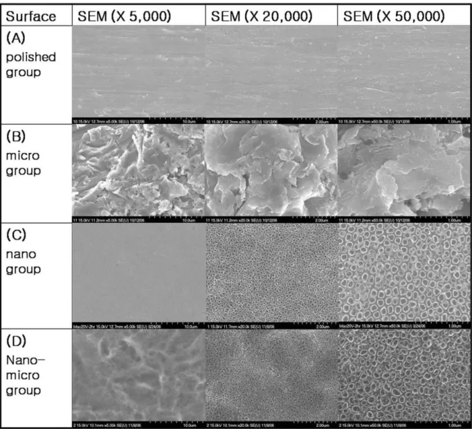
FE-SEM (Field Emission Scanning Electron Microscope) images for the experimental groups is shown in Fig. 3.
After anodizing, the surface roughness of the nano group was significantly increased, but the surface roughness of nano-micro group was significantly decreased (Table I).
The decrease in roughness of the nano-micro group is
probably due to the smoothing of the sharp edge shown in Fig. 3.
2. Wettability test
Table II shows the average and standard deviations of contact angle for the distilled water, plasma, and rhBMP-2
solution after dropping. In order to compare the results more easily, histograms are drawn for the overall results (Fig. 4).
Nano-micro group had the lowest contact angle value in distilled water and the order of contact angle values is as follow; nano-micro group > nano group > micro group >
and polished group (Fig. 5). After anodic oxidation treatment, titanium surface has given much smaller contact
Fig. 3. FE-SEM images of specimen. (A) polished group; it showed smooth surface on the SEM image. (B) micro group; it showed rough- ened surface in micro unit on the titanium surface by Al2O3. (C) nano group; It showed similar smooth surface to that of polished surface at low magnification (× 5,000). But at high magnification (× 50,000), it showed nano-sized tube formation on the smooth titanium surface.
(D) nano-micro group; It showed roughened surface in micro units as that of microsurface at low magnification (× 5,000). But at high magnification (× 50,000), it showed nano-sized tube formation on the microsurface.
angle values. There were statistically significant differences in one-way ANOVA (P<0.001), and statistical significance except between micro group and nano group in the Tukey’s HSD. In the experiment of plasma, the results of contact angle showed the ascending order; nano group > nano- micro group > micro group > polished group. The result had also shown statistically significant differences (P<0.001) and there were no statistically significant differences in groups between micro group and nano group, micro group and nano-micro group, and nano group and nano-micro group. However, the other groups were statistically significant differences. The contact angle of rhBMP-2 showed the similar tendency with distilled water. The contact angle values of rhBMP-2 gave statistical significance except between micro group and nano-micro group.
3. In vivo study for osseointegration
Figure 6 shows a light microscopy image of implant section stained with haematoxylin and eosin. Three weeks
later, the bone was healed well surrounding the titanium implant without fibrous tissue engagement and complication of immunologic reaction. At longitudianl section, new bone formation was observed along the implant surface in a cancellous bone marrow space as well as a cortical bone. There was little difference between the nano group implants and the nano-micro implants. The cross section of implants showed the similar tendency with longitudinal section of implant.
4. rhBMP-2 delivery
After soaking disks in the concentration of 250 ng/㎖ of rhBMP-2 for 48 hours, the excess solution of rhBMP-2 were removed. After that, they were lyophilized for 24 hours, and then the rhBMP-2 on the surface of titanium was reserved for 72 hours by PBS (phosphate buffered saline).
All the extracted solution was analyzed by ELISA.
The amount of rhBMP-2 solution on the titanium surfaces was measured by ELISA (Fig. 7). The concentration of rhBMP-2 solution on the different surface-treated titanium
Table I. Surface roughness of Ti specimens
Titanium surface Roughness (μm)
polished group 0.35±0.05
micro group 1.95±0.10
nano group 0.52±0.04
nano-micro group 1.57±0.10
The data are listed as mean values ± standard deviations (n=10).
Table II. Sessile drop measurement (contact angle) on different titanium surfaces
polished group micro group nano group nano-micro group
Distilled water 55.3±6.2bcd 33.6±5.9acd 27.6±5.4ad 18.9±6.1abc
Plasma 59.5±8.9bcd 43.3±7.6ac 36.1±6.3a 41.4±3.3a
RhBMP-2/PBS 58.5±7.2bcd 33.7±7.4ac 16.2±2.9abd 12.8±2.6ac
Data were expressed as mean and standard deviation. a is statistically significance with polished group, b with micro group, c with nano group and d with nano-micro group respectively.
Fig. 4. Contact angle on different surface with 3 different medium after dropping.
Fig. 5. Contact angle measurement by image analysis system with distilled water, plasma and rhBMP-2 solution.
Fig. 7. ELISA results of the specimens.
Table III. The concentration of rhBMP-2 on the titanium surfaces with different treatments
Titanium surface Concentration of rhBMP-2 solution (pg/㎖)
polished group 48.4 ± 12.5
micro group 63.0 ± 46.0
nano group 260.7 ± 57.4
nano-micro group 492.1 ± 15.5
Fig. 6. A light microscopy image of the longitudinal section of implant. (A) nano group implant (× 20). (B) nano group implant (× 40). (C) nano-micro group implant (× 20). (D) nano-micro group implant (× 40).
(A)
(C) (D)
(B)
was measured (Fig. 8). Table III shows the concentration of rhBMP-2 on the titanium surfaces with different treatments.
After 3 days reactions, nano-micro group (492.1±15.5 pg/
㎖) delivered the most amount of rhBMP-2, followed by nano group (260.7±57.4 pg/㎖), micro group (63.0±46.0 pg/㎖), and polished group (48.4±12.5 pg/㎖). Ten times more amount of rhBMP-2 was delivered from the nano- micro group than on the polished group. After anodic oxidation, all the surfaces delivered more amount of rhBMP-2. The amount of transferable rhBMP-2 showed statistical significance except between polished group and micro group.
DISCUSSION
Many studies have introduced numerous techniques to improve the osseointegration of implants16. Albrektsson defined osseointegration as the clinically stable state of implants and surrounding bone directly contacted without intervening fibrous connective tissues, and emphasized the importance of texture, design, surface treatment methods, bone quality, surgical techniques, and loading condition of implants for osseointegration17.
Recently, various surface treatment techniques for improvement of osseointegration of implants have been introduced18,19. Machined surfaces have also been accepted as one of the stable surfaces by long-term clinical reports and animal experiments20. Nevertheless, various other surface treatment techniques have been researched for more effective and faster osseointegration. Recently, anodic oxidation has been the most actively studied for surface
treatment technique. Typically, TiUnite� (Nobel Biocare, Sweden) has an anodized titanium surface. Under high voltage (200~300 V), anodic oxidation produced a crater appearances on the titanium surface. Anodized implant surfaces was improved the wettability, enhanced osteoblast attachment and activity, and consequently improved osseointegration22-24. Under low voltage (20 V), anodized titanium surfaces included increment of titanium oxide layers and formation of nanotubular structure in them. The nanotubular structures created on the surface of implants have about 200 nm in diameter and 500 nm in length21. Such tubular structures are expected to the firmer osseointegration because processes of osteoblast was attached to the nanotubular structure. And it is also expected to serve as reservoirs of bioactive materials such as bone growth factors and bone morphogenic proteins25.
During surgical procedure for implant installation, clinicians can experience the difference of the wettability of tissue fluid according to surface treatments when the fixture contacts with the tissue fluid. After implant installation, the fixture surface reacts with the tissue fluid to make molecular biological changes. The fixture with more roughened surface and the higher surface energy reacts with more tissue fluid to increase the migration and arrangement of osteogenic cells, which is advantageous for osseointe- gration4. Yanagisawa found that the contact angles of materials affected both the cell attachment and spreading rates26. With small contact angles (high wettability), the cell attachment rate was high, while it was low when the contact angles were large, i.e., wettability was low4. Thus, they concluded that wettability of biomaterials was an important parameter for biological effect at the cell level26. Grinnell et al. also suggested that wettability on the surfaces of biomaterials was reported to affect cell attachment considerably. The reason is believed to be that microvilla and filopodia, which work at the early stage of cell attachment, can be more easily permeable to the energy barrier between the material and the cells themselves12. It has been pointed out that the cell attachment to a material closely relates to wettability of its surface. For this reason, the focus of biomaterials development has shifted to the control of wettability of the material surface and attachment of tissue to the implant site.
In this study, Sessile drop method25, a static contact angle Fig. 8. The concentration of rhBMP-2.
analysis technique, was applied to investigate the wettability differences among the four surface treatment techniques (polished group, micro group, nano group, nano-micro group). The order of surface contact angle of specimens was nano-micro group > nano group > micro group > polished group. Regardless of initial contact angle medium, the same trend could be observed in the experimental system. Thus, nano-micro surface, made from the procedure of blasting to produce micro-roughness followed by anodic oxidation to make nano-sized tubes, had the lowest contact angle value and showed the highest hydrophilicity. It was considered that nano size tubes by anodic oxidation enhanced the wettability of titanium by changing its surface energy.
Therefore, it was assumed that nano-micro surface improved cell migration and differentiation and then resulted in the faster rate of osseointegration process.
Through the efforts of biotechnology, bone growth factor has been cloned. The osteoconductivity of metallic implants used in dentistry and orthopedic surgery can be enhanced by coating their surfaces with a layer of bone matrix-like material16,22. Sachse reported that application of nonglycosylated BMP-2 coated on solid implants may heal bone tissue faster even in aged-compromised individuals27. Liu also reported that the incorporation of BMP-2 into biomimetic calcium phosphate coatings yielded highly biocompatible, osteoconductive and osteoinductive properties28. So far the research of rhBMP-2 is conducted qualitatively in radiographical and histological method.
There are little about the quantitatively analysis of rhBMP- 2. The problems of applying rhBMP-2 clinically were still lack of studying quantitative analysis.
The size, surface area, wettability, and 3-dimensional form of nano-size tubes should be contributed to deliver or release BMP slowly on the titanium surface in higher dose.
Nano-size tubes produced by anodic oxidation technique will be expected to effectively serve as a BMP carrier system. This study was to evaluate the amount of delivered rhBMP-2 on the titanium implants quantitatively. Nano- micro group reserved more amount of rhBMP-2 than any other group surfaces. The result is assumed that the surface area increases as the pores are formed on the titanium surface and the nano-size tube seems to work as a reservoir of rhBMP-2. However, furthermore researches of the long- term releasing effect of rhBMP-2 were needed. And the
quantity of BMP-2 liberated in this manner is probably also negligible and almost certainly lies below the osteoinductive threshold. Since the time and the concentration used in this study were not enough to induce bone, further experiments should be conducted to measure delivery capacity with increased rhBMP-2 concentration and releasing time.
It is crucial that any new biomaterial used for orthopedic applications must demonstrate appropriate biocompatibility.
Thus in this study, biocompatibility of titanium with nano- size tube was investigated by implanting of titanium with nano-size tube at rat tibia in vivo and it can also be applied dental implants.
The rat tibia model allowed unicortical fixation of implants, with the most portion of each implant exposed to the medullary space of the tibia25,26. In this manner, the model revealed trabecular or woven bone formation at the implant in this space. Unlike larger animal models in which relatively large portion of the implant may oppose cortical bone at placement, this model can efficiently display surface effects on osteogenesis.
After 3 weeks, implants were retrieved and biocompatibility was evaluated by histology of the tissue surrounding the implant. In this study, there was no fibrous tissue engagement in the tissues surrounding the titanium implant with nano-size tube. Titanium is known to be biocompatible, and therefore should not cause any undesirable immune response in vivo. Thus, these preliminary in vivo results suggested that the nanotubular surfaces by anodic oxidation did not cause any adverse immune response under in vivo conditions. For the reason of this experiment being a pilot study, it was strongly suggested that quantitatively and qualitatively experiments should be designed. Further evaluation is needed for bone to implant contact quantitatively with rhBMP-2 delivery.
CONCLUSION
Within the parameters of this study design, the following conclusions may be suggested.
1. The best wettability was shown on the nano-micro group, and it was followed by nano group, micro group, and polished group.
2. In the histology, all implants showed good healing and
new bone formation was observed along the implant surface.
3. After 3 days, nano-micro group delivered the most amount of rhBMP-2, followed by nano group, micro group and polished group.
4. It indicated that anodic oxidation on blasting surface produce functionally graded nano-micro porous structure and enhance hydrophilicity of the surface and osseointegration.
5. The nano-micro porous structure could be a useful carrier of osteogenic molecules like rhBMP-2.
REFERENCES
1. Lampin M, Warocquier C, Legris C, Degrange M, Sigot- Luizard MF. Correlation between substratum roughness and wettability, cell adhesion, and cell migration. J Biomed Mater Res 1997;36:99?108.
2. Cho CS, Kobayashi A, Goto M, Park KH, Akaide T.
Difference in adhesion and proliferation of fibroblast be- tween Langmuir-Blodgett films and cast surfaces of poly (gamma-benzyl L-glutamate)/ poly(ethylene oxide) di- block copolymer. J Biomed Mater Res 1996;32:425?32.
3. Smith DC, Pilliar RM, Metson JB, McIntyre NS. Dental implant materials. II. Preparative procedures and surface spectroscopic studies. J Biomed Mater Res 1991;25:1069- 84.
4. Lim YJ, Oshida Y. Initial contact angle measurements on dental materials. Bio-Medical Materials and Engineering.
2001;11:325-41.
5. Mekayarajjananonth T, Winkler S. Contact angle measure- ment on dental implant biomaterials. J Oral Implantology 1999;25:230-6.
6. Berglundh T, Abrahamsson I, Lang NP, Lindhe J. De novo alveolar bone formation adjacent to endosseous implants.
A model study in the dog. Clin Oral Impl Res 2003;14:251-62.
7. Schenk RK, Buser D, Hardwick WR, Dahlin C. Healing pattern of bone regeneration in membrane-protected de- fects. A histologic study in the canine mandible. Int. J. Oral
& Maxillo. Impl. 1994;9:13-29.
8. Ameen AP, Short RD, Johns R, Schwach G. The surface analysis of implant materials. 1. The surface composition of a titanium dental implant material. Clin Oral Implants Res 1993;4:144-50.
9. Baier RE, Shafrin EG, Zisman WA. Adhesion: mecha- nisms that assist or impede it. Science 1968;162:1360-8.
10. Yang B, Uchida M, Kim HM, Zhang X, Kokubo T.
Preparation bioactive titanium metal via anodic oxidation treatment. Biomaterials 2004;25:1003-10.
11. Balaur E, Macak JM, Taveira L, Schmuki P. Tailoring the wettability of TiO2 nanotube layers. Electrochemistry Communications 2005;7:1066-70.
12. Grinnell F. Cellular adhesiveness and extracellular substra- ta. Int Rev Cytol 1978;53:65-144.
13. Aronov D, Rosen R, Ron EZ, Rosenman G. Tunable hy- droxyapatite wettability: Effect on adhesion of biological molecules. Process Biochemistry 2006;41:2367-72.
14. Hosseinkhani H, Hosseinkhani M, Khademhosseini A, Kobayashi H. Bone regeneration through controlled release of bone morphogenetic protein-2 from 3-D tissue engi- neered nano-scaffold. J Controlled Release 2007;117:380- 6.
15. Woo BH, Fink BF, Page R, Schrier JA, Jo YW, Jiang G, Deluca M, Vasconez HC, Deluca PP. Enhancement of bone growth by sustained delivery of recombinant human bone morphogenetic protein-2 in a polymeric matrix.
Pharmaceutical Research. 2001;18:1747-53.
16. Esposito M, Hirsch JM, Lekholm U, Thomsen P. Failure patterns of four ossointergrated oral implant systems. J Mater Sci Mat Med 1997;8:843-7.
17. Albrektsson T, Dahl E, Erbom LI. Osseointegrated oral im- plants. A Swedish multicenter study of 8139 consecutively inserted Nobelpharma implants. J Periodontal 1988;59:287-96.
18. Keller JC, Dougherty WJ, Grotendorst GR, Wrightman JP.
In vitro cell attachment to characterized cpTi surfaces. J Adhesion 1989;28:115?33.
19. Keller JC, Draughn RA, Wrightman JP, Dougherty WJ.
Characterization of sterilized CP titanium implant surfaces.
Int J Oral Maxillofac Implants 1990;5:360-9.
20. Eckert SE, Parein A, Myshin HL, Padilla JL. Validation of dental implant systems through a review of the literature supplied by system manufacturers. J Prosthet Dent 1997;77:271-9.
21. Kim HS, Park SW, Lee KM. Mechanism of Formation and Characteristics of TiO2 Nanotube on Titanium by Anodizing. Department of Prosthodantics, Chonnam National University Graduate School, 2007.8.
22. Montanaro L, Arciola CR, Campoccia D, Cervellati M. In vitro effects on MG63 osteoblast-like cells following con- tact with two roughness-differing fluorohydroxyapatite- coated titanium alloys. Biomaterials 2002;23:3651-9.
23. Rodriguez R, Kim KH, Ong JL. In vitro osteoblast re- sponse to anodized titanium and anodized titanium fol- lowed by hydrothermal treatment. J Biomed Mater Res 2003;65:352-8.
24. Zhu X, Ong JL, Kim SY, Kim KH. Surface characteristics and structure of anodic oxide films containing Ca and P on a titanium implant material. J Biomed Mater Res 2002;60:333-8.
25. Clokie CM, Warshawsky H. Morphologic and radioauto- graphic studies of bone formation in relation to titanium
implants using the rat tibia as a model. Int J Oral Maxillofac Implants 1995;10:155-65.
26. Yanagisawa I, Sakuma H, Shimura M. Effects of “wettabil- ity”of biomaterials on culture cells. J Oral Implantol 1989;15:168?77.
27. Sachse A. Osteointegration of hydroxyapatite-titanium im-
plants coated with nonglycosylated recombinant human bone morphogenetic protein-2 (BMP-2) in aged sheep.
Bone 2005;37:699-710.
28. Liu Y. BMP-2 liberated from biomimetic implant coatings induces and sustains direct ossification in an ectopic rat model. Bone 2005;36:745?57.
WETTABILITY AND DRUG DELIVERY OF FUNCTIONALLY GRADED NANO-MICRO POROUS TITANIUM SURFACE
Kwi-Dug Yun1, DDS, Mong-Sook Vang2, DDS, PhD, Hong-So Yang2, DDS, PhD, Sang-Won Park3*, DDS, PhD, Ha-Ok Park4, DDS, PhD, Hyun-Pil Lim5, DDS
1Graduate student, Department of Prosthodontics, College of Dentistry, Chonnam University
2Professor, Department of Prosthodontics, College of Dentistry, Chonnam University
3Associate professor, Department of Prosthodontics, College of Dentistry, Chonnam University
4Assistant professor, Department of Prosthodontics, College of Dentistry, Chonnam University
5Clinical instruction, Department of Prosthodontics, College of Dentistry, Chonnam University
STATEMENT OF PROBLEM: It is known that an anodic oxidation technique, one of the methods for the implant surface treatment, re- markably increased surface area, enhanced wettability and accelerated the initial bone healing. Purpose: This study was performed to evalu- ate the wettability of anodized titanium surface which has a nanotubular structure, to assess osseointegration after the placement of implant with nano-size tubes on tibia of rats and to analyze quantitatively transferable rhBMP-2 on each surface. MATERIAL AND METHOD:
Four different kinds of surface-treated titanium discs (polished (machined surface) group, micro (blasting surface) group, nano (anodized- machined surface) group, and nano-micro (anodized-blasting surface) group) were fabricated (n=10). Three different media were chosen to measure the surface contact angles; distilled water, plasma and rhBMP-2 solution. After a single drop (0.025 ㎖) of solution, the picture was taken with the image camera, and contact angle was measured by using image analysis system. For the test of osseointegration, 2 kinds of anodized surface (anodized-machined surface, anodized-blasting surface) implants having 2.0 ㎜ in diameter and 5.0 ㎜ in length insert- ed into the tibia of Wistar rats. After 3 weeks, tibia were harvested and the specimens were stained with hematoxylin and eosin for histolog- ical analysis. To test the possibility of drug delivery, after soaking sample groups in the concentration of 250 ng/㎖ of rhBMP-2 for 48 hours, the excess solution of rhBMP-2 were removed. After that, they were lyophilized for 24 hours, and then the rhBMP-2 on the surface of titanium was resolved for 72 hours in PBS. All the extracted solution was analyzed by ELISA. One-way analysis of variance (ANOVA) was performed on the data. RESULTS: The wettability is improved by anodic oxidation. The best wettability was shown on the nano-mi- cro group, and it was followed by nano group, micro group, and polished group. In the histological findings, all implants showed good heal- ing and the new bone formation were observed along the implant surface. After 3 days, nano-micro group delivered the most amount of rhBMP-2, followed by nano group, micro group, and polished group. CONCLUSION: It indicated that anodic oxidation on blasting sur- face produce functionally graded nano-micro porous structure and enhance hydrophilicity of the surface and osseointegration. The findings suggest that the nano-micro porous structure could be a useful carrier of osteogenic molecules like rhBMP-2.
KEY WORDS: Wettability, Drug delivery, Nano-micro porous, Titanium surface
Corresponding Author: Sang-Won Park
Department of Prosthodontics, School of Dentistry, Chonnam National University Graduate School Hak 1 dong, Dong gu, Gwangju, 504-190, Korea +82-62-220-4420: e-mail, [email protected] Received June 9, 2008 Last Revison June 17, 2008 Accepted June 20, 2008.