287 책임저자:정용자, 608-736, 부산광역시 남구 대영 3동 110-1
경성대학교 약학대학
Tel: 051-610-4887, Fax: 051-628-6540 E-mail: [email protected]
접수일:2007년 11월 21일, 게재승인일:2007년 12월 19일
Correspondence to:Yong-Za Chung
College of Pharmacy, Kyung Sung University, 110-1, Daeyeong 3-dong, Nam-gu, Busan 608-736, Korea
Tel: +82-51-620-4887, Fax: +82-51-628-6540 E-mail: [email protected]
The Leaf Part of Capsicum annuum L. var. angulosum Mill Reduces the Hepatic Detoxification Capacity
Won Sup Lee1, Mi-jin Seo2, Soo-Young Kwak2, Un-Joo Lee2 and Yong-Za Chung2
1Department of Internal Medicine, Gyeong Sang National University College of Medicine and Gyeong-Sang Institute of Health Sciences, Jinju 660-751, 2College of Pharmacy, Kyung Sung University,
Busan 608-736, Korea
We previously demonstrated that the leaf part of Capsicum annuum L. var. angulosum Mill (leaf of green peppers, CA-L) has anti-tumor activity on the cancer cells. In this study, we evaluated the changes in hepatic enzymes especially relating to the detoxification for the evaluation of its effects on the liver, because it is recently reported that deficiency of hepatic enzymes relating to the detoxification can be a predisposing factor for the drug-induced hepatitis, and the other adverse clinical effects on rats and mice. We found that daily administration of the extract of CA-L (ECA-L) to the rats and mice for 2 weeks lead to decreases in glutathione and glutathione S-transferase levels of the liver in dose-dependent manners in rats and mice (p=0.029, p=0.022, respectively). In addition, the γ-GTP levels in the liver were increased 2 weeks after administration of ECA-L in rats and mice in dose-dependent manners. No other adverse clinical effects on rats and mice were found except on the liver. Our study suggests that 2-weeks administration of ECA-L to the rats and mice should decreases in hepatic detoxification capacity in dose-dependent manners. In this report, it can be assumed that more than 2 weeks administration of ECA-L should be a risk factor for drug-induced hepatitis, especially in combination with potential hepatotoxic drugs. (Cancer Prev Res 12, 287-295, 2007)
Key Words: Capsicum annuum L. var. angulosum Mill, Detoxification, Glutathione, Glutathione S- transferase, Hepatotoxicity, Leaf
INTRODUCTION
Phytochemicals or food substances in edible or medicinal plants are known to safely modulate physiological function and enhance anti-cancer activity.1∼3) With the growth of ecological movements, natural products and herbal remedies have become more popular. However, we have sometimes met the patients with severe drug-induced hepatitis at the emergency room after taking herbal medication, more frequently in the combination of chemotherapeutic drugs or other potential hepatotoxic drugs.
Actually in the France, seven cases of drug-induced hepatitis were reported after formal approval for germander as herbal medicine.4) Even though herb medication has been suspected
to predispose or induce the hepatitis, it is difficult to identify the causal plant extracts because herbal medicine is usually prescribed in combination of unpurified many different herbs at a time and sometimes used in combination with conven- tional drugs which have a potential hepatotoxicity. Recently, it has been reported that deficiency of hepatic enzymes relating to the detoxification can be a predisposing factor for the drug-induced hepatitis.5,6) Therefore, identifying the effects of each plant extract on the liver is crucial to avoid unexpected severe drug-induced hepatitis and to improve the awareness of the possibility.
We previously demonstrated that the extract of leaf part of Capsicum annuum L. var. angulosum Mill (leaf of green peppers, CA-L) had anti-tumor activity on the cancer cells.7,8) However,
it is not known whether extract of leaf part of Capsicum annuum L. var. angulosum Mill, even though it is edible, has other adverse effects on the normal organs especially in the liver. For the evaluation of its adverse effects, we therefore assessed the changes in hepatic enzymes especially relating to the detoxification as well as the other adverse clinical effects on rats and mice.
MATERIALS AND METHODS
1. Sample preparation
The leaf part of Capsicum annuum L. var. angulosum Mill CA-L was harvested in the September. It was washed with distilled water. It was homogenized and rapidly filtered through 4 layers of gauze. And then it was refiltered by 0.45 cellulose acetate membrane syringe filter (Drummond, PA, USA). The pre- paration for the treatment was extracted at the concentration of 1 g of sample/ 1 ml of final preparation by adjusting the final preparation volume with distilled water. The extract of leaf part of Capsicum annuum L. var. angulosum Mill was lyophilized. To calculate the yield of the powder, we measured the weight. The final weight of power from 10 g of ECA-L was 1.35 mg. We used the lyophilized form of extract of leaf part of Capsicum annuum L. var. angulosum Mill.
2. Animals
Thirty male of IcR mice (4 weeks old) weighting between 20.4 g and 22.0 g and 24 male Sprague-Dawley rats (6 weeks old) weighting between 202.5 g and 212.5 g were purchased from Hyochang Inc (Pusan, Korea).
These were housed in a controlled environment (23±1oC for conditioning under a 12 h light/dark cycle). 24 rats and 30 mice were randomly assigned to three groups: control, a group treated daily at the dose of 10 ug of extract of leaf part of Capsicum annuum L. var. angulosum Mill per body weight (g), and a group treated daily at the dose of 20ug of extract of leaf part of Capsicum annuum L. var. angulosum Mill per body weight (g). Eight rats and ten mice were assigned to each group. The half of each group were examined on the day 7 after the treatment and the others on day 14 regarding body weight, weight of the liver, hepatic enzyme activity, the number of lymphocyte from spleen, and other clinical factors.
The median values on these items were calculated for each group in each animal. For the examination, the rats and mice
were anesthetized, and the abdominal and thoracic cavity was opened. Any rats or mice displaying gross pathologic findings of organs which seemed congenital anomaly were excluded.
The liver was homogenized and stored at 20oC for deter- mination of the extent of lipid peroxidation. Arterial blood was withdrawn for the changes of serum hepatic enzymes.
3. Determination of glutathione (GSH) concen- tration in the liver tissue
According to the Ellman method,9) glutathione concentration was determined from the absorbance of supernatant at λ=412 nm, which was separated from 0.5 ml of the liver homogenate treated with 0.5 ml of 2% sulfosalicylic acid after centrifu- gation at 2,500 rpm for 10 min. The concentration was calcu- lated by standard measurement curve.
4. Determination of glutathione S-transferase (GST) activity
In accordance with Habig method,10) 10 ul of cytosolic fraction of the liver homogenate was incubated at 25oC for 5 min with the mixture containing 2.9 ml of 0.1M potassium phosphate buffer (pH 6.5), and 75 ul of reduced glutathione.
It was allowed to react for 2 min with 25ul of 1-chloro-2,4- dinitrobenzene added as substrate and halted with 20%
trichloroacetic acid. The mixture was centrifuged at 3,000 rpm for 10 min. Finally, GST activity was determined from its absorbance at λ=340 nm. The absorbance constant of 1-chloro- 2,4-dinitrobenzene (9.6 mM-1Cm-1) was used to calculate its activity.
5. Determination of γ-glutamylcystein synthe- tase (γ-GCS) activity
According to Meister & Richman method,11) 1ml of solution consisting of 0.1M Tris HCl buffer (pH 8.0), 8.9 mM L-glutamic acid, 0.94 mM EDTA, 3.2 mM MgCl2, 1.35 mM ATP and 1 mM L-α-Aminobutylic acid was reacted with by adding 0.1 ml of cytosolic fraction of the liver homogenate at 37oC for 10 min and terminated it by adding 10% trichlo- roacetic acid. And then molybdic acid and aminonaphthol sulfonic acid was added to it. The mixture was centrifuged at 3,000 rpm for 15 min. The absorbance of the supernatant read at λ= 600 nm. The basic unit was expressed as Pi nmole/mg protein/min.
6. Measurement of lipid peroxidation (LPO) in the liver tissue
Lipid peroxidation was assayed by measuring the malodi- aldehyde concentration according to the method described by Ohkawa et al.12) The liver tissue was weighed and homogenized with being treated with 4 times weight of 0.1 M potassium phosphate buffer (pH 7.4). After incubation of the liver homogenate for 3 hours, it was treated with equal volume of buffer solution, which contained 8.1% sodium dodecyl sulfate, 20% acetate buffer (pH 3.5), and 0.8% thiobarbituric acid (for color reaction). The mixture was heated at 95oC for 1 hour and cooled at room temperature. After 5ml of mixture of n-BuOH and Pyridine in the ratio of 15:1 was added to it, the homogenate was centrifuged at 3,000 rpm for 15 min. The absorbance of supernatant read at λ=532 nm. The con- centration was expressed as malondialdehyde nmole/gram of liver tissue.
7. Determination of γ-glutamyl transpeptidase (γ-GTP) activity
The enzymatic assay procedures used here involve determi- nation of the formation of the 5-aminoslicyclic acid. A number of γ-Glutamyl derivatives can function as γ-Glutamyl donors.
The method here involves the use of γ-glutamyl-3-carboxy- 4-hydroxy anilide; γ-Glutamyl transpeptidase catalyzes the following reaction.
γ-glutamyl-3-carboxy-4-hydroxy anilide+glycylglycine →γ-Glutamylglycylglycine+5-aminoslicyclic acid.
The reaction of mixtures were started in the 1ml of solution consisting of 0.26 mg of γ-glutamyl-3-carboxy-4-hydroxy anilide, 2.26 mg of glycylglycine and trisaminomethane solu- tion (7.87 mg/ml) adding 20 ul of serum at 37oC for 20 min.
The solution was allowed to react for 10 min with 3 ml of reagents (the mixture of 85 ml of 168 mg/dl p-Xylenol and 20 ml of 1,600 mg/dl sodium metaperiodate) at 25oC. And then, the formation of 5-aminoslicyclic acid was determined by measuring the absorbance at λ=600 nm and the enzymatic activity was calculated.
8. Determination of monoamine oxidase (MAO) activity in the liver and serum
Incubation of 10 ul of mitochondrial fraction from the liver homogenates or 20 ul of serum from mice and rats was performed at 37oC for 10 min by adding 0.1 ml of 1 M potassium phosphate buffer (pH 7.4), and 0.2 ml of 5.7 mM tyramine. The reaction was halted by addition of 0.1 ml of 10% sodium tungstenate, and 0.1 ml of 2N H2SO. And then it was centrifuged to separate the supernatant, and 0.2 ml of 0.4N NaOH, 1 ml of phenol and 7 ml of 0.1M Na2HPO4
were added to supernatant. The absorbance of the supernatant read at λ=660 nm.
9. Mouse splenocytes
The splenocytes of rats and mice were prepared as described below. The spleens of rats and mice were removed and placed in a Petri dish containing 2 ml of medium (containing 2%
bovine serum). The spleens were cut and made into small pieces. And repeatedly pipetting up and down with a Pasteur pipette was done to dissociate the aggregated cells. Through this process, many splenocyte released from spleen. Finally, the suspension was filtered through a very loose pledget of cotton- wool. Erythrocytes, contaminants in the splenocytes suspension, were removed by treatment of 0.83 w/v% ammonium chloride.
Most of macrophages and granulocytes were also removed by drawing the cell suspension into a syringe with glass wool filter. Dye exclusion tests (0.5% Trypan blue) were routinely performed on cell suspension and the lymphocytes were counted.
10. Statistical analysis
Values presented are medians. Differences among values were evaluated by a Kruskal-Wallis test. A value of p<0.05 was considered to be significant.
RESULTS
1. The biochemical effects of the extract of leaf part of Capsicum annuum L. var. angulosum Mill on the liver
1) Glutathione concentration, GST, and γ-GCS activity in the liver: GST is an enzyme involved in synthesis of glutathione conjugates to play an important role in cell
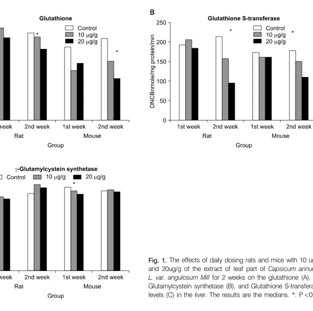
Fig. 1. The effects of daily dosing rats and mice with 10 ug/g and 20ug/g of the extract of leaf part of Capsicum annuum L. var. angulosum Mill for 2 weeks on the glutathione (A), γ- Glutamylcystein synthetase (B), and Glutathione S-transferase levels (C) in the liver. The results are the medians. *: P<0.05 defenses. The synthesis of glutathione is catalyzed by a two
step reaction involving both the rate limiting step enzyme, GST, and γ-GCS. In addition, GST has also clear-cut antioxi- dant functions. Therefore, we checked GSH concentration, GST, and γ-GCS activity in the liver.
As shown in Fig. 1, glutathione and GST levels in the liver were decreased in dose-dependent manners 2 weeks after the daily treatment of the extract of leaf part of Capsicum annuum L. var. angulosum Mill in the rats (p=0.029 and p=0.012, respectively) and mice (p=0.022 and 0.004, respectively). The trend of decrease in glutathione and GST levels is similar in both rats and mice. γ-GCS levels were decreased 1 week after the daily treatment of the extract of leaf part of Capsicum annuum L. var. angulosum Mill by statistically significant degrees in the rats and the mice (p=0.026 and p=0.039, respectively), but not 2 week after the administration of the extract of leaf
part of Capsicum annuum L. var. angulosum Mill in rats and mice (p=0.087 and p=0.994, respectively).
2) Lipid oxidation in the liver tissue and serum γ- GTP: Lipid oxidation is to measure the oxidative activity of xenobiotics and γ-GTP is related lipid oxidation.13) The serum γ-GTP levels were increased in dose-dependent manners 2 weeks after the daily treatment of the extract of leaf part of Capsicum annuum L. var. angulosum Mill in the rats and the mice (p= 0.017 and p=0.002, respectively) (Fig. 2B). The lipid peroxidation levels in the liver were increased in dose- dependent manners 2 weeks after the treatment of the extract of leaf part of Capsicum annuum L. var. angulosum Mill in the mice (p= 0.006). In the rats the lipid peroxidation levels seemed to be increased on the increase of dose, but the relationship was not statistically significant (p=0.16).
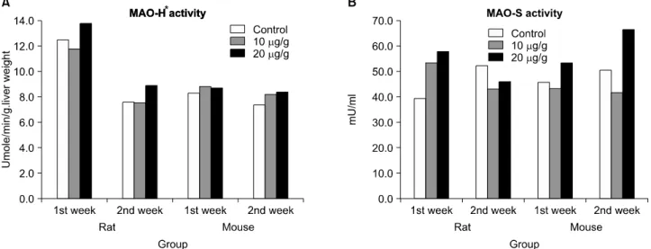
3) MAO levels of serum and the liver tissue:
Fig. 2. The effects of daily dosing rats and mice with 10ug/g and 20ug/g of the extract of leaf part of Capsicum annuum L.
var. angulosum Mill for 2 weeks on the lipid peroxidation levels in the liver (A) and the serum γ-glutamyl transpeptidase levels (B). The level of lipid peroxidation was expressed as malondialdehyde nmole/gram of liver tissue. The results are the medians.
*: P<0.05
Fig. 3. The effects of daily dosing rats and mice with 10ug/g and 20ug/g of the extract of leaf part of Capsicum annuum L.
var. angulosum Mill for 2 weeks on the monoamine oxidase (MAO) levels in mitochondrial fraction of the liver (A) and in the sera (B). The results are the medians. No statistically significant differences were noted among the groups. *MAO-H: MAO in mitochondrial fraction of the hepatocytes.
Cytochrmome P450 is involved in biotransformation of xenobiotics and MAO did.14) The elevation of serum MAO (MAO-S) is frequently found in liver cirrhosis.15) Therefore, we also checked the level of MAO-S and MAO level in mito- chondrial fraction of the hepatocytes (MAO-H). No significant differences in MAO-S and MAO-H levels among treatment groups were observed for the 2 weeks treatment in the both animals (Fig. 3).
2. The other effects of the extract of leaf part of Capsicum annuum L. var. angulosum Mill on the rats and mice
1) Clinical observations, body weight and food/
water intake: During the course of the experiment no treat- ment-related signs of toxicological significance in clinical appea- rance were observed in ether animals. There were no differences in food and water consumption between the treated and control
Fig. 4. The effects of daily dosing rats and mice with 10 ug/g and 20 ug/g of the extract of leaf part of Capsicum annuum L. var. angulosum Mill for 2 weeks on the body weights. The results are the medians. No statistically significant differences were noted among the groups.
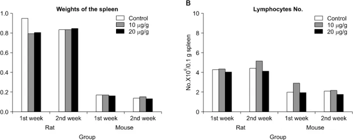
Fig. 6. The effects of daily dosing rats and mice with 10 ug/g and 20 ug/g of the extract of leaf part of Capsicum annuum L.
var. angulosum Mill for 2 weeks on the weights of the spleen (A) and the number of lymphocyte in the spleen (B). The results are the medians. No statistically significant differences were noted among the groups.
Fig. 5. The effects of daily dosing rats and mice with 10ug/g and 20ug/g of the extract of leaf part of Capsicum annuum L. var. angulosum Mill for 2 weeks on the liver weights. The results are the medians.
*: P<0.05 groups in the 14-day study. No statistically significant differ-
ences in the weights of rats and mice were detected among the groups (Fig. 4).
2) Weights and gross findings of the organs: No gross abnormalities of toxicological significance were noted in either animals when necropsied at the end of the treatment period (day 7, and day 14 after treatment). The weights of the liver
were increased 2 weeks after daily dosing of 10 ug/g and 20ug/g of the extract of leaf part of Capsicum annuum L. var.
angulosum Mill in the rats (p=0.041) whereas no statistically significant differences in the weights of liver in the mice among the groups (Fig. 5).
3) Weights of spleen and lymphocyte count in the spleen: In oriental medicine, it is accepted that spleen is an immune organ and herb medicinal therapy should increase immunogenicty. Until now, there is no standard tool to
measure immunogenicity. Therefore, we measured the weights and lymphocyte count with the spleen to check whether the extract of leaf part of Capsicum annuum L. var. angulosum Mill has some effects on the spleen. There were no statistically significant differences in the number of lymphocyte in the spleen and the weights of the spleen (Fig. 6).
DISCUSSION
We found that administration of extract of leaf part of Capsicum annuum L. var. angulosum Mill to the rats and mice for 2 weeks led to decreases in glutathione, and GST levels of the liver in dose-dependent manners. In addition, the γ- GTP levels in the liver were increased 2 weeks after administration of the extract of leaf part of Capsicum annuum L. var. angulosum Mill in rats and mice in dose-dependent manners (Fig. 2). This is the first report showing that the edible plant extract, the extract of leaf part of Capsicum annuum L. var. angulosum Mill, can reduce the hepatic detoxification capacity and suggesting long-term administration of the extract of leaf part of Capsicum annuum L. var. angulosum Mill should induce hepatic damages like that of alcohol does.
Glutathione plays critical roles in defending cells from the oxidant damages. Glutathione can directly scavenge free radicals or act as a substrate for GSTs during the detoxification of various toxic compounds and electrophilic compounds.16) The depletion of glutathione in the liver means the depletion of hepatic detoxification capacity. Actually clinical case was reported indicating that the depletion of glutathione can be a predisposing factor for drug-induced hepatitis.5,6) GST is also directly related to hepatic detoxification capacity16) and the depletion can be a risk factor for drug-induced hepatitis.17) The increase in lipid peroxidation in the liver is also related to lipid conjugation with various electrophiles, indicating the oxidant activity of the extract of leaf part of Capsicum annuum L. var. angulosum Mill. Until recently, it was thought that plant extracts which has been used as dietary foods should be safe and have antioxidant activity in stead of oxidant activity. It was reported that capsaicin, curcumin and resevatrol, although they are well known compounds with antioxidant activity which are isolated from fruits or edible plants, have prooxidant activity which leads to cancer cell apoptosis.18,19)
In our previous reports, the extract of leaf part of Capsicum annuum L. var. angulosum Mill had antitumor activity. In respect
of the antitumor activity of the extract of leaf part of Capsicum annuum L. var. angulosum Mill, it would make a sense that the extract of leaf part of Capsicum annuum L. var. angulosum Mill has some harmful effects on the normal tissue like prooxidant activity.
From these results, it is tempting to speculate that the extract of leaf part of Capsicum annuum L. var. angulosum Mill resulted in the depletion of glutathione and GST and the increase of lipid peroxidation via oxidant activity of the extract of leaf part of Capsicum annuum L. var. angulosum Mill. Indeed, Fig. 1 shows that the depletion of glutathione and GST and the increase of lipid peroxidation 2 weeks after administration of the extract of leaf part of Capsicum annuum L. var. angulosum Mill. This suggests that the extract of leaf part of Capsicum annuum L. var. angulosum Mill should have oxidant activity.
However, there is another explanation for the depletion of glutathione and increase in lipid peroxidation even though this mechanism can not explain how the depletion of GST and γ- GCS happened. The increase of γ-GTP is observed in chronic alcoholic and some medication such as barbiturates, and phenytoin without any elevation in other hepatic enzyme related to the hepatic damages. γ-GTP is related hydrolysis of extracellular glutathione. γ-GTP mediated metabolism of glutathione is liable to expose cellular membranes to expose cellular membranes to the onset of lipid peroxidation.20) Actually it is reported that elevation in γ-GTP stimulated lipid peroxidation.13) Increase of lipid peroxidation in the liver is related to the depletion of glutathione. Our results seem consistent with the above explanation. To verify the exact mechanism by which the administration of extract of leaf part of Capsicum annuum L. var. angulosum Mill for 2 weeks results in the depletion of glutathione, GST and elevation of γ-GTP and lipid peroxidaton in the liver of the rats and mice, further studies are required.
Another lack of this study is that the sample size of this study was not large enough to give us concrete values, because it is likely that some of these differences are chance occurrences.
However, in this report the same results were found in the rats and the mice. This suggests that our results should be reliable.
The clinical meaning is that taking the extract of leaf part of Capsicum annuum L. var. angulosum Mill makes the liver susceptible to drug-induced hepatitis via depletion in hepatic detoxification capacity especially in combination with potential hepatotoxic drugs. It can be assumed that more than 2 weeks
administration of the extract of leaf part of Capsicum annuum L. var. angulosum Mill should be a risk factor for drug-induced hepatitis, especially in combination with potential hepatotoxic drugs.
CONCLUSION
In summary, we previously reported the antitumor activity of the extract of leaf part of Capsicum annuum L. var. angulosum Mill. In this study we evaluated the effects of the extract of leaf part of Capsicum annuum L. var. angulosum Mill on the normal liver, especially detoxification systems as well as other adverse effects in the rats and the mice. Our study suggests that in rats and mice 2-weeks administration of the extract of leaf part of Capsicum annuum L. var. angulosum Mill should decrease glutathione, and GST levels in the liver in dose- dependent manners leading to reduction in hepatic detoxi- fication capacity, and increase serum γ-GTP levels in the same manners. No other adverse clinical effects on rats and mice were found except on their liver.
ACKNOWLEDGMENT
The authors thank Dr. Yune-Sik Kang at department preventive medicine in Gyeong Sang National University College of Medicine for his valuable advices on statistical analysis for these data. The authors thank Dr. Jong-Won Choi in College of Pharmacy, Kyung Sung University for the arrangement of the participation of Mi-jin Seo in this study.
This study was supported by the research fund of Kyung Sung University (2005) and in part Korean Cancer Research Institute.
REFERENCES
1) Sandur SK, Ichikawa H, Sethi G, Ahn KS, Aggarwal BB.
Plumbagin (5-hydroxy-2-methyl-1,4-naphthoquinone) suppresses NF-kappa B activation and NF-kappa B-regulated gene products through modulation of p65 and I-kappa B alpha kinase activation, leading to potentiation of apoptosis induced by cytokine and chemotherapeutic agents. J Biol Chem 281, 17023-17033, 2006.
2) Ding M, Feng R, Wang SY, Bowman L, Lu Y, Qian Y, Castranova V, Jiang BH, Shi X. Cyanidin-3-glucoside, a natural product derived from blackberry, exhibits chemo-
preventive and chemotherapeutic activity. J Biol Chem 281, 17359-17368, 2006.
3) Motohashi N, Wakabayashi H, Kurihara T, Takada Y, Maruyama S, Sakagami H, Nakashima H, Tani S, Shirataki Y, Kawase M, Wolfard K, Molnár J. Cytotoxic and multidrug resistance reversal activity of a vegetable, ‘Anastasia Red’, a variety of sweet pepper. Phytother Res 17, 348-352, 2003.
4) Larrey D, Vial T, Pauwels A, Castot A, Biour M, David M, Michel H. Hepatitis after germander (Teucrium chamaedrys) administration: another instance of herbal medicine hepato- toxicity. Ann Intern Med 117, 129-132, 1992.
5) Tokatli A, Kalkanoğlu-Sivri HS, Yüce A, Coşkun T.
Acetaminophen-induced hepatotoxicity in a glutathione synth- etase-deficient patient. Turk J Pediatr 49, 75-76, 2007.
6) Akai S, Hosomi H, Minami K, Tsuneyama K, Katoh M, Nakajima M, Yokoi T. Knock down of gamma-gluta- mylcysteine synthetase in rat causes acetaminophen-induced hepatotoxicity. J Biol Chem 282, 23996-4003, 2007.
7) Chung YZ. Induction of cancer cell apoptosis by the extract of Capsicum annuum L. var. angulosum Mill sorted according to the parts in hepatoma cells and MCF-7 cells. Yakhak Hoeji 47, 57-68, 2003.
8) Chung YZ. Apoptotic effects of some plants on MCF-7 mammary gland adenoma cells. J Life Science 14, 61-66, 2004.
9) Ellman GL. Tissue sulfhydryl group. Arch Biochem Biophys 82, 70-77, 1959.
10) Habig WH, Pabst MJ, Jakoby WB. Glutathione S- trans- ferase: the frist enzymatic step in mercapturic acid formation.
J Biol Chem 249, 7130-7139, 1974.
11) Richman RG and Meister A. Regulation of γ-Glutamy- lcystein synthetase by nonallosteric feedback inhibition by glutathione. J Biol Chem 250, 1422-1426, 1975.
12) Ohkawa H, Ohishi N, Yaki K. Assay for lipid peroxide in animal tissues by thiobarbituric acid reaction. Anal Biochem 95, 351-358, 1979.
13) Paolicchi A, Tongiani R, Tonarelli P, Comporti M, Pompella A. gamma-Glutamyl transpeptidase-dependent lipid peroxida- tion in isolated hepatocytes and HepG2 hepatoma cells. Free Radic Biol Med 22, 853-860, 1997.
14) Benedetti MS. Biotransformation of xenobiotics by amine oxidases. Fundam Clin Pharmacol 15, 75-84, 2001.
15) Ono T, Eto K, Sakata Y, Takeda M. A new colorimetric assay for monoamine oxidase in serum and its clinical application.
J Lab Clin Med 85, 1022-1031, 1975.
16) Masella R, Di Benedetto R, Vari R, Filesi C, Giovannini C.
Novel mechanisms of natural antioxidant compounds in biological systems: involvement of glutathione and gluta- thione-related enzymes. J Nutr Biochem 16, 577-586, 2005.
17) Luo JC, Cheng TJ, Kuo HW, Chang MJ. Abnormal liver function associated with occupational exposure to dimethylfor- mamide and glutathione S-transferase polymorphisms. Bioma- rkers 10, 464-474, 2005.
18) Galati G, Sabzevari O, Wilson JX, O'Brien PJ. Prooxidant
activity and cellular effects of the phenoxyl radicals of dietary flavonoids and other polyphenolics. Toxicology 177, 91-104, 2002.
19) Yoshino M, Haneda M, Naruse M, Htay HH, Tsubouchi R, Qiao SL, Li WH, Murakami K, Yokochi T. Prooxidant activity of curcumin: copper-dependent formation of 8-
hydroxy-2'-deoxyguanosine in DNA and induction of apo- ptotic cell death. Toxicol In Vitro 18, 783-789, 2004.
20) Stark AA, Zeiger E, Pagano DA. Glutathione metabolism by gamma-glutamyltranspeptidase leads to lipid peroxidation:
characterization of the system and relevance to hepatocarcino- genesis. Carcinogenesis 14, 183-189, 1993.