D I A B E T E S & M E T A B O L I S M J O U R N A L D I A B E T E S & M E T A B O L I S M J O U R N A L
This is an Open Access article distributed under the terms of the Creative Commons Attribution Non-Commercial License (http://creativecommons.org/licenses/by-nc/4.0/) which permits unrestricted non-commercial use, distribution, and reproduction in any medium, provided the original work is properly cited.
Inhibition of Serotonin Synthesis Induces Negative
Hepatic Lipid Balance
Jun Namkung1,2, Ko Eun Shong1,3, Hyeongseok Kim1, Chang-Myung Oh1, Sangkyu Park1,4, Hail Kim1,3 1Graduate School of Medical Science and Engineering, Korea Advanced Institute of Science and Technology, Daejeon, 2Department of Biochemistry, Yonsei University Wonju College of Medicine, Wonju,
3Biomedical Science and Engineering Interdisciplinary Program, Korea Advanced Institute of Science and Technology, Daejeon, 4Department of Biochemistry, Catholic Kwandong University College of Medicine, Gangneung, Korea
Background: Hepatic steatosis is caused by metabolic stress associated with a positive lipid balance, such as insulin resistance and obesity. Previously we have shown the anti-obesity effects of inhibiting serotonin synthesis, which eventually improved insulin sensitivity and hepatic steatosis. However, it is not clear whether serotonin has direct effect on hepatic lipid accumulation. Here, we showed the possibility of direct action of serotonin on hepatic steatosis.
Methods: Mice were treated with para-chlorophenylalanine (PCPA) or LP-533401 to inhibit serotonin synthesis and fed with high fat diet (HFD) or high carbohydrate diet (HCD) to induce hepatic steatosis. Hepatic triglyceride content and gene expression profiles were analyzed.
Results: Pharmacological and genetic inhibition of serotonin synthesis reduced HFD-induced hepatic lipid accumulation. Fur-thermore, short-term PCPA treatment prevented HCD-induced hepatic steatosis without affecting glucose tolerance and brown-ing of subcutaneous adipose tissue. Gene expression analysis revealed that the expressions of genes involved in de novo lipogene-sis and triacylglycerol synthelipogene-sis were downregulated by short-term PCPA treatment as well as long-term PCPA treatment. Conclusion: Short-term inhibition of serotonin synthesis prevented hepatic lipid accumulation without affecting systemic insulin sensitivity and energy expenditure, suggesting the direct steatogenic effect of serotonin in liver.
Keywords: Diabetes mellitus; Fatty liver; Lipogenesis; Obesity; Serotonin
Corresponding authors: Sangkyu Park https://orcid.org/0000-0001-5525-2860
Department of Biochemistry, Catholic Kwandong University College of Medicine, 24 Beomil-ro 579beon-gil, Gangneung 25601, Korea
E-mail: [email protected]
Hail Kim https://orcid.org/0000-0002-6652-1349
Graduate School of Medical Science and Engineering, Korea Advanced Institute of Science and Technology, 291 Daehak-ro, Yuseong-gu, Daejeon 34141, Korea
INTRODUCTION
Nonalcoholic fatty liver disease (NAFLD) is common liver dis-ease and occurs due to the lipid accumulation in the liver. It is exacerbated from steatosis through steatohepatitis to cirrhosis and hepatocellular carcinoma [1,2]. NAFLD is closely associat-ed with obesity, which induces positive hepatic lipid balance leading to lipid accumulation and pathological metabolic dis-turbances in the liver [3-5]. Because excessive energy intake leads to positive lipid balance and obesity, regulation of
sys-temic energy metabolism is a possible therapeutic approach against NAFLD [6-8].
Serotonin (5-hydroxytryptamine) is a neurotransmitter in-volved in the regulation of mood, appetite, and stress responses in the brain [9]. Recently, peripheral serotonin is emerging as a regulator of systemic energy homeostasis. Peripheral serotonin system is functionally separated from central serotonin system because serotonin cannot cross blood brain barrier (BBB) [10]. Serum level of serotonin was elevated in mice fed high fat diet (HFD) [11]. Increasing systemic serotonin activity by knock-https://doi.org/10.4093/dmj.2017.0084
ing out serotonin transporter caused severe obesity, insulin re-sistance, and hepatic steatosis [12,13]. Inhibition of serotonin synthesis in periphery reduced obesity by increasing energy expenditure and decreasing energy storage [14,15]. Genetic inhibition of tryptophan hydroxylase 1 (Tph1), a rate-limiting enzyme for serotonin synthesis in periphery, in adipose tissues resulted in the insulin-sensitizing and anti-obesity effects by increasing energy expenditure [14]. Thus, peripheral serotonin works as an obesity hormone which leads to positive energy and lipid balance.
Most of body serotonin is synthesized and secreted from en-terochromaffin cells in the gut. Since gut-derived serotonin (GDS) which is released from gut is stored in platelets or me-tabolized in liver, plasma free serotonin level in peripheral blood is very low. However, considering that liver is the first organ encountering GDS via portal vein and free serotonin levels in portal blood is relatively higher, liver can be the target of GDS. Although inhibition of serotonin synthesis in periph-eral tissues reduced hepatic lipid accumulation [15], it has not been tested if serotonin has direct effects on liver. In this study, we explored the functional relationship between hepatic ste-atosis and serotonin by using pharmacological and genetic models of serotonin inhibition and have shown that serotonin has direct effects on hepatic lipid accumulation.
METHODS
Animal experimentsThe experimental protocol for this study was approved by the Institutional Animal Care and Use Committee at the Korea Advanced Institute of Science and Technology (KA2011-29).
Tph1 floxed (Tph1fl/fl) mice [16] were crossed with
Adiponec-tin-Cre mice [17] to generate fat-specific Tph1-knockout (Tph1
FKO). 5-Hydroxytryptamine receptor 3A (Htr3a) knockout (KO) mice (B6.129X1-Htr3atm1jul/J) were purchased from the
Jackson Laboratory (Bar Harbor, ME, USA). Sequences of primers used in mouse genotyping are given in Table 1. C57BL/6J mice were purchased from the Charles River Japan (Yokohama, Japan). Mice were housed on 12-hour light-dark cycle in climate controlled specific pathogen-free barrier facili-ty. Diet and water were provided ad libitum. At 8 or 12 weeks of age, mice were fed a standard chow diet (SCD), HFD (60% fat calories), or high carbohydrate diet (HCD, 70% carbohy-drate calories). Animal diets used in this study were purchased from Research Diets (New Brunswick, NJ, USA). Para-chloro-phenylalanine (PCPA, Sigma-Aldrich, St. Louis, MO, USA) was dissolved in phosphate-buffered saline (PBS). PBS or 300 mg/kg PCPA was daily administered by intraperitoneal injec-tion. LP-533401 (Dalton Pharma Services, Toronto, ON, Can-ada) was dissolved in polyethylene glycol 400 (Sigma-Aldrich) and 5% dextrose (40:60 v/v). Vehicle or 30 mg/kg LP-533401 was administered daily by oral gavage.
Histological analysis
Preparation of tissue sections and H&E staining were per-formed as previously described [18,19]. Liver tissues and in-guinal white adipose tissues were harvested, fixed in 4% (w/v) paraformaldehyde and embedded in paraffin. Five-µm-thick tissue sections were deparaffinized, rehydrated, and stained with H&E.
Quantification of hepatic triglyceride levels
Liver tissues were homogenized using FastPrep-24 (MP Bio-medicals, Santa Ana, CA, USA) in 5% nonyl phenoxypolye-thoxylethanol (NP-40; Sigma-Aldrich). Aliquot of homoge-nates were used for measurement of protein concentrations Table 1. Sequences of primers used in mouse genotyping
Mouse Primers (5´ to 3´) Product size, bp
Tph1fl/fl GGATCCTAACCGAGTGTTCC GCACACCACCAACTCTTTCC Wild type: 350 Floxed: 450 Adiponectin-Cre TGCCATGTGAGTCTGCCTTT AACCAGCGTTTTCGTTCTGC Cre+: 700 Htr3a KO TGGATGTGGAATGTGTGCGAG AACAGCTATGCAGAAATGAAGTT GGCTGACTGCGTAGAATAAAGG Wild type: 400 Knockout: 210 Tphfl/fl, tryptophan hydroxylase 1 floxed; Htr3a, 5-hydroxytryptamine receptor 3A; KO, knockout.
with BCA Protein Assay Kit (Thermo Fisher Scientific, Waltham, MA, USA). The remaining homogenates were heat-ed to 95°C for 5 minutes and coolheat-ed at room temperature; this cycle was repeated to solubilize all hepatic fat. After centrifuga-tion, supernatants were used for measurement of hepatic tri-glyceride (TG) levels. Tritri-glyceride Reagent (Sigma-Aldrich) or PBS was added, and samples were incubated at 37°C for 30 minutes to hydrolyze hepatic TG into glycerol. Samples were then incubated with Free Glycerol Reagent (Sigma-Aldrich) at 37°C for 5 minutes for colorimetric assay of hydrolyzed TG levels. Differences in absorbance at 540 nm for hydrolyzed or non-hydrolyzed TG were quantified using a glycerol standard. Hepatic TG contents were normalized by protein amounts of
liver tissues.
Glucose and insulin tolerance tests
For the glucose tolerance test, overnight-fasted mice were in-traperitoneally injected with 2 g/kg D-glucose (Sigma-Al-drich) in PBS. For the insulin tolerance test, 0.75 U/kg human insulin (Humulin R; Lilly, Indianapolis, IN, USA) was intra-peritoneally injected into mice after fasting for 6 hours. Blood samples were then obtained from the tail vein at 0, 15, 30, 45, 60, 90, and 120 minutes after injection. Glucose concentrations were measured using a Gluco DR Plus glucometer (Allmedi-cus, Anyang, Korea).
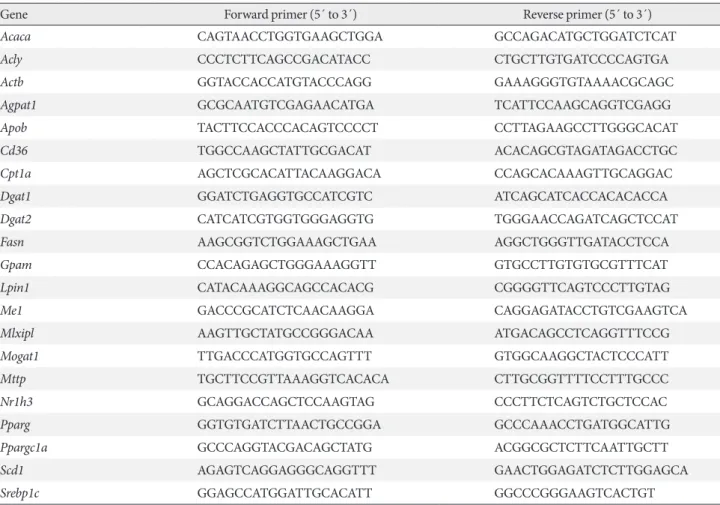
Table 2. Sequences of primers used in quantitative real-time polymerase chain reaction
Gene Forward primer (5´ to 3´) Reverse primer (5´ to 3´)
Acaca CAGTAACCTGGTGAAGCTGGA GCCAGACATGCTGGATCTCAT
Acly CCCTCTTCAGCCGACATACC CTGCTTGTGATCCCCAGTGA
Actb GGTACCACCATGTACCCAGG GAAAGGGTGTAAAACGCAGC
Agpat1 GCGCAATGTCGAGAACATGA TCATTCCAAGCAGGTCGAGG
Apob TACTTCCACCCACAGTCCCCT CCTTAGAAGCCTTGGGCACAT
Cd36 TGGCCAAGCTATTGCGACAT ACACAGCGTAGATAGACCTGC
Cpt1a AGCTCGCACATTACAAGGACA CCAGCACAAAGTTGCAGGAC
Dgat1 GGATCTGAGGTGCCATCGTC ATCAGCATCACCACACACCA
Dgat2 CATCATCGTGGTGGGAGGTG TGGGAACCAGATCAGCTCCAT
Fasn AAGCGGTCTGGAAAGCTGAA AGGCTGGGTTGATACCTCCA
Gpam CCACAGAGCTGGGAAAGGTT GTGCCTTGTGTGCGTTTCAT
Lpin1 CATACAAAGGCAGCCACACG CGGGGTTCAGTCCCTTGTAG
Me1 GACCCGCATCTCAACAAGGA CAGGAGATACCTGTCGAAGTCA
Mlxipl AAGTTGCTATGCCGGGACAA ATGACAGCCTCAGGTTTCCG
Mogat1 TTGACCCATGGTGCCAGTTT GTGGCAAGGCTACTCCCATT
Mttp TGCTTCCGTTAAAGGTCACACA CTTGCGGTTTTCCTTTGCCC
Nr1h3 GCAGGACCAGCTCCAAGTAG CCCTTCTCAGTCTGCTCCAC
Pparg GGTGTGATCTTAACTGCCGGA GCCCAAACCTGATGGCATTG
Ppargc1a GCCCAGGTACGACAGCTATG ACGGCGCTCTTCAATTGCTT
Scd1 AGAGTCAGGAGGGCAGGTTT GAACTGGAGATCTCTTGGAGCA
Srebp1c GGAGCCATGGATTGCACATT GGCCCGGGAAGTCACTGT
Acaca, acetyl-CoA carboxylase alpha; Acly, ATP citrate lyase; Actb, actin beta; Agpat1, 1-acylglycerol-3-phosphate O-acyltransferase 1; Apob, apolipoprotein B; Cpt1a, carnitine palmitoyltransferase 1a; Dgat1, diacylglycerol O-acyltransferase 1; Dgat2, diacylglycerol O-acyltransferase 2; Fasn, fatty acid synthase; Gpam, glycerol-3-phosphate acyltransferase; Lpin1, lipin 1; Me1, malic enzyme 1; Mlxipl, MLX interacting protein-like (ChREBP, carbohydrate response element binding protein); Mogat1, monoacylglycerol O-acyltransferase 1; Mttp, microsomal triglyceride transfer protein; Nr1h3, nuclear receptor subfamily 1, group H, member 3 (LXR, liver X receptor); Pparg, peroxisome proliferator activated re-ceptor gamma; Ppargc1a, Pparg coactivator 1 alpha; Scd1, stearoyl-CoA desaturase 1; Srebp1c, sterol regulatory element binding transcription factor 1c.
Quantitative reverse transcription polymerase chain reaction analysis
TRIzol reagent (Ambion, Carlsbard, CA, USA) was used for total RNA extraction from harvested tissues. To eliminate ge-nomic DNA, RNA was treated with TURBO DNase (Ambi-on). Complementary DNA was generated with Superscript III Reverse Transcriptase (Invitrogen, Carlsbad, CA, USA) from 1 μg of total RNA. Real-time quantitative reverse transcription polymerase chain reaction was performed with Fast SYBR Green Master Mix (Applied Biosystems, Foster City, CA, USA) and a ViiA 7 real-time PCR system (Applied Biosystems) ac-cording to the manufacturer’s instructions. Expressional pro-files were quantified based on the relative delta delta Ct (threshold cycle) method [20] with β-actin as a reference gene.
The sequences of primers are given in Table 2. Statistical analysis
Data are presented as the mean±standard error of mean. The values were compared using Student t-test or one-way analysis of variance. Differences with P values of less than 0.05 were considered statistically significant.
RESULTS
Serotonin inhibition protects against HFD-induced hepatic steatosis
In order to determine the effects of serotonin inhibition on di-et-induced hepatic steatosis, we used several models of
sero-Fig. 1. Serotonin inhibition protected against high fat diet (HFD)-induced hepatic steatosis. Eight-week-old mice were fed a
stan-dard chow diet (SCD) or HFD for 12 weeks with vehicle, para-chlorophenylalanine (PCPA), or LP-533401 treatment. (A) H&E staining of liver sections from SCD- or HFD-fed mice with vehicle or PCPA treatment. (B) Quantification of hepatic triglyceride (TG) levels in PCPA-treated mice (n=6). (C-E) H&E staining of liver sections (left) and quantification of hepatic TG levels (right) from HFD-fed mice treated with LP-533401 (C), fat-specific Tph1-knockout (Tph1 FKO) mice (D), and 5-hydroxytryptamine re-ceptor 3A (Htr3a) knockout (KO) mice (E) (n=6). Representative images are shown. Scale bars, 50 μm. Tphfl/fl, tryptophan
hy-droxylase 1 floxed. aP<0.05, bP<0.01, cP<0.001. 500 400 300 200 100 0 500 400 300 200 100 0 1,000 800 600 400 200 0 400 300 200 100 0 TG (m g/g p ro tein) TG (m g/g p ro tein) TG (m g/g p ro tein) TG (m g/g p ro tein) HFD HFD SCD HFD HFD Vehicle LP-533401 WT Htr3a KO Vehicle PCPA Tph1fl/fl Tph1 FKO a c b a Vehicle Vehicle Tph1fl/fl WT PCPA LP-533401 Tph1 FKO Htr3a KO SCD HFD E B D A C
tonin inhibition on HFD. Pharmacologically, serotonin syn-thesis can be blocked by Tph inhibitor, PCPA, or LP-533401, which targets the rate-limiting step of serotonin synthesis. Firstly, we examined the livers of mice fed an HFD and treated with PCPA. Histological analysis revealed that HFD-induced hepatic steatosis was blocked by PCPA treatment (Fig. 1A). Accordingly, hepatic TG content was significantly decreased by PCPA treatment (Fig. 1B). PCPA can cross BBB and is known to increase food intake by inhibiting serotonin produc-tion in the brain [21]. To further confirm the effects of periph-eral serotonin inhibition, we investigated the effect of LP-533401, a peripheral Tph inhibitor which cannot cross the BBB, on hepatic steatosis [22]. Similar to the results with PCPA, LP-533401 also decreased HFD-induced hepatic TG accumulation (Fig. 1C). We also tested Tph1 FKO and Htr3a KO mice, which exhibit increased energy expenditure on HFD [14]. Hepatic TG content was decreased in the liver of both
Tph1 FKO and Htr3a KO mice (Fig. 1D and E). Thus,
inhibi-tion of peripheral serotonin either by decreased synthesis or reduced activity, protects from HFD-induced hepatic steatosis. PCPA blocks positive hepatic lipid balance on HFD
Hepatic steatosis results from a positive hepatic lipid balance, which occurs through integration of major five components:
de novo lipogenesis, TG synthesis, fatty acid (FA) uptake, FA
oxidation, and very low density lipoprotein (VLDL) secretion [4,23]. Each component is again determined by the amount of substrate, allosteric regulation by metabolites, and amount and activity of enzymes of their pathways. To identify the compo-nents regulated by serotonin, we investigated gene expression profiles of enzymes involved in hepatic lipid metabolism. PCPA decreased the expression of most genes related to de
novo lipogenesis, TG synthesis, and FA uptake (Fig. 2A-C).
Thus, PCPA blocks the induction of a positive hepatic lipid balance by HFD. In contrast, the expression of genes involved in FA oxidation and VLDL secretion was not changed by PCPA treatment (Fig. 2D and E). Because most genes in the af-fected pathway were downregulated, we hypothesized that these changes may reflect the regulation of upstream transcrip-tion factors. Indeed, PCPA treatment decreased the expression of lipogenic transcription factors such as Pparg (peroxisome proliferator activated receptor gamma), Nr1h3 (Lxrα; nuclear receptor subfamily 1, group H, member 3 [LXR, liver X recep-tor]), and Ppargc1a (Pparg coactivator 1 alpha) (Fig. 2F).
Short-term intervention of PCPA protects against hepatic steatosis independently from energy expenditure and insulin sensitivity
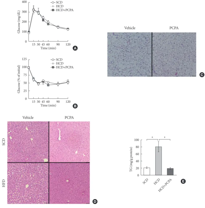
The alleviation of HFD-induced hepatic steatosis by PCPA could be largely attributed to the decreased flux of steatogenic substrates from adipose tissues resulting from the increased energy expenditure in adipose tissues [14,15]. In this case, most of gene expressions in liver of mice fed with HFD were supposed to be similar with those of mice fed with SCD. How-ever, some of the gene expressions were downregulated by PCPA even in liver of mice fed with SCD. These data strongly suggested the direct action of serotonin on liver where several serotonin receptors are expressed [24]. In order to determine the direct effects of serotonin on hepatic steatosis regardless of systemic insulin resistance and energy expenditure, we em-ployed short-term intervention with an HCD and PCPA for 2 weeks. Notably, the short-term HCD had no effect on glucose tolerance (Fig. 3A) or insulin sensitivity (Fig. 3B), and short-term PCPA treatment did not induce beige adipogenesis in in-guinal white adipose tissue (Fig. 3C), which is the characteris-tic of long-term intervention with PCPA [14]. Similar to the HFD model, the HCD induced hepatic TG accumulation, and PCPA decreased hepatic steatosis, resulting in decreased he-patic TG content (Fig. 3D and E).
Short-term intervention of PCPA decreases de novo lipogenesis and TG synthesis
We have shown that the inhibition of hepatic lipid accumula-tion by PCPA was not entirely attributed to the increased ener-gy expenditure in adipose tissues but to the inhibition of direct action of serotonin on liver. In order to know more precise mechanism of serotonin on hepatic lipid accumulation, we checked gene expression profiles of lipid metabolism in the liv-ers of PCPA-treated mice. Among components of hepatic lipid balance, genes of de novo lipogenesis and TG synthesis were upregulated by HCD, which were reversed by PCPA (Fig. 4A and B), similar to the results in HFD-fed PCPA-treated mice. However, there were no changes in genes encoding compo-nents of FA uptake, FA oxidation, and VLDL secretion (Fig. 4C-E). Because lipogenic genes were downregulated, we also investigated upstream transcription factors. Expression levels of Pparg, Srebp1c (sterol regulatory element binding transcrip-tion factor 1c), and Mlxipl (MLX interacting protein-like [Chrebp, carbohydrate response element binding protein]), which are lipogenic transcription factors, were significantly decreased
Fig. 2. Para-chlorophenylalanine (PCPA) treatment suppressed the positive hepatic lipid balance. Eight-week-old mice were fed a
standard chow diet (SCD) or high fat diet (HFD) for 12 weeks and treated with vehicle or PCPA treatment. Hepatic expressional profiles of genes related to de novo lipogenesis (A), triglyceride synthesis (B), fatty acid (FA) uptake (C), FA oxidation (D), very low density lipoprotein secretion (E), and transcription factors (F) were assessed by quantitative reverse transcription polymerase chain reaction (n=6). Acaca, acetyl-CoA carboxylase alpha; Fasn, fatty acid synthase; Acly, ATP citrate lyase; Me1, malic enzyme 1;
Scd1, stearoyl-CoA desaturase 1; Gpam, glycerol-3-phosphate acyltransferase; Agpat1, 1-acylglycerol-3-phosphate
O-acyltransfer-ase 1; Lpin1, lipin 1; Mogat1, monoacylglycerol O-acyltransferO-acyltransfer-ase 1; Dgat1, diacylglycerol O-acyltransferO-acyltransfer-ase 1; Dgat2, diacylglycer-ol O-acyltransferase 2; Cpt1a, carnitine palmitoyltransferase 1a; Mttp, microsomal triglyceride transfer protein; Apob, apdiacylglycer-olipopro- apolipopro-tein B; Pparg, peroxisome proliferator activated receptor gamma; Ppargc1a, Pparg coactivator 1 alpha; Srebp1c, sterol regulatory el-ement binding transcription factor 1c; Mlxipl, MLX interacting protein-like (ChREBP, carbohydrate response elel-ement binding protein); Nr1h3, nuclear receptor subfamily 1, group H, member 3 (LXR, liver X receptor). aP<0.05, bP<0.01, cP<0.001.
4 3 2 1 0 1.5 1.0 0.5 0 2.0 1.5 1.0 0.5 0 2.0 1.5 1.0 0.5 0 1.5 1.0 0.5 0 8 6 4 2 0 2.0 1.5 1.0 0.5 0 1.5 1.0 0.5 0 6 4 2 1 4 3 2 1 0 1.5 1.0 0.5 0 1.5 1.0 0.5 0 2.5 2.0 1.5 1.0 0.5 0 1.5 1.0 0.5 0 4 3 2 1 0 1.5 1.0 0.5 0 1.5 1.0 0.5 0 1.5 1.0 0.5 0 1.5 1.0 0.5 0 1.5 1.0 0.5 0 Re lat iv e exp res sio n Re lat iv e exp res sio n SCD SCD SCD SCD SCD SCD SCD SCD SCD SCD SCD SCD SCD SCD SCD SCD SCD SCD SCD SCD Gpam Acaca Agpat1 Cd36
Pparg Ppargc1a Srebp1c Mlxipl Nr1h3
Cpt1a Mttp Apob Fasn Lpin1 Acly Mogat1 Me1 Dgat1 Scd1 Dgat2 HFD HFD HFD HFD HFD HFD HFD HFD HFD HFD HFD HFD HFD HFD HFD HFD HFD HFD HFD HFD Vehicle PCPA b b b a a a b a a B E D C a b c a a a a Re lat iv e exp res sio n Re lat iv e exp res sio n Re lat iv e exp res sio n Re lat iv e exp res sio n a b a a b b a A F a
Fig. 3. Short-term treatment with para-chlorophenylalanine (PCPA) in the context of high carbohydrate diet (HCD) protected
against hepatic steatosis independently from energy expenditure and insulin sensitivity. Twelve-week-old mice were fed a stan-dard chow diet (SCD) or HCD for 2 weeks and treated with vehicle or PCPA treatment. Intraperitoneal glucose tolerance tests (A) and insulin tolerance tests (B) were performed (n=4). (C) H&E staining of inguinal white adipose tissue sections from HCD-fed mice with vehicle or PCPA treatment. (D) H&E staining of liver sections from SCD- or HCD-fed mice with vehicle or PCPA treatment. (E) Quantification of hepatic triglyceride levels in PCPA-treated mice (n=6). Representative images are shown. Scale bars, 50 μm. aP<0.05. Vehicle PCPA C 100 80 60 40 20 0 TG (m g/g p ro tein) SCD HCD HCD+PCP A a a E Vehicle PCPA SCD HFD D 125 100 75 50 25 0 G lucos e (% o f ini tia l) B 15 30 45 60 90 120 Time (min) SCD HCD HCD+PCPA 400 300 200 100 0 G lucos e (m g/dL) A 15 30 45 60 90 120 Time (min) SCD HCD HCD+PCPA
(Fig. 4F). Collectively, these data suggested that PCPA induced negative hepatic lipid balance by decreasing de novo lipogene-sis and TG synthelipogene-sis via downregulation of lipogenic
tran-scription factors independently from insulin sensitivity and energy expenditure.
Fig. 4. Para-chlorophenylalanine (PCPA) treatment suppressed the positive hepatic lipid balance via downregulation of Pparg, Srebp1c, and Mlxipl. Twelve-week-old mice were fed a standard chow diet (SCD) or high carbohydrate diet (HCD) for 2 weeks with
vehicle or PCPA treatment. Hepatic expressional profiles of genes related to de novo lipogenesis (A), triglyceride synthesis (B), fatty acid (FA) uptake (C), FA oxidation (D), very low density lipoprotein secretion (E), and transcription factors (F) were assessed by quantitative reverse transcription polymerase chain reaction (n=6). Acaca, acetyl-CoA carboxylase alpha; Fasn, fatty acid synthase;
Acly, ATP citrate lyase; Me1, malic enzyme 1; Scd1, stearoyl-CoA desaturase 1; Gpam, glycerol-3-phosphate acyltransferase; Agpat1,
1-acylglycerol-3-phosphate O-acyltransferase 1; Lpin1, lipin 1; Mogat1, monoacylglycerol O-acyltransferase 1; Dgat1, diacylglycer-ol O-acyltransferase 1; Dgat2, diacylglycerdiacylglycer-ol O-acyltransferase 2; Cpt1a, carnitine palmitoyltransferase 1a; Apob, apdiacylglycer-olipoprotein B;
Mttp, microsomal triglyceride transfer protein; Pparg, peroxisome proliferator activated receptor gamma; Ppargc1a, Pparg
coacti-vator 1 alpha; Srebp1c, sterol regulatory element binding transcription factor 1c; Mlxipl, MLX interacting protein-like (ChREBP, carbohydrate response element binding protein); Nr1h3, nuclear receptor subfamily 1, group H, member 3 (LXR, liver X receptor).
aP<0.05, bP<0.001. 2.0 1.5 1.0 0.5 0 4 3 2 1 0 1.5 1.0 0.5 0 1.5 1.0 0.5 0 3 2 1 0 2.0 1.5 1.0 0.5 0 2.5 2.0 1.5 1.0 0.5 0 2.0 1.5 1.0 0.5 0 2.0 1.5 1.0 0.5 0 1.5 1.0 0.5 0 2.5 2.0 1.5 1.0 0.5 0 1.5 1.0 0.5 0 2.0 1.5 1.0 0.5 0 2.5 2.0 1.5 1.0 0.5 0 2.5 2.0 1.5 1.0 0.5 0 1.5 1.0 0.5 0 1.5 1.0 0.5 0 2.0 1.5 1.0 0.5 0 1.5 1.0 0.5 0 2.0 1.5 1.0 0.5 0 Re lat iv e exp res sio n Re lat iv e exp res sio n Re lat iv e exp res sio n Re lat iv e exp res sio n Re lat iv e exp res sio n Re lat iv e exp res sio n Gpam Acaca Agpat1 Cd36
Pparg Ppargc1a Srebp1c Mlxipl Nr1h3
Cpt1a Apob Mttp Fasn Lpin1 Acly Mogat1 Me1 Dgat1 Scd1 Dgat2 SCD HCD HCD+PCPA a a a a b a a a a a B E D C a a A F a a a
DISCUSSION
HFD, HCD, and ethanol intake are known to induce fatty liver both in human and rodent. These metabolic stresses induce a positive lipid balance in the liver either by increasing de novo lipogenesis and FA uptake or decreasing FA oxidation and VLDL secretion. The alteration of lipid balance can be induced by insulin resistance which induces upregulation of lipogenic gene expressions and increased the flux of steatogenic sub-strates, free FAs on liver [4]. In this study, we found that phar-macological or genetic inhibition of serotonin production alle-viated hepatic steatosis and decreased hepatic TG accumula-tion in HFD- or HCD-induced hepatic steatosis models.
PCPA is known to irreversibly inhibit Tph, the rate-limiting enzyme in serotonin synthesis, and lead to systemic serotonin depletion [25-27]. Serotonin is known to exert its anorexigenic effects via Htr2c in the central nervous system [28]. Thus intra-cranial injection of PCPA increases food intake and obesity [21]. In contrast, intraperitoneal injection of PCPA exerts anti-obesity effects independently from food intake [14]. Thus, pe-ripheral serotonin system is functionally as well as anatomical-ly separated from central serotonin system on metabolism. We found that both systemic inhibition of serotonin production by PCPA and selective inhibition of peripheral serotonin produc-tion by LP-533401 suppressed HFD-induced hepatic fatty changes. Therefore, peripheral serotonin is thought be in-volved in diet-induced hepatic steatosis.
Gene expression profiles of hepatic lipid metabolism showed that PCPA suppressed the HFD-induced positive hepatic lipid balance by downregulation of genes involved in de novo lipo-genesis, TG synthesis, and FA uptake, as in a previous study of white adipose tissue [14]. Because insulin resistance increases hepatic steatosis [29] and PCPA ameliorates insulin resistance in the context of an HFD [14], the anti-steatotic effects of PCPA may reflect both the direct effects of decreased serotonin to hepatic serotonin receptors and the indirect effects on other organs, such as adipose tissue and skeletal muscle. To identify the direct effects of serotonin, we used short-term intervention with PCPA with an HCD. Similar to the results in the HFD-fed model, PCPA could prevent HCD-induced hepatic steatosis. This mechanism was independent from energy expenditure and insulin sensitivity. Thus, serotonin could directly regulate hepatic lipid metabolism.
We found decreased expressions of genes associated with de
novo lipogenesis and TG synthesis by short-term intervention
with PCPA, including downregulation of Mlxipl (carbohy-drate-responsive element-binding protein), a major lipogenic transcription factor that functions in response to high glucose [30], as well as lipogenic Srebp1c and Pparg [31]. Together with other reports that serotonin increased fat content in primary hepatocytes [32,33], our data strongly suggested that serotonin can be a direct lipogenic stimulus in the liver via regulation of lipogenic transcription factors, as has been observed in adi-pose tissue. Although causative serotonin receptor and its sig-naling pathway are not identified, we show the direct action of serotonin on hepatic lipid metabolism. Models of serotonin inhibition showed anti-steatogenic effects on diet-induced he-patic steatosis (Fig. 1), but these are sum of direct effects from decreased hepatic serotonin action and indirect effects from increased energy expenditure and enhanced insulin sensitivity. We tried to distinguish those two mechanisms, and found that short-term intervention of PCPA treatment on HCD didn’t change energy expenditure and insulin sensitivity. Thus we firstly identified direct effects of serotonin inhibition on liver in
vivo. Additional studies using liver-specific serotonin receptor
knockout are required to elucidate the detailed mechanism how serotonin signaling is associated with diet-induced hepat-ic steatosis. Selective modulation of serotonin pathway might introduce new therapeutic approach against hepatic steatosis.
CONFLICTS OF INTEREST
No potential conflict of interest relevant to this article was re-ported.
ACKNOWLEDGMENTS
We thank all the members of the Integrated Lab of Metabolism, Obesity, and Diabetes (iMOD) at KAIST for their helpful dis-cussions and technical support. This work was supported by a grant from the Korean Diabetes Association (grant number: 2017S-1 to Jun Namkung) and grants from the National Re-search Foundation of Korea (NRF) funded by Ministry of Sci-ence, ICT & Future Planning (grant numbers: NRF-2014M3A9 D8034464 to Hail Kim and NRF-2016R1C1B1011688 to Jun Namkung).
REFERENCES
steatosis to cirrhosis. Hepatology 2006;43(2 Suppl 1):S99-112. 2. Baffy G, Brunt EM, Caldwell SH. Hepatocellular carcinoma in
non-alcoholic fatty liver disease: an emerging menace. J Hepa-tol 2012;56:1384-91.
3. Adams LA, Waters OR, Knuiman MW, Elliott RR, Olynyk JK. NAFLD as a risk factor for the development of diabetes and the metabolic syndrome: an eleven-year follow-up study. Am J Gastroenterol 2009;104:861-7.
4. Kawano Y, Cohen DE. Mechanisms of hepatic triglyceride ac-cumulation in non-alcoholic fatty liver disease. J Gastroenterol 2013;48:434-41.
5. Tchernof A, Despres JP. Pathophysiology of human visceral obesity: an update. Physiol Rev 2013;93:359-404.
6. Galgani J, Ravussin E. Energy metabolism, fuel selection and body weight regulation. Int J Obes (Lond) 2008;32 Suppl 7:S109-19.
7. Golabi P, Bush H, Younossi ZM. Treatment strategies for non-alcoholic fatty liver disease and nonnon-alcoholic steatohepatitis. Clin Liver Dis 2017;21:739-53.
8. Samuel VT, Shulman GI. Nonalcoholic fatty liver disease as a nexus of metabolic and hepatic diseases. Cell Metab 2018;27: 22-41.
9. Berger M, Gray JA, Roth BL. The expanded biology of sero-tonin. Annu Rev Med 2009;60:355-66.
10. Watanabe H, Rose MT, Aso H. Role of peripheral serotonin in glucose and lipid metabolism. Curr Opin Lipidol 2011;22:186-91.
11. Kim HJ, Kim JH, Noh S, Hur HJ, Sung MJ, Hwang JT, Park JH, Yang HJ, Kim MS, Kwon DY, Yoon SH. Metabolomic analysis of livers and serum from high-fat diet induced obese mice. J Proteome Res 2011;10:722-31.
12. Chen X, Margolis KJ, Gershon MD, Schwartz GJ, Sze JY. Re-duced serotonin reuptake transporter (SERT) function causes insulin resistance and hepatic steatosis independent of food in-take. PLoS One 2012;7:e32511.
13. Uceyler N, Schutt M, Palm F, Vogel C, Meier M, Schmitt A, Lesch KP, Mossner R, Sommer C. Lack of the serotonin trans-porter in mice reduces locomotor activity and leads to gender-dependent late onset obesity. Int J Obes (Lond) 2010;34:701-11.
14. Oh CM, Namkung J, Go Y, Shong KE, Kim K, Kim H, Park BY, Lee HW, Jeon YH, Song J, Shong M, Yadav VK, Karsenty G, Kajimura S, Lee IK, Park S, Kim H. Regulation of systemic en-ergy homeostasis by serotonin in adipose tissues. Nat Com-mun 2015;6:6794.
15. Crane JD, Palanivel R, Mottillo EP, Bujak AL, Wang H, Ford RJ, Collins A, Blumer RM, Fullerton MD, Yabut JM, Kim JJ, Ghia JE, Hamza SM, Morrison KM, Schertzer JD, Dyck JR, Khan WI, Steinberg GR. Inhibiting peripheral serotonin synthesis reduces obesity and metabolic dysfunction by promoting brown adipose tissue thermogenesis. Nat Med 2015;21:166-72. 16. Yadav VK, Ryu JH, Suda N, Tanaka KF, Gingrich JA, Schutz G,
Glorieux FH, Chiang CY, Zajac JD, Insogna KL, Mann JJ, Hen R, Ducy P, Karsenty G. Lrp5 controls bone formation by inhib-iting serotonin synthesis in the duodenum. Cell 2008;135:825-37.
17. Eguchi J, Wang X, Yu S, Kershaw EE, Chiu PC, Dushay J, Estall JL, Klein U, Maratos-Flier E, Rosen ED. Transcriptional control of adipose lipid handling by IRF4. Cell Metab 2011;13:249-59. 18. Fischer AH, Jacobson KA, Rose J, Zeller R. Cutting sections of
paraffin-embedded tissues. CSH Protoc 2008;2008:pdb. prot4987.
19. Fischer AH, Jacobson KA, Rose J, Zeller R. Hematoxylin and eosin staining of issue and cell sections. CSH Protoc 2008;2008: pdb.prot4986.
20. Livak KJ, Schmittgen TD. Analysis of relative gene expression data using real-time quantitative PCR and the 2 (-delta delta C (T)) Method. Methods 2001;25:402-8.
21. Breisch ST, Zemlan FP, Hoebel BG. Hyperphagia and obesity following serotonin depletion by intraventricular p-chlorophe-nylalanine. Science 1976;192:382-5.
22. Liu Q, Yang Q, Sun W, Vogel P, Heydorn W, Yu XQ, Hu Z, Yu W, Jonas B, Pineda R, Calderon-Gay V, Germann M, O’Neill E, Brommage R, Cullinan E, Platt K, Wilson A, Powell D, Sands A, Zambrowicz B, Shi ZC. Discovery and characterization of novel tryptophan hydroxylase inhibitors that selectively inhibit serotonin synthesis in the gastrointestinal tract. J Pharmacol Exp Ther 2008;325:47-55.
23. Marchesini G, Petta S, Dalle Grave R. Diet, weight loss, and liv-er health in nonalcoholic fatty livliv-er disease: pathophysiology, evidence, and practice. Hepatology 2016;63:2032-43.
24. Sumara G, Sumara O, Kim JK, Karsenty G. Gut-derived sero-tonin is a multifunctional determinant to fasting adaptation. Cell Metab 2012;16:588-600.
25. Koe BK, Weissman A. P-chlorophenylalanine: a specific deple-tor of brain serotonin. J Pharmacol Exp Ther 1966;154:499-516.
26. Sanders-Bush E, Sulser F. P-chloroamphetamine: in vivo inves-tigations on the mechanism of action of the selective depletion of cerebral serotonin. J Pharmacol Exp Ther 1970;175:419-26.
27. Engelman K, Lovenberg W, Sjoerdsma A. Inhibition of sero-tonin synthesis by para-chlorophenylalanine in patients with the carcinoid syndrome. N Engl J Med 1967;277:1103-8. 28. Nonogaki K, Strack AM, Dallman MF, Tecott LH.
Leptin-inde-pendent hyperphagia and type 2 diabetes in mice with a mu-tated serotonin 5-HT2C receptor gene. Nat Med 1998;4:1152-6.
29. Browning JD, Horton JD. Molecular mediators of hepatic ste-atosis and liver injury. J Clin Invest 2004;114:147-52.
30. Postic C, Dentin R, Denechaud PD, Girard J. ChREBP, a tran-scriptional regulator of glucose and lipid metabolism. Annu Rev Nutr 2007;27:179-92.
31. Pettinelli P, Videla LA. Up-regulation of PPAR-gamma mRNA expression in the liver of obese patients: an additional reinforc-ing lipogenic mechanism to SREBP-1c induction. J Clin Endo-crinol Metab 2011;96:1424-30.
32. Rozenblit-Susan S, Chapnik N, Froy O. Metabolic effect of flu-voxamine in mouse peripheral tissues. Mol Cell Endocrinol 2016;424:12-22.
33. Osawa Y, Kanamori H, Seki E, Hoshi M, Ohtaki H, Yasuda Y, Ito H, Suetsugu A, Nagaki M, Moriwaki H, Saito K, Seishima M. L-tryptophan-mediated enhancement of susceptibility to nonalcoholic fatty liver disease is dependent on the mammali-an target of rapamycin. J Biol Chem 2011;286:34800-8.