560
©The Korean Society of Food Science and Technology
Bacillus cereus에 대한 actinonin의 항균 효과
정동윤·염수진·유연철·김종헌·이병휘·장훈녕·정희곤*
충남대학교 식품공학과
Antimicrobial activities of actinonin against Bacillus cereus
Dongyun Jung, Su-Jin Yum, Yeon-Cheol Yu, Jong-Heon Kim, Byung-Hwi Lee, Hoon-Nyung Jang, and Hee Gon Jeong*
Department of Food Science and Technology, Chungnam National University
Abstract The objective of this study was to investigate the anti-Bacillus cereus activity of actinonin. Actinonin inhibited the growth of B. cereus in a dose dependent manner. The growth-inhibitory activity of actinonin was evaluated using a broth micro-dilution method, and minimum inhibitory concentration (MIC) and agar disk diffusion tests. B. cereus showed high susceptibility to actinonin in a concentration-dependent manner and MIC was determined to be 0.192µg/mL. Additionally, 1 and 2 mM actinonin induced formation of B. cereus inhibition zones. In addition, as compared to B. cereus alone, B. cereus added with 10µM actinonin showed a lower level of cytotoxicity in HeLa cells in vitro. Thus, this study revealed that actinonin could be a potential source of a natural antimicrobial agent or a pharmaceutical component against B. cereus. Keywords: Actinonin, Bacillus cereus, antimicrobial agent
서
론
식품위생기술과 의학의 발달에도 불구하고 전 세계적으로 식 품으로 인한 질병이나 위해는 꾸준히 일어나고 있으며, 특히 식 중독 발병은 최근 식생활 패턴의 변화, 기후 변화, 면역력이 약 화된 노령 인구의 증가 등으로 인하여 꾸준한 증가추세를 보이 고 있다(1). 따라서 식중독 발병을 야기하는 식중독균(foodborne pathogen)을 억제하는 것은 식품위생상 가장 중요한 과제 중 한 가지로 대두되고 있으며, 현재 이러한 식중독균을 억제하기 위한 치료방법으로 항생제가 빈번히 사용되고 있다. 그러나 항생제를 장기간 사용할 경우 항생제 내성, 장기의 손상, 면역 과민 반응 등 부작용이 나타난다고 보고되고 있을 뿐만 아니라(2-4), 유기농 식품, 무보존료 제품이 전반적으로 선호되고 있는 사회적 분위기 때문에 천연항균제에 대한 소비자의 요구도는 지속적으로 높아 지고 있는 실정이다(5). 이로 인해, 기존 항균제들이 가지는 부작 용을 나타내지 않는 천연물질로 구성된 새로운 항생물질을 찾아 내는 연구가 활발히 진행되고 있으며, 지금까지 밝혀진 천연 항 생물질 중 actinonin은 일부 병원성 미생물들의 생장을 효과적으 로 억제하는 것으로 알려져 있다(6-7). 주로 토양에서 분리된 Streptomyces나 Actinomyces spp.에 의해 생성되는 pseudo-tripeptide인 actinonin은 세포의 단백질 합성 과 정에서 polypeptide의 N-말단에 생성되는 N-formylmethionine을 제거하는 peptide deformylase (PDF)의 억제제(inhibitor)로서 작용하 는 것으로 알려져 있다(8-10). 인간의 경우 암세포의 미토콘드리 아에 존재하는 human peptide deformylase (Homo sapiens PDF or HsPDF)의 기능을 억제함으로써 항암에 효과가 있는 것으로 보고되어 있으며(7,11), 세균의 경우 역시 세균의 생장 및 대사에 필요한 단백질의 올바른 합성을 저해하여 이들의 생장을 억제 할 수 있는 천연항균물질로서 알려져 있다(6,12-13). 그러나 actinonin 의 병원성 미생물에 대한 항균 효과 연구는 아직까지 미흡한 실 정이며, 특히 Staphylococcus aureus와 병원성 대장균(pathogenic Escherichia coli) 등 일부 식중독균에 대해서는 오히려 항균활성 을 나타내지 않는다는 보고가 있다(14). 식중독의 대표적인 원인균 중 하나인 Bacillus cereus는 중온성, 호기성, 그람 양성으로 알려져 있으나, 때때로 혐기적 환경에서 도 생존하며 포자(spore) 형성을 통해 다양한 환경에 적응할 수 있다(15). 장류, 발효유 등을 포함하는 여러 식품에서 검출되며 열에 불안정한 diarrheal toxin에 의한 설사형 식중독(diarrheal foodborne illness)과 cereulide라 불리는 열에 안정한 emetic toxin 에 의한 구토형 식중독(emetic foodborne illness)을 각각 야기한다 (16-18). 하지만 이러한 B. cereus의 위험성에도 불구하고 B. cereus 에 의한 오염을 제어하기 위한 연구는 아직까지 미흡한 실정이 다. 따라서, 본 연구는 정확히 규명되지 않은 천연항균물질인 actinonin의 식중독균에 대한 항균활성 특성을 파악하기 위하여 B. cereus를 대상으로 연구를 진행하였다.
재료 및 방법
Actinonin 용액 제조 및 실험 균주와 배양 조건실험에 적용하기 위한 actinonin (Abcam, Cambridge, UK) solution은 5 mg의 분말 형태인 actinonin을 DMSO (dimethyl sul-foxide)에 녹여 50 mM 농도의 solution으로 만들어 사용하였다. 본 *Corresponding author: Hee Gon Jeong, Department of Food
Sci-ence and Technology, College of Agriculture and Life SciSci-ences, Chungnam National University, Daejeon 34134, Korea
Tel: 82-42-821-6726 Fax: 82-42-821-8785 E-mail: [email protected]
Received July 19, 2016; revised September 19, 2016; accepted September 23, 2016
연구에 사용된 표준균주인 Bacillus cereus ATCC 27348은 Luria-Bertani (LB; Difco Inc., Detroit, MI, USA) 배지에서 37oC의 호 기적인 조건으로 배양하였다.
Actinonin에 의한 B. cereus 생장 억제 측정
먼저 actinonin의 B. cereus에 대한 억제 효과 여부를 파악하고 자 actinonin이 첨가된 LB 액체 배지에서의 B. cereus 생장 변화 를 측정하였다. 계대배양된 B. cereus가 흡광도(optical density, OD600 nm) 0.8의 대수기(log-phase)에 이르렀을 때, 90 µM의 actino-nin을 첨가한 실험군과 DMSO를 첨가한 대조군의 각 흡광도를 2 시간 간격으로 8시간 동안 측정하여 생장곡선을 작성하였다.
Actinonin의 항균활성 측정
Actinonin의 B. cereus에 대한 항균활성은 세가지 방법으로 측 정되었다. 먼저, Liu 등, Kim 등, Brudzynski 등(19-21)에서 제시 된 broth micro-dilution method를 응용하여 actinonin의 농도에 따 른 B. cereus에 대한 항균활성을 측정하였다. 흡광도 값 0.8까지 배양한 B. cereus의 colony forming unit (CFU)를 측정하였고 7.8 log CFU/mL를 96-well plate에 분주하고, 각 well에 actinonin (0, 2, 20, 50, 100, 500, 750µM)을 첨가한 뒤, 37oC, 200 rpm에서 8 시간 동안 진탕배양기(Daewon Science, Bucheon, Korea)에서 진 탕하였다. 그 후 시료를 취하여 PBS (phosphate-buffered saline)를 사용하여 10진 희석 한 후 LB 고체 배지에 도말하였다. 이후 37oC의 incubator에서 18시간 배양 후 형성된 집락수를 계수하여 colony forming unit (log CFU/mL)로 나타내었다. 또한 같은 조건 으로 B. cereus를 actinonin과 함께 96-well plate의 각 well에 분 주 후 37oC에서 8시간 동안 정치 시킨 후 plate의 각 well에 처 리된 배양액을 취하여 10진 희석 후 LB 고체 배지에 도말하고 18시간동안 37oC의 incubator에서 배양 후 집락수를 계수하였다. 두번째로, National Committee for Clinical Laboratory Standards (22)의 방법에 따른 agar disk diffusion test를 시행함으로써 직경 0.8 cm의 paper disk들을 16시간 동안 진탕 배양 된 B. cereus가 도말된 Mueller-Hinton (Difco Inc.) 고체배지 표면에 위치시켜 각 paper disk들에 actinonin (0, 0.02, 0.1, 0.2, 1, 2 mM)을 첨가하고 18시간 배양한 후 억제환의 지름을 측정하였다. 세번째로, Chaieb 등, Paulo 등, Gutiérrez-Larraínzar 등(23-25)에서 이용된 broth micro-dilution MIC (minimum inhibitory concentration) test를 이 용하여 B. cereus에 대한 actinonin의 MIC를 측정하였다. 첫번째 항균활성 측정 실험과 같은 조건으로 하여 배양된 B. cereus가 들 어있는 96-well plate의 각 well에 0, 0.0625, 0.125, 0.25, 0.5, 1, 2µM의 actinonin을 첨가한 뒤, 37oC에서 18시간 동안 정치 시켰 다. 그 후 각 well의 흡광도(OD600 nm)를 측정하여 DMSO가 함유
된(0 µM actinonin) B. cereus 반응액의 흡광도보다 낮은 흡광도를 가진 B. cereus 반응액이 함유하는 actinonin의 농도를 MIC로 측 정하였다.
동물세포 배양 및 세포 독성 측정
본 실험에 사용된 동물세포 HeLa 세포(cervical cancer cell line) 는 미생물자원센터(Korean Collection for Type Cultures, Jeongeup, Korea)에서 구입하여 사용하였다. 세포 배양액으로는 10% FBS (Fetal Bovine Serum; Gibco, Grand Island, NY, USA), 1% pen-icillin-streptomycin (Gibco)이 첨가된 DMEM (Dulbecco’s Modi-fied Eagles Medium; Gibco)이 사용되었다. 37oC, 5% CO2 배양기 에서 세포가 cell culture flask (SPL Life Sciences, Pocheon, Korea)의 70-80% 가량 자라나는 3-4일 간격으로 계대배양하면서
실험에 사용하였다. Actinonin에 의한 B. cereus의 HeLa 세포에 대한 세포 독성 억제는 Park 등(26)의 방법에 따라 측정되었다. 먼저, 0.5% trypsin-EDTA (10X)로 T75 cell culture flask의 80% 가량 자라난 세포를 걷어내어 24-well plate의 각 well들에 DMEM 과 함께 2 mL씩 분주하여 37oC, 5% CO
2 배양기에서 16시간 동
안 배양을 하였다. 이후 phenol red가 함유되지 않은 DMEM (Gibco)으로 각 well의 세포 배지를 바꾸고, B. cereus를 MOI (Multiplicity Of Infection) 20으로 세포에 감염시켰다. 감염된 세 포들에 DMSO와 10 µM actinonin을 첨가하여 45분마다 135분까 지 CytoTox96 non-radioactive cytotoxicity kit (Promega, Madi-son, WI, USA)를 이용하여 파괴된 세포로부터 나오는 lactate dehydrogenase (LDH) 양을 흡광도 (OD490 nm) 측정으로 알아냈다. 통계분석
모든 실험 단계는 3반복 실시하였으며 결과의 통계분석은 SAS (Statistical Analysis System; SAS Institute Inc, Cary, NC, USA) 프로그램을 통하여 one-way analysis of variance (ANOVA) 방법 을 이용하여 p<0.05 유의 수준에서 통계분석을 하였다.
결과 및 고찰
Actinonin의 생장억제 효과
Actinonin의 B. cereus에 대한 생장억제 여부는 흡광도 측정을 Fig. 1. Growth inhibition effect of actinonin against Bacillus cereus. (A) Actinonin (C19H35N3O5)is a pseudo-tripeptide hydroxamate
inhibitor derived from L-prolinol and its n-pentyl side chain binds hydrophobic subsite of peptide deformylase (PDF), which inhibits the activity of PDF that removes N-formyl groups before the action of methionine aminopeptidase (MAP) in N-terminal methionine excision (NME) pathway from eubacteria. (B) B. cereus was incubated at 37oC for 16 h and subcultured in 20 mL of LB broth media until its optical density (OD600 nm) reached 0.8. Then, growth of the B. cereus group and the B. cereus added with 90µM of actinonin group was identifiedby measuring their optical density every two hours until eight hours after actinonin treatment. Results are the means of three observations±standard deviations.
통해 확인되었다. 90 µM의 actinonin이 포함된 LB 배지에서 배양 된 B. cereus는 DMSO가 처리된 배지에서 배양된 B. cereus에 비 해 대수기부터 정지기에 이르기까지 지속적인 생장 저해 효과를 확인할 수 있었고, 8시간 이후에는 흡광도가 약 20% 감소한 것 이 관찰되었다(Fig. 1).
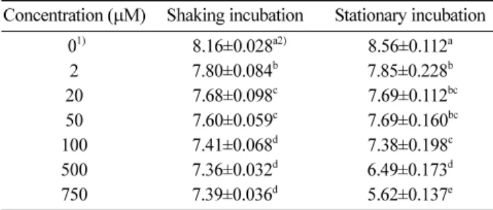
Margolis 등(14)은 actinonin이 S. aureus, pathogenic E. coli의 생장을 억제 시키지 못한다고 보고한 반면, Chen 등(6)은 actinonin 에 의해 S. epidermidis, Haemophilus influenza, Moraxella catarrhalis, Neisseria gonorrheae 등의 생장이 억제되었다고 보고하였다. 이는 S. aureus나 pathogenic E. coli의 peptide deformylase (PDF)와는 달리 S. epidermidis, H. influenza, M. catarrhalis, N. gonorheae 등이 가지고 있는 PDF의 활성이 actinonin에 의해 저해됨으로써 나타나는 현상 때문인 것으로 규명되었다(6,14). 따라서 이후 actinonin의 B. cereus에 대한 항균활성 기작 연구에 있어 B. cereus의 PDF와 actinonin과의 관계 규명이 중요하다고 판단된다. Actinonin 농도에 따른 B. cereus에 대한 항균활성 Actinonin의 농도별 항균활성을 평가하기 위해 첫번째로 broth micro-dilution method를 응용하여 LB에서 B. cereus의 생장 억제 효과를 측정하였다. 7.8 log CFU/mL 수준으로 LB 액체배지에 접 종된 B. cereus를 2, 20, 50, 100, 500, 750 µM 농도의 actinonin 으로 처리하여 항균활성을 측정한 결과 Table 1과 같이 나타났 다. 진탕 조건에서의 B. cereus는 actinonin 농도 2 µM부터 유의 적 감소가 확인되었고, 20 µM에서는 약 0.12 log CFU/mL의 감 소가 확인되었으며, 50 µM과 100 µM에서는 각각 0.08, 0.19 log CFU/mL의 감소가 관찰 되었다(p<0.01). 그러나 100 µM 이상인 500µM, 750 µM에서의 항균활성은 100 µM에서의 항균활성과 유 의적인 차이가 없음을 알 수 있었다. 반면, 정치 조건에서의 B. cereus는 농도 2 µM부터 유의적 감소를 보이기 시작하였고 20, 50µM에서는 0.87 log CFU/mL, 100 µM에서는 1.18 log CFU/ mL의 감소가 확인되었다. 특히 100 µM 이후의 500, 750 µM에서 진탕 조건과는 다르게 각각 2.07, 2.94 log CFU/mL의 더 많은 감 소량을 보였다(p<0.01) (Table 1).
항균 활성을 측정하기 위한 두 번째 실험으로 agar disk diffu-sion test를 실시하였다. 0.02, 0.1, 0.2, 1, 2 mM의 actinonin을 함 유한 paper disk를 B. cereus가 도말된 Mueller-Hinton 고체배지에 위치시킨 후 관찰한 결과 1 mM과 2 mM actinonin에 의해서 억 제환이 형성된 것을 확인하였다(Fig. 2). 1, 2 mM actinonin이 처 리된 각 배지들에서 약 1.32, 1.75 mm 크기의 억제환들을 형성시 킨 것을 확인하였고, 억제환들의 크기가 농도에 따라 유의적으로 증가한 것을 알 수 있었다.
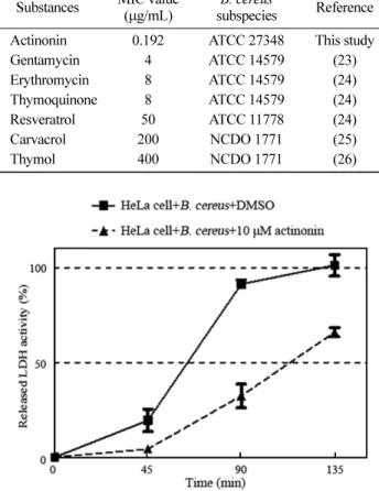
마지막으로 broth micro-dilution MIC test를 통해 actinonin의 B. cereus에 대한 MIC를 측정하였으며, 이를 선행 연구들에서의 Table 1. Antimicrobial susceptibility of Bacillus cereus to
actinonin depending on the concentration of actinonin Concentration (µM) Shaking incubation Stationary incubation
01) 08.16±0.028a2) 8.56±0.112a 2 7.80±0.084b 7.85±0.228b 20 7.68±0.098c 07.69±0.112bc 50 7.60±0.059c 07.69±0.160bc 100 7.41±0.068d 7.38±0.198c 500 7.36±0.032d 6.49±0.173d 750 7.39±0.036d 5.62±0.137e Control: The same amount of DMSO was added to the control of each strains.
All values are mean±standard deviation of three repeated process. Different superscripts indicate highly significant difference (p<0.01). Unit: Log CFU/mL
Fig. 2. Growth inhibition of Bacillus cereus by actinonin treatment in agar disk diffusion test. (A) Examples of inhibition zones for Bacillus cereus formed by actinonin in agar disk diffusion test; (B) Diameters of inhibition zones of B. cereus depending on concentration of actinonin.; Mueller-Hinton agar plates were inoculated with 100µL of B. cereus. Disk papers were placed on the each plate and inoculated with different concentrations of actinonin (0, 0.02, 0.1, 0.2, 1, 2 mM). The plates were incubated at 37oC for 16 h, then inhibition zones were determined. DMSO was added to the control group (0 mM). Double stars indicate p values <0.01 and ND indicates that inhibition zone was not detected.
천연 순수 분리물질들의 B. cereus에 대한 MIC와 비교를 하였다 (23-25). Chaieb 등(23)은 gentamycin, erythromycin, thymoquinone 의 MIC가 각각 4, 8, 8 µg/mL 라고 보고하였다. Paulo 등(24)의 연구에서는 resveratrol의 MIC가 50 µg/mL로 측정되었다. Carvac-rol, thymol의 MIC는 각각 200, 400 µg/mL 로서 Gutiérrez-Lar-raínzar 등(25)의 연구에서 보고되었다. 그러나 본 연구에서 같은 방법을 통해 얻어진 actinonin의 MIC는 0.192 µg/mL (0.5 µM)로 서 기존의 연구되어 온 천연추출물보다 낮은 MIC 값이 측정되 었으며, 이는 actinonin이 B. cereus에 대한 항균 활성이 다른 천 연추출물에 비해 우수함을 의미한다(Table 2). Actinonin에 의한 B. cereus의 세포 독성 저해 효과 10µM의 actinonin을 MOI 20의 B. cereus와 함께 HeLa 세포에 투여하여 시간에 따른 released LDH activity를 측정함으로써 actinonin의 세포 독성 억제율을 측정하였다. B. cereus와 DMSO 가 첨가된 대조군에서의 released LDH activity는 시간 경과에 비 례하여 증가하여 45분에서 약 19%, 90분에서 약 91%를 나타내 었고, 135분에서 최대치를 보인 반면, B. cereus와 actinonin이 첨 가된 실험군에서는 측정 초기 시간부터 대조군에 비해 released LDH activity가 감소하여 반응 시간 45분에는 약 4%, 90분에는 약 32%, 135분에는 약 65%를 나타내었다(Fig. 3).
요
약
본 연구에서는 지금까지 알려진 바와 달리 항균활성의 적용범 위가 모호한 actinonin을 대표적 식중독균인 B. cereus에 적용함 으로써 식중독균에 대한 항균 효과에 대해 연구하였다. 계대배양 된 대수기의 B. cereus에 actinonin을 처리하여 생장 저해 여부를 확인하였고, 96-well plate를 이용한 broth micro-dilution 방법과 agar disk diffusion 방법을 통해 actinonin의 B. cereus에 대한 항 균활성을 측정해본 결과, actinonin의 농도에 비례하여 B. cereus 의 생장이 저해되는 것으로 나타났다. 또한, actinonin의 처리에 따른 B. cereus의 세포 독성 측정 결과 actinonin이 B. cereus의 세 포 독성 또한 억제하는 것을 확인하였다. 따라서, actinonin은 천 연항균물질로서 B. cereus에 의한 식품 오염을 억제하고 식중독 예방 및 잠재적 치료제로서 적용 될 가능성이 있는 것으로 관찰 되었으며, 나아가 다른 식중독균들에 대한 적용 가능성 탐색과 관련한 연구가 필요할 것으로 생각된다.감사의 글
본 연구는 2013 충남대학교 신임교원학술연구비로부터 지원을 받아 수행된 연구이며 이에 감사드립니다.References
1. Newell DG, Koopmans M, Verhoef L, Duizer E, Aidara-Kane A, Sprong H, Opsteegh M, Langelaar M, Threfall J, Scheutz F, van der Giessen J, Kruse H. Food-borne diseases - the challenges of 20 years ago still persist while new ones continue to emerge. Int. J. Food Microbiol. 139: S3-S15 (2010)
2. Ricke SC, Kundinger MM, Miller DR, Keeton JT. Alternatives to antibiotics: Chemical and physical antimicrobial interventions and foodborne pathogen response. Poult. Sci. 84: 667-675 (2005) 3. Unemo M, Del Rio C, Shafer WM. Antimicrobial resistance
expressed by Neisseria gonorrhoeae: A major global public health problem in the 21st century. Microbiol. Spectr. 4: 10.1128/ microbiolspec.EI10-0009-2015 (2016)
4. Walsh C, Fanning S. Antimicrobial resistance in foodborne patho-gens-A cause for concern?. Curr. Drug Targets 9: 808-815 (2008) 5. Cho KH, Park SG. Antibacterial effects on Bacillus
stearothermo-philus by adding natural grapefruit seed extracts in soymilk. J. Korean Ind. Eng. Chem. 16: 139-143 (2004)
6. Chen DZ, Patel DV, Hackbarth CJ, Wang W, Dreyer G, Young DC, Margolis PS, Wu C, Ni ZJ, Trias J, White RJ, Yuan Z. Acti-nonin, a naturally occurring antibacterial agent, is a potent deformylase inhibitor. Biochemistry 39: 1256-1262 (2000) 7. Lee MD, She YH, Soskis MJ, Borella CP, Gardner JR, Hayes
PA, Dy BM, Heaney ML, Philips MR, Bornmann WG, Sirotnak FM, Scheinberg DA. Human mitochondrial peptide deformylase, a new anticancer target of actinonin-based antibiotics. J. Clin. Invest. 114: 1107-1116 (2004)
8. Yekkour A, Meklat A, Bijani C, Toumatia O, Errakhi R, Lebrihi A, Mathieu F, Zitouni A, Sabaou N. A novel hydroxamic acid-containing antibiotic produced by a Saharan soil-living Streptomy-ces strain. Lett. Appl. Microbiol. 60: 589-596 (2015)
9. Pratt LM, Beckett RP, Davies SJ, Launchbury SB, Miller A, Spavold ZM, Todd RS, Whittaker M. Asymmetric synthesis of BB-3497-A potent peptide deformylase inhibitor. Bioorg. Med. Chem. Lett. 11: 2585-2588 (2001)
10. Bashiardes G, Bodwell GJ, Davies SG. Asymmetric-synthesis of (-)-actinonin and (-)-epi-actinonin. J. Chem. Soc. Perkin Trans. 1: 459-469 (1993)
11. Sayama K, Goto Y, Iguchi T, Takeda Y, Matsuzawa A. Effects of an antibiotic protease inhibitor, actinonin on the growth within collagen gels of non-metastatic and metastatic mouse mammary tumors of the same origin. Cancer Lett. 94: 171-177 (1995) Table 2. Minimum inhibitory concentration of various natural
antimicrobial compounds on B. cereus Substances MIC value
(µg/mL)
B. cereus
subspecies Reference Actinonin 0.192 ATCC 27348 This study
Gentamycin 4 ATCC 14579 (23) Erythromycin 8 ATCC 14579 (24) Thymoquinone 8 ATCC 14579 (24) Resveratrol 50 ATCC 11778 (24) Carvacrol 200 NCDO 1771 (25) Thymol 400 NCDO 1771 (26)
Fig. 3. Determination of lactate dehydrogenase (LDH) release from HeLa cell infected with Bacillus cereus in the presence or absence of actinonin. The HeLa cells were infected by B. cereus at MOI 20, and supplemented with DMSO or 10µM of actinonin. LDH release from the cells was determined after 45, 90, and 135 min of the infection by measuring optical density (OD490 nm). The results are means of three observations±standard deviations.
12. Adams JM, Capecchi MR. N-formylmethionyl-sRNA as the initi-ator of protein synthesis. Proc. Natl. Acad. Sci. 55: 147-55 (1966)
13. Mazel D, Pochet S, Marliere P. Genetic characterization of polypeptide deformylase, a distinctive enzyme of eubacterial translation. EMBO J. 13: 914-23 (1994)
14. Margolis PS, Hackbarth CJ, Young DC, Wang W, Chen D, Yuan Z, White R, Trias J. Peptide deformylase in Staphylococcus aureus: Resistance to inhibition is mediated by mutations in the formyltransferase gene. Antimicrob. Agents Ch. 44: 1825-31 (2000)
15. Oh MH, Ham JS, Cox JM. Diversity and toxigenicity among members of the Bacillus cereus group. Int J. Food Microbiol. 152: 1-8 (2012)
16. Agata N, Ohta M, Mori M, Isobe M. A novel dodecadepsipep-tide, cereulide, is an emetic toxin of Bacillus cereus. FEMS Microbiol. Lett. 129: 17-20 (1995)
17. Ehling-Schulz M, Fricker M, Scherer S. Bacillus cereus, the causative agent of an emetic type of food-borne illness. Mol. Nutr. Food Res. 48: 479-487 (2004)
18. Granum PE. Bacillus cereus and its toxins. Soc. Appl. Bacteriol. Symp. Ser. 23: 61S-66S (1994)
19. Liu H, Zhao Y, Zhao D, Gong T, Wu Y, Han H, Xu T, Peschel A, Han S, Qu D. Antibacterial and anti-biofilm activities of thiaz-olidione derivatives against clinical staphylococcus strains. Emerg. Microbes. Infect. 4: 10.1038/emi.2015.1 (2015)
20. Kim JJ, In YW, Oh SW. Antimicrobial activity of citral against Salmonella Typhimurium and Staphylococcus aureus. Korean J. Food Sci. Technol. 43: 791-794 (2011)
21. Brudzynski K, Abubaker K, Wang T. Powerful bacterial killing by buckwheat honeys is concentration-dependent, involves com-plete DNA degradation and requires hydrogen peroxide. Front. Microbiol. 3: 242 (2012)
22. Clinical and Laboratory Standards Institute (CLSI). Performance Standards for Antimicrobial Susceptibility Testing; Twenty-Forth Informational Supplement. CLSI document M100-S24, Wayne, PA, USA. (2014)
23. Chaieb K, Kouidhi B, Jrah H, Mahdouani K, Bakhrouf A. Anti-bacterial activity of thymoquinone, an active principle of Nigella sativa and its potency to prevent bacterial biofilm formation. BMC Complement. Altern. Med. 11: 29 (2011)
24. Paulo L, Ferreira S, Gallardo E, Queiroz JA, Domingues F. Anti-microbial activity and effects of resveratrol on human pathogenic bacteria. World J. Microbiol. Biotechnol. 26: 1533-1538 (2010) 25. Gutiérrez-Larraínzar M, Rúa J, Caro I, de Castro C, de Arriaga
D, García-Armesto MR, del Valle P. Evaluation of antimicrobial and antioxidant activities of natural phenolic compounds against foodborne pathogens and spoilage bacteria. Food Control. 26: 555-563 (2012)
26. Park KS, Ono T, Rokuda M, Jang MH, Okada K, Idia T, Honda T. Functional characterization of two type III secretion systems of Vibrio parahaemolyticus. Infect. Immun. 72: 6659-6665 (2004)