INTRODUCTION
Maternal chorioamnionitis has been known to result in a lung maturation response. The incidence of respiratory dis- tress syndrome in preterm infants decreased when they were exposed to maternal chorioamnionitis (1). In animal study, maternal chorioamnionitis increased the size of surfactant pool and the volume of potential gas space (2). However, maternal chorioamnionitis has also been associated with ab- normal lung development. The exposure to maternal cho- rioamnionitis increased the incidence of bronchopulmonary dysplasia (BPD) in preterm infants (1, 3), and resulted in alveolar simplification and impaired microvascular develop- ment (4, 5), which are histologic hallmarks of new BPD in the post-surfactant era (6).
Transforming growth factor-beta (TGF-β) is an impor- tant regulator of cellular proliferation and differentiation. It has been considered to be crucial for the normal lung devel- opment and implicated in the development of BPD. Dur- ing branching morphogenesis in the lung development, the expression of TGF-βwas reduced (7, 8). Overexpression of
TGF-βinhibited airway branching (8, 9), while inhibition of TGF-βenhanced airway branching in vitro (9-11). Com- pared to branching morphogenesis, neither the role nor the status of expression of TGF-βduring alveolarization is well known. Several researchers reported that overexpression of TGF-β1 inhibited alveolarization in neonatal rat (12, 13), and inhibition of TGF-βresulted in an emphysematous change in adult mouse (14, 15). However, the role of TGF-βin nor- mal lung development, especially during alveolarization in human preterm infants, remains unclear.
TGF-βis also known as an important mediator of fibro- sis. Kotecha et al. reported an increased level of TGF-β1 in bronchoalveolar lavage (BAL) fluid of preterm infants who developed BPD later (16). In animal model, enhanced endoge- nous production or overexpression of TGF-β1 resulted in pulmonary pathologic changes seen in human BPD, which are enlarged alveolar sacs, poor secondary septation, thick and hypercellular septa, and abnormal capillary development (12, 13, 17). However, the role of TGF-βin the injury response of the lungs in preterm infants also remains unclear.
The objective of our study was to examine the effect of
609
Chang Won Choi, Beyong Il Kim, Kyoung Eun Joung, Jin-A Lee, Yun Kyoung Lee, Ee-Kyung Kim, Han-Suk Kim, June Dong Park, and Jung-Hwan Choi
Department of Pediatrics, Seoul National University College of Medicine, Seoul, Korea
Address for correspondence Beyong Il Kim, M.D.
Department of Pediatrics, Seoul National University Bundang Hospital, 300 Gumi-dong, Bundang-gu, Seongnam 463-707, Korea
Tel : +82.31-787-7282, Fax : +82.31-787-4054 E-mail : [email protected]
*This work was supported by the Fund of S. N. U.
Pediatric Alumni (1999-01).
DOI: 10.3346/jkms.2008.23.4.609
Decreased Expression of Transforming Growth Factor-beta1 in Bronchoalveolar Lavage Cells of Preterm Infants with Maternal Chorioamnionitis
Maternal chorioamnionitis has been associated with abnormal lung development.
We examined the effect of maternal chorioamnionitis on the expression of trans- forming growth factor-beta1 (TGF-ββ1) in the lungs of preterm infants. A total of 63 preterm (≤≤34 weeks) infants who were intubated in the delivery room were prospec- tively enrolled. Their placentas were examined for the presence of chorioamnioni- tis. Bronchoalveolar lavage (BAL) fluid and cells were obtained shortly after birth.
TGF-ββ1 was measured in BAL fluid and TGF-ββ1 mRNA expression was determined by reverse transcription polymerase chain reaction (RT-PCR) in BAL cells. TGF-ββ1 mRNA expression in BAL cells showed a positive correlation with gestational age (r=0.414, p=0.002). TGF-ββ1 mRNA expression was significantly decreased in the presence of maternal chorioamnionitis (0.70±±0.12 vs. 0.81±±0.15, p=0.007). Adjust- ment for gestational age, birth weight, and delivery mode did not nullify the signifi- cance. TGF-ββ1 mRNA expression was marginally significantly decreased in preterm infants who developed bronchopulmonary dysplasia (BPD) later (0.75±±0.11 vs.
0.82±±0.15, p=0.055). However, adjustment for gestational age, patent ductus arteriosus (PDA), and maternal chorioamnionitis nullified the significance. These results might be an indirect evidence that maternal chorioamnionitis may inhibit normal lung development of fetus.
Key Words : Transforming Growth Factor-beta; Chorioamnionitis; Bronchopulmonary Dysplasia
Received : 10 July 2007 Accepted : 15 December 2007
maternal chorioamnionitis on the expression of TGF-β1, the isoform most implicated in fibrosis among its three isoforms whose biological properties are nearly identical, and com- pare the extent of TGF-β1 expression between preterm infants who develop BPD later and who do not.
MATERIALS AND METHODS Study design
A prospective cohort study was done to examine the effect of maternal chorioamnionitis on the expression of TGF-β1 in BAL cells of preterm infants. The cohort consisted of preterm (gestational age <34 weeks) infants who were admitted to neonatal intensive care unit (NICU) at Seoul National Uni- versity Children’s Hospital and Seoul National University Bundang Hospital (after May 2003) between March 1999 and February 2004 and who met the following criteria: 1) endotracheal intubation at delivery room, and 2) absence of major congenital anomalies. During the study period, a total of 79 preterm infants were enrolled. Among these preterm infants, 7 infants whose parents’ consent was not obtained and 9 infants to whom BAL was not done or done after the first 24 hr after birth due to death or clinical instability were excluded from the analysis. Finally 63 preterm infants were analyzed. Of these, 8 preterm infants died before 36 weeks postmenstrual age when the assessment for the development of BPD was made. The institutional review board at Seoul National University Hospital approved the study protocol (H-0603-230-172), and informed consent was obtained from a parent before enrollment.
Bronchoalveolar lavage
Endotracheal intubation in the delivery room was done only when clinically indicated according to the neonatal resuscitation program (18). BAL fluid and cells were obtain- ed by the method described by Kotecha (19) shortly after birth as soon as the preterm infants became stable, but before surfactant replacement therapy when indicated. Briefly, with the baby supine, two aliquots of sterile saline solution, 1 mL/
kg (maximum 2 mL), were instilled via endotracheal tube, and then immediately sucked back, and the returned BAL fluid was collected in a suction trap. BAL fluid was centrifug- ed for 10 min at 1,000 g within 10 min after acquisition. The supernatant and sediment, which is a cell fraction, were con- tained to separate polypropylene tubes, and stored at -70℃ and -160℃in liquid nitrogen, respectively. The stored super- natant and sediment were used for the measurement of TGF- β1 concentration and TGF-β1 mRNA reverse transcription polymerase chain reaction (RT-PCR), respectively.
Determination of TGF-ββ1 and TGF-ββ1 mRNA in BAL fluid and cells
The TGF-β1 concentration in BAL fluid was measured using a commercially available ELISA kit (Quantikine�human TGF-β1 immunoassay, R&D Systems Inc., Minneapolis, MN, U.S.A.). The sensitivity of the test was <7 pg/mL. Interas- say and intra-assay coefficients of variation were 7.3% and 11.7%, respectively. The frozen BAL fluid sediment in liq- uid nitrogen was pulverized and homogenized using Qia- shredder�(Quiagen Inc., Valencia, CA, U.S.A.). The extrac- tion of mRNA from the homogenates was done using Olig- otex�direct mRNA kit (Quiagen Inc.). Reverse transcription of TGF-β1 mRNA was done using MuLV reverse transcrip- tase (PerkinElmer Inc., Waltham, MA, U.S.A.) and RNA PCR kit�(PerkinElmer Inc.). The sense and antisence primers for human TGF-β1 cDNA were designed by PrimerSelect� version 3.11 (DNAStar Inc., Madison, WI, U.S.A.). The primer for forward reaction was 5′GCCCTGGATACCAA- CTATTGCT3′, and the primer for reverse reaction was 5′ AGGCTCCAAATATAGGGGCAGG3′. PCR was done using automatic PCR cycler (Gene Cycler�, Bio-Rad Labo- ratories, Hercules, CA, U.S.A.). The size of the PCR prod- uct was 161 bp. For internal standard, PCR was also done for β-actin. PCR products were loaded and analyzed on 1%
agarose gel with a 10-fold diluted 100 bp DNA ladder (Pro- mega Corp. Madison, WI, U.S.A.) as a DNA marker. Image analysis was performed using ‘Band leader’ software version 2.01. The signal intensity of TGF-β1 mRNA was expressed relative to that of β-actin.
Definition of maternal chorioamnionitis and bronchopul- monary dysplasia
Maternal chorioamnionitis was defined histologically as
≥grade 2 inflammation of the amnion or choriodecidua or
≥grade 1 necrotizing funisitis according to the grading sys- tem suggested by Salafia et al. (20). BPD was defined as a state of chronic oxygen requirement at 36 weeks postmen- strual age plus a total oxygen duration of ≥28 day with a consistent chest radiographic finding, which is persistent hazy opacification or a cyst-like pattern of density and lucency.
Statistical analysis
Statistical analysis was done using SPSS version 11.5 (SPSS Inc., Chicago, IL, U.S.A.). Comparison of means between groups was performed using one-way analysis of variance (ANOVA) test or Mann-Whitney U-test. Comparison of fre- quencies between groups was done using 2or Fisher’s exact test. To investigate the relationship between TGF-β1 or TGF- β1 mRNA in BAL fluid and cells and maternal chorioam- nionitis or BPD, while eliminating the effects of potential confounding variables, multivariate logistic regression anal-
ysis was performed. A p value of <0.05 was considered sta- tistically significant.
RESULTS
Clinical profiles of preterm infants with and without mater- nal chorioamnionitis
Gestational age and the rate of Cesarean section delivery were significantly lower in preterm infants with maternal chorioamnionitis. There were no significant differences in birth weight, Apgar scores at 1 and 5 min, sex ratio, prema- ture rupture of membrane, antenatal steroid therapy, and intrauterine growth restriction. Respiratory distress syndrome (RDS) was significantly less prevalent, while BPD was more
Maternal chorioamnioni-
tis (n=23)
No maternal chorioamnioni-
tis (n=40) p
Birth weight (g) 1,096±418 1,328±514 0.070 Gestational age (week) 27.6±3.0 30.1±2.6 <0.001 Apgar score at 1 min 4.0±2.4 4.3±2.4 0.748 Apgar score at 5 min 5.7±2.2 5.8±2.5 0.845
Male (%) 14 (61) 17 (43) 0.196
Cesarean section (%) 13 (57) 38 (95) <0.001
PROM (%) 8 (35) 7 (18) 0.137
Antenatal steroid (%) 17 (74) 20 (50) 0.110
IUGR (%) 1 (4) 6 (15) 0.407
RDS (%) 11 (48) 31 (78) 0.026
PDA (%) 14 (61) 24 (60) 1.000
BPD (%) 16 (70) 15 (38) 0.019
Sepsis (%) 4 (17) 9 (23) 0.753
PVL (%) 1 (4) 1 (3) 1.000
TGF-β1 in BAL fluid (pg/mL) 85.5±49.1 87.1±67.2 0.925 TGF-β1 mRNA in BAL cells 0.70±0.12 0.81±0.15 0.007
(relative to β-actin)
Table 1. Clinical profiles, TGF-β1 in BAL fluid and TGF-β1 mRNA in BAL cells of preterm infants with and without maternal chori- oamnionitis
Data shown as mean±SD.
TGF-β1, transforming growth factor-beta1; BAL, bronchoalveolar lavage;
PROM, premature rupture of membrane; IUGR, intrauterine growth restriction; RDS, respiratory distress syndrome; PDA, patent ductus arteriosus; BPD, bronchopulmonary dysplasia; PVL, periventricular leuko- malacia.
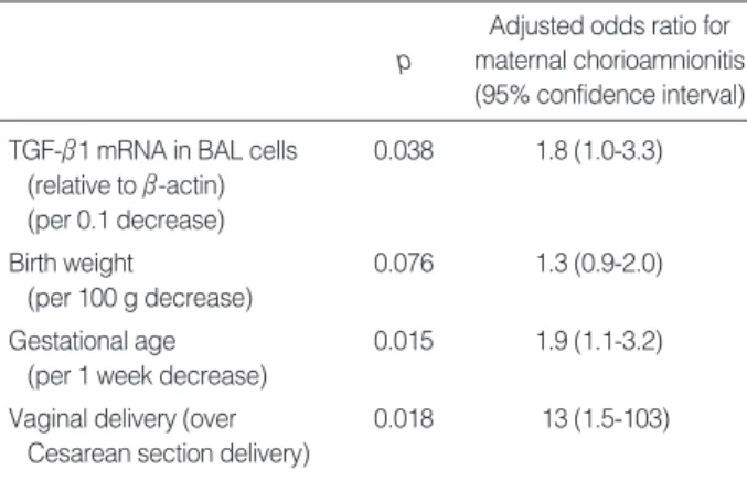
Adjusted odds ratio for maternal chorioamnionitis (95% confidence interval) p
TGF-β1 mRNA in BAL cells 0.038 1.8 (1.0-3.3) (relative to β-actin)
(per 0.1 decrease)
Birth weight 0.076 1.3 (0.9-2.0)
(per 100 g decrease)
Gestational age 0.015 1.9 (1.1-3.2)
(per 1 week decrease)
Vaginal delivery (over 0.018 13 (1.5-103) Cesarean section delivery)
Table 2. Adjusted odds ratio of each significant variable for the presence of maternal chorioamnionitis assessed by logistic regression analysis
TGF-β1, transforming growth factor-beta1; BAL, bronchoalveolar lavage.
TGF-beta1 mRNA (relative to beta-actin)
1.1
1.0
0.9
0.8
0.7
0.6
0.5
0.4
22 24 26 28 30 32 34 36
Gestational age (week)
r=0.412 p=0.014
r=0.414 p=0.002 Chorioamnionitis
Fig. 1. The correlations between TGF-β1 mRNA in BAL cells and gestational age (A) and birth weight (B). (A) A significant positive corre- lation is shown in total population (r=0.414, p=0.002) and in the subgroup without maternal chorioamnionitis (r=0.412, p=0.014), while poor correlation in the subgroup with maternal chorioamnionitis. (B) A significant positive correlation is shown in total population (r=0.372, p=0.005) and in the subgroup without maternal chorioamnionitis (r=0.481, p=0.003), while poor correlation in the subgroup with maternal chorioamnionitis.
A
TGF-beta1 mRNA (relative to beta-actin)
1.1 1.0 0.9 0.8 0.7 0.6 0.5 0.4 0.3
0 500 1,000 1,500 2,000 2,500
Birth weight (g) B
r=0.372 p=0.005 r=0.481 p=0.003 No chorioamnionitis Total population Chorioamnionitis No chorioamnionitis
prevalent in preterm infants with maternal chorioamnioni- tis. There were no significant differences in echocardiogra- phy-documented patent ductus arteriosus, culture-proven sepsis, and periventricular leukomalacia (Table 1).
TGF-ββ1 and TGF-ββ1 mRNA in BAL fluid and cells in pre- term infants with and without maternal chorioamnionitis
The concentrations of TGF-β1 in BAL fluid were lower in preterm infants with maternal chorioamnionitis, although statistically insignificant. Signal intensities of TGF-β1 mRNA expressed relative to that of β-actin were significantly lower in preterm infants with maternal chorioamnionitis (0.70± 0.12 vs. 0.81±0.15, p=0.007) (Table 1).
Multivariate logistic regression analysis including con- founders such as birth weight, gestational age and Cesarean section delivery in the logistic model revealed an indepen- dent relationship between TGF-β1 mRNA in BAL cells and maternal chorioamnionitis (adjusted odds ratio 1.8 per 0.1 decrease of signal intensity of TGF-β1 mRNA expressed rela- tive to that of β-actin, 95% confidence interval 1.0-3.3) (Table 2).
The correlations between TGF-ββ1 mRNA in BAL cells and gestational age and birth weight
Signal intensity of TGF-β1 mRNA expressed relative to β-actin demonstrated a moderate positive correlation with gestational age (r=0.414, p=0.002) and birth weight (r=
0.372, p=0.005). However, in preterm infants with mater- nal chorioamnionitis, there were no correlations between them. In preterm infants without maternal chorioamnioni- tis, there were also moderate correlations between signal intensity of TGF-β1 mRNA and birth weight and gesta- tional age (Fig. 1).
There were no correlations between the concentration of TGF-β1 in BAL fluid and birth weight or gestational age (data not shown).
Clinical profiles of preterm infants who developed BPD and who did not
Birth weight, gestational age, and Apgar scores at 1 and 5 min were significantly lower in preterm infants who devel- oped BPD later. Maternal chorioamnionitis, echocardiogra- phy-documented patent ductus arteriosus (PDA) and cul- ture-proven sepsis were more prevalent in preterm infants who developed BPD later. The higher prevalence of sepsis in preterm infants who developed BPD later was thought
BPD (n=28)
No BPD
(n=27) p
Birth weight (g) 997±235 1,649±397 <0.001 Gestational age (week) 28.1±2.3 31.8±1.9 <0.001 Apgar score at 1 min 3.7±2.0 5.3±2.4 0.007 Apgar score at 5 min 5.3±1.7 6.8±2.3 0.009
Male (%) 15 (54) 13 (48) 0.688
Cesarean section (%) 23 (82) 22 (82) 0.949 Maternal chorioamnionitis (%) 15 (54) 5 (19) 0.007
PROM (%) 6 (21) 6 (22) 0.943
Antenatal steroid (%) 19 (68) 13 (48) 0.139
IUGR (%) 3 (11) 2 (7) 0.670
RDS (%) 17 (61) 20 (74) 0.291
PDA (%) 24 (86) 12 (44) 0.001
Sepsis (%) 11 (39) 0 (0) <0.001
PVL (%) 2 (7) 0 (0) 0.157
TGF-β1 in BAL fluid (pg/mL) 89.9±71.9 91.4±55.1 0.936 TGF-β1 mRNA in BAL cells 0.75±0.11 0.82±0.15 0.055
(relative to β-actin)
Table 3. Clinical profiles, TGF-β1 in BAL fluid and TGF-β1 mRNA in BAL cells of preterm infants who developed bronchopul- monary dysplasia later and who did not
Data shown as mean±SD.
Eight preterm infants died before 36 weeks postmenstrual age when the assessment for the development of BPD was made and were exclud- ed from the analysis.
TGF-β1, transforming growth factor-beta1; BAL, bronchoalveolar lavage;
PROM, premature rupture of membrane; IUGR, intrauterine growth restriction; RDS, respiratory distress syndrome; PDA, patent ductus arteriosus; BPD, bronchopulmonary dysplasia; PVL, periventricular leukomalacia.
MCA (n=20) BPD
(n=15)
No BPD (n=5)
No MCA (n=35) BPD
(n=13)
No BPD (n=22) TGF-β1in BAL 83.8±47.4 137.7±70.3 96.4±93.1 86.9±53.4
fluid (pg/mL)
p 0.171 0.709
TGF-β1mRNA in 0.71±0.12 0.66±0.16 0.79±0.10 0.86±0.13 BAL cells
(relative to β-actin)
p 0.496 0.097
BPD (n=28) MCA (n=15)
No MCA (n=13)
No BPD (n=27) MCA
(n=5)
No MCA (n=22) TGF-β1in BAL 83.8±47.4 96.4±93.1 137.7±70.3 86.9±53.4
fluid (pg/mL)
p 0.659 0.221
TGF-β1mRNA in 0.71±0.12 0.79±0.10 0.66±0.16 0.86±0.13 BAL cells
(relative to β-actin)
p 0.106 0.007
Table 4. TGF-β1 in BAL fluid and TGF-β1 mRNA in BAL cells by the presence of maternal chorioamnionitis and the development of bronchopulmonary dysplasia
Data shown as mean±SD.
TGF-β1, transforming growth factor-beta1; BAL, bronchoalveolar lavage;
MCA, maternal chorioamnionitis; BPD, bronchopulmonary dysplasia.
to be consequential rather than causative of BPD consider- ing the temporal relationship between them. There were no significant differences in sex ratio, the rate of Cesarean sec- tion delivery, premature rupture of membrane, antenatal steroid therapy, intrauterine growth restriction, RDS and periventricular leukomalacia (Table 3).
TGF-ββ1 and TGF-ββ1 mRNA in BAL fluid and cells and later development of BPD
The concentrations of TGF-β1 in BAL fluid did not differ significantly between preterm infants who developed BPD later and who did not (Table 3). Signal intensities of TGF-β1 mRNA expressed relative to β-actin in BAL cells were mar- ginally significantly lower in preterm infants who developed BPD later (0.75±0.11 vs. 0.82±0.15, p=0.055) (Table 3).
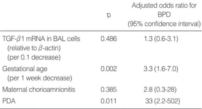
Subsequent subgroup analyses by the presence of mater- nal chorioamnionitis and the development of BPD demon- strated no significant differences in the concentration of TGF- β1 in BAL fluid and signal intensities of TGF-β1 mRNA expressed relative to β-actin in BAL cells, except for preterm infants who did not develop BPD (Table 4). Multivariate logistic regression analysis including confounders such as gestational age, chorioamnionitis and PDA which were sig- nificant variables in the univariate analysis in the logistic model failed to reveal an independent relationship between TGF-β1 mRNA in BAL cells and the later development of BPD (Table 5).
DISCUSSION
Pulmonary fibrosis was a prominent feature in preterm infants who died of BPD (21, 22). Moreover, markers of fibro- sis such as hydroxyproline, fibronectin, and desmosine were increased in lung tissue, tracheal fluid, and urine of preterm infants who developed BPD (23-25). TGF-βhas been known
to be an important mediator of fibrosis. Accordingly, TGF- βhas been implicated in the pathogenesis of BPD. Kotecha et al. (16) demonstrated increased levels of TGF-β1 in BAL fluid in preterm infants who developed BPD later. Howev- er, as with the introduction of antenatal steroid, surfactant replacement therapy, and gentle ventilatory strategies, the incidence of classic BPD in which fibrosis is a predominant feature has decreased (26). Instead, a milder new type of BPD has been increasing in preterm infants who were not exposed to significant hyperoxia and baro/volutrauma (27). This type of BPD in the post-surfactant era has different pathologic characteristics from that of classic BPD. It lacks fibrosis, air- way injury, and emphysema which are predominant features of classic BPD. Instead, an arrest in alveolar and microvas- cular development is the pathologic hallmark of this type of BPD in the post-surfactant era (6).
Recently, there has been growing evidence that antenatal inflammatory process may inhibit normal alveolarization (4, 5, 28). In epidemiologic and clinical studies, antenatal inflam- mation or maternal chorioamnionitis was related to the devel- opment of BPD (1, 29-31). However, there have been few studies as to the status of TGF-βexpression in the lungs when the fetus is exposed to maternal chorioamnionitis. The exact role of TGF-βin normal alveolarization phase in human lung development is not known clearly, although its role in branching morphogenesis is well known in in-vitro studies (7-11). Considering that an increased TGF-βlevel has been associated with the development of BPD, it could be reason- ably postulated that TGF-βwould be increased in preterm infants with maternal chorioamnionitis which has also been related to the development of BPD. Recently, Kunzmann et al. (32), demonstrated elevated TGF-β1 mRNA and protein expression in the lungs by experimental maternal chorioam- nionitis in preterm neonatal sheep model. In contrast, the expression of connective tissue growth factor, a down-stream mediator of some of the pro-fibrotic effects of TGF-β1, decreas- ed. In our study, however, the expression of TGF-β1 mRNA and protein decreased in BAL cells and fluid obtained short- ly after birth from the preterm infants who were exposed to maternal chorioamnionitis, although the latter lacked sta- tistical significance. These results are opposing to those of Kunzmann et al. and also to common expectation. Some speculations may be done as to our unexpected results. Firstly, in our study, the profile of BAL cells was not analyzed. There- fore, decreased expression of TGF-β1 mRNA could be sim- ply a reflection of different cell profiles. If the proportion of cell types in BAL fluid obtained from each preterm infant differed by the presence of airway inflammation or maturity, the interpretation of the measured value of a specific mRNA which is expressed differently by airway cell type would be problematic. In our study, there is a possibility that the pro- portion of three major cell types of BAL cell which are alve- olar macrophages, neutrophils, and epithelial cells may be affected by maternal chorioamnionitis and gestational age.
Adjusted odds ratio for BPD
(95% confidence interval) p
TGF-β1 mRNA in BAL cells 0.486 1.3 (0.6-3.1) (relative to β-actin)
(per 0.1 decrease)
Gestational age 0.002 3.3 (1.6-7.0)
(per 1 week decrease)
Maternal chorioamnionitis 0.385 2.8 (0.3-28)
PDA 0.011 33 (2.2-502)
Table 5. Adjusted odds ratio of each significant variable for the development of bronchopulmonary dysplasia assessed by logistic regression analysis
BPD, bronchopulmonary dysplasia; BAL, bronchoalveolar lavage; PDA, patent ductus arteriosus.
However, TGF-β1 is known to be markedly expressed in alveolar macrophages, and to a lesser degree, in neutrophils and epithelial cells (16). If it is the case, the validity of TGF- β1 mRNA value in BAL cells by RT-PCR method would be unaffected. Secondly, the method of BAL can matter. We used non-bronchoscopic BAL due to infeasibility of bron- choscopy for study preterm infants because of their small body size. Therefore, there may be a concern that the cells from distal airway and alveoli were not obtained sufficient- ly. However, Kotecha (33) reported that the cell profiles of non-bronchoscopic BAL cells from each lung were very sim- ilar, thus very representative of lung environment. Grigg et al. (34) also demonstrated that a sufficient amount of distal airway cells was obtained by non-bronchoscopic BAL. There- fore, decreased expression of TGF-β1 mRNA in non-bron- choscopic BAL cells in our study may be thought to be rep- resentative of actual alveolar environment. However, further studies that will analyze the cell profiles of BAL cells are required to eradicate the controversy on the representative- ness of true alveolar environment of our data. Moreover, the lack of statistical significance of TGF-β1 in BAL fluid in com- parison to that of its m-RNA expression in BAL cell was observed in our study. This disparity between TGF-β1 m- RNA and its protein was not observed in other recent study that dealt with the same molecule and might have originat- ed from the small sample size and/or post-transcriptional modifications (32). However, its elucidation requires further studies.
In this study, the expression of TGF-β1 in BAL cells show- ed a significant positive correlation with gestational age and birth weight. It increased as gestational age and birth weight increased. When maternal chorioamnionitis was present, the expression of TGF-β1 in BAL cells significantly decreased inde- pendently of gestational age and birth weight. If our data are acceptable as a reflection of actual alveolar environment, it could be suggested that the expression of TGF-β1 in the lungs increases as a function of maturity, and that maternal chorioamnionitis exerts a negative influence on this normal increase of TGF-β1 expression at the later phase of human lung development. These findings may be thought to be an indi- rect evidence showing the possibility that maternal chorioam- nionitis might inhibit normal lung development of fetus, although the exact role and the expression status of TGF-β1 in saccular phase are not known clearly. Be that as it may be, however, this assumption is not supported by our results in that there was no significant relationship between the expres- sion of TGF-β1 in BAL cells and the development of BPD.
There should be another caution in interpreting our results.
The study subjects in our study were preterm infants who needed tracheal intubation in delivery room. However, they were relatively larger preterm infants with a mean birth weight of 1,243±491 g and a gestational age of 29.5±3.1 weeks who can usually be managed successfully without intubation in delivery room nowadays. Therefore, there is a possibility
that the preterm infants enrolled to our study might have suffered some antenatal insults such as maternal chorioam- nionitis and perinatal asphyxia. Actually, the incidence of maternal chorioamnionitis is as high as 37% and the 5-min Apgar score was as low as 5±2, each of which is higher and lower than 27% and 7±2, respectively, of our previous epi- demiologic study enrolled preterm infants who have similar birth weight and gestational age irrespective of whether tra- cheal intubation was done or not in delivery room (3). This difference supports the likelihood of a higher proportion of antenatally adversely affected preterm infants in our present study. Therefore, hasty generalization of our results should not be made.
In conclusion, positive correlation of TGF-β1 mRNA expres- sion in BAL cells obtained from preterm infants shortly after birth with maturity and decreased expression of TGF-β1 mRNA in the presence of maternal chorioamnionitis suggest a possi- bility that maternal chorioamnionitis might inhibit the nor- mal lung development of fetus. However, further studies will be necessary to delineate the representativeness of non-bron- choscopic BAL cells of alveolar environment and search for better easily obtainable pulmonary specimens that have a good reflection of alveolar environment in human preterm infants.
REFERENCES
1. Watterberg KL, Demers LM, Scott SM, Murphy S. Chorioamnioni- tis and early lung inflammation in infants in whom bronchopulmonary dysplasia develops. Pediatrics 1996; 97: 210-5.
2. Bry K, Lappalainen U, Hallman M. Intraamniotic interleukin-1 accel- erates surfactant protein synthesis in fetal rabbit and improves lung stability after premature birth. J Clin Invest 1997; 99: 2992-9.
3. Choi CW, Kim BI, Park JD, Koh YY, Choi JH, Choi JY. Risk fac- tors for the different types of chronic lung disease of prematurity according to the preceding respiratory distress syndrome. Pediatr Int 2005; 47: 417-23.
4. Willet KE, Jobe AH, Ikegami M, Newnham J, Brennan S, Sly PD.
Antenatal endotoxin and glucocorticoid effects on lung morphome- try in preterm lambs. Pediatr Res 2000; 48: 782-8.
5. Bry K, Lappalainen U. Pathogenesis of bronchopulmonary dyspla- sia: the role of interleukin 1beta in the regulation of inflammation mediated pulmonary retinoic acid pathways in transgenic mice.
Semin Perinatol 2006; 30: 121-8.
6. Husain NA, Siddiqui NH, Stocker JT. Pathology of arrested acinar development in postsurfactant bronchopulmonary dysplasia. Hum Pathol 1998; 29: 710-7.
7. Sakurai H, Nigam SK. Transforming growth factor-beta selectively inhibits branching morphogenesis but not tubulogenesis. Am J Phys- iol 1997; 272(1 Pt 2): F139-46.
8. Liu J, Tseu I, Wang J, Tanswell K, Post M. Transforming growth factor beta2, but not beta1 and beta3, is critical for early rat lung branching. Dev Dyn 2000; 217: 343-60.
9. Zhao J, Bu D, Lee M, Slavkin HC, Hall FL, Warburton D. Abroga- tion of transforming growth factor-beta type II receptor stimulates embryonic mouse lung branching morphogenesis in culture. Dev Biol 1996; 180: 242-57.
10. Zhao J, Lee M, Smith S, Warburton D. Abrogation of Smad3 and Smad2 or of Smad4 gene expression positively regulates murine embryonic lung branching morphogenesis in culture. Dev Biol 1998;
194: 182-95.
11. Zhao J, Shi W, Chen H, Warburton D. Smad7 and Smad6 differen- tially modulate transforming growth factor beta-induced inhibition of embryonic lung morphogenesis. J Biol Chem 2000; 275: 23992-7.
12. Gauldie J, Galt T, Bonniaud P, Robbins C, Kelly M, Warburton D.
Transfer of active form of transforming growth factor-beta 1 gene to newborn rat lung induces changes consistent with bronchopul- monary dysplasia. Am J Pathol 2003; 163: 2575-84.
13. Vicencio AG, Lee CG, Cho SJ, Eickelberg O, Chuu Y, Haddad GG, Elias JA. Conditional overexpression of bioactive transforming growth factor-beta1 in neonatal mouse lung: a new model for bron- chopulmonary dysplasia? Am J Respir Cell Mol Biol 2004; 31:
650-6.
14. Morris DG, Huang X, Kaminski N, Wang Y, Shapiro SD, Dolganov G, Glick A, Sheppard D. Loss of integrin alpha(v)beta6-mediated TGF-beta activation causes Mmp12-dependent emphysema. Nature 2003; 422: 169-73.
15. Bonniaud P, Kolb M, Galt T, Robertson J, Robbins C, Stampfli M, Lavery C, Margetts PJ, Roberts AB, Gauldie J. Smad3 null mice develop airspace enlargement and are resistant to TGF-beta-medi- ated pulmonary fibrosis. J Immunol 2004; 173: 2099-108.
16. Kotecha S, Wangoo A, Silverman M, Shaw RJ. Increase in the con- centration of transforming growth factor beta-1 in bronchoalveolar lavage fluid before development of chronic lung disease of prematu- rity. J Pediatr 1996; 128: 464-9.
17. Vicencio AG, Eickelberg O, Stankewich MC, Kashgarian M, Had- dad GG. Regulation of TGF-beta ligand and receptor expression in neonatal rat lung exposed to chronic hypoxia. J Appl Physiol 2002;
93: 1123-30.
18. Kattwinkel J. Textbook of neonatal resuscitation. 5th ed. Elk Grove Village American Academy of Pediatrics and American Heart Asso- ciation; 2006; 5: 1-42.
19. Kotecha S, Chan B, Azam N, Silverman M, Shaw RJ. Increase of interleukin-8 and soluble intercellular adhesion molecule-1 in bron- choalveolar lavage fluid from premature infants who develop chron- ic lung disease. Arch Dis Child Fetal Neonatal Ed 1995; 72: F90-6.
20. Salafia CM, Weigl C, Silberman L. The prevalence and distribution of acute placental inflammation in uncomplicated term pregnancies.
Obstet Gynecol 1989; 73: 383-9.
21. Stocker JT. The respiratory tract. In: Srocker JT, Dehner LP, eds.
Pediatric pathology. Philadelphia: JB Lippincott, 1992: 533-41.
22. Anderson WR, Engel RR. Cardiopulmonary sequelae of reparative stages of bronchopulmonary dysplasia. Arch Pathol Lab Med 1983;
107: 603-8.
23. Hislop AA, Wigglesworth JS, Desai R, Aber V. The effects of preterm delivery and mechanical ventilation on human lung growth. Early Hum Dev 1987; 15: 147-64.
24. Gerdes JS, Yoder MC, Douglas SD, Paul M, Harris MC, Polin RA.
Tracheal lavage and plasma fibronectin: relationship to respiratory distress syndrome and development of bronchopulmonary dyspla- sia. J Pediatr 1986; 108: 601-6.
25. Bruce MC, Wedig KE, Jentoft N, Martin RJ, Cheng PW, Boat TF, Fanaroff AA. Altered urinary excretion of elastin crosslinks in pre- mature infants who develop bronchopulmonary dysplasia. Am Rev Respir Dis 1985; 131: 568-72.
26. Jobe AH, Bancalari E. Bronchopulmonary dysplasia. Am J Respir Crit Care Med 2001; 163: 1723-9.
27. Charafeddine L, D’Angio CT, Phelps DL. Atypical chronic lung disease patterns in neonates. Pediatrics 1999; 103: 759-65.
28. Ueda K, Cho K, Matsuda T, Okajima S, Uchida M, Kobayashi Y, Minakami H, Kobayashi K. A rat model for arrest of alveolariza- tion induced by antenatal endotoxin administration. Pediatr Res 2006; 59: 396-400.
29. Jonsson B, Tullus K, Brauner A, Lu Y, Noack G. Early increase of TNF alpha and IL-6 in tracheobronchial aspirate fluid indicator of subsequent chronic lung disease in preterm infants. Arch Dis Child Fetal Neonatal Ed 1997; 77: F198-201.
30. Kim BI, Lee HE, Choi CW, Jo HS, Choi EH, Koh YY, Choi JH.
Increase in cord blood E-selectin and tracheal aspirate neutrophils at birth and the development of new bronchopulmonary dysplasia. J Perinat Med 2004; 32: 282-7.
31. Choi CW, Kim BI, Kim HS, Park JD, Choi JH, Son DW. Increase of interleukin-6 in tracheal aspirate at birth: a predictor of subse- quent bronchopulmonary dysplasia in preterm infants. Acta Paedi- atr 2006; 95: 38-43.
32. Kunzmann S, Speer CP, Jobe AH, Kramer BW. Antenatal inflam- mation induced TGF-beta1 but suppressed CTGF in preterm lungs.
Am J Physiol Lung Cell Mol Physiol 2007; 292: L223-31.
33. Kotecha S. Bronchoalveolar lavage of newborn infants. Pediatr Pul- monol Suppl 1999; 18: 122-4.
34. Grigg J, Arnon S, Silverman M. Fractional processing of sequential bronchoalveolar lavage fluid from intubated babies. Eur Respir J 1992; 5: 727-32.