INTRODUCTION
The incidence of pulmonary disease caused by nontubercu- lous mycobacteria (NTM) in HIV-negative patients is increas- ing worldwide (1-3). A substantial proportion of these pati- ents has no predisposing risk factors, such as pre-existing lung disease or demonstrable immunodeficiency. These patients are predominantly elderly women with no history of smok- ing who complain of chronic cough and sputum (1-3). Chest high-resolution computed tomography (HRCT) scans reveal characteristic findings of bilateral multifocal bronchiolitis (well-defined small nodules and branching centrilobular nod- ules, or tree-in-bud pattern) and bronchiectasis (4-9). Mycobac- terium avium complex (MAC) and Mycobacterium abscessus are well-recognized organisms that cause these forms of NTM lung disease (4-9).
Although NTM infection is the most common cause of bilateral bronchiectasis combined with tree-in-bud pattern of bronchiolitis, these computed tomography (CT) finding are not specific for NTM lung disease. Indeed, the clinical and CT findings of diffuse panbronchiolitis (DPB) are similar to
those reported for patients with NTM lung disease. DPB is a chronic inflammatory lung disease of unknown cause which is prevalent in East Asia, including Japan and Korea (10).
Patients with DPB have chronic cough, sputum, and dysp- nea. In addition, the CT findings in patients with DPB are diffuse, small, round and linear opacities, dilatation of the small bronchi and bronchioles, and bronchial wall thicken- ing (11, 12). Therefore, the clinical symptoms of patients with the nodular bronchiectatic form of NTM lung disease or DPB are often nonspecific and radiographic findings are very similar in both disease. Recently, our group found that the most common identifiable cause of CT findings of bilat- eral bronchiectasis and bronchiolitis was NTM lung disease;
and DPB was the second most common cause (7).
Despite the similarity between the clinical and radiograph- ic features of NTM lung disease and DPB, the treatment of these two diseases is very different. DPB is highly respon- sive to treatment with low-dose macrolide therapy (10, 13), while the treatment of NTM lung disease requires the use of multiple antibiotics, including macrolides, for a prolonged duration (1, 2). If patients with NTM lung disease were given
427
Hye Yun Park1, Gee Young Suh1, Man Pyo Chung1, Hojoong Kim1, O Jung Kwon1, Myung Jin Chung2, Tae Sung Kim2, Kyung Soo Lee2, and Won-Jung Koh1
Division of Pulmonary and Critical Care Medicine, Departments of Medicine1and Radiology2, Samsung Medical Center, Sungkyunkwan University School of Medicine, Seoul, Korea
Address for correspondence Won-Jung Koh, M.D.
Division of Pulmonary and Critical Care Medicine, Department of Medicine, Samsung Medical Center, Sungkyunkwan University School of Medicine, 50 Irwon-dong, Gangnam-gu, Seoul 135-710, Korea Tel : +82.2-3410-3429, Fax : +82.2-3410-6542 E-mail : [email protected]
*This work was supported by the Korea Science and Engineering Foundation (KOSEF) grant funded by the Korea government (MEST) (R11-2002-103).
DOI: 10.3346/jkms.2009.24.3.427
Comparison of Clinical and Radiographic Characteristics between Nodular Bronchiectatic Form of Nontuberculous Mycobacterial Lung Disease and Diffuse Panbronchiolitis
The nodular bronchiectatic form of nontuberculous mycobacterial (NTM) lung dis- ease and diffuse panbronchiolits (DPB) show similar clinical and radiographic find- ings. The present study was performed to clarify the clinicoradiographic similarities as well as the differences between NTM lung disease and DPB. The initial clinico- radiographic features of 78 patients with the nodular bronchiectatic form of NTM lung disease (41 patients with Mycobacterium avium complex infection and 37 pa- tients with Mycobacterium abscessus infection) were compared with those of 35 patients with DPB. Old age, female sex, a history of tuberculosis treatment, and hemoptysis were related to NTM lung disease while exertional dyspnea, coarse crackles, history of sinusitis, obstructive abnormalities in pulmonary function tests, and hypoxemia were related to DPB. The number of lobes involved with bronchi- olitis and bronchiectasis on chest computed tomography were more numerous in DPB patients. There is considerable overlap in the clinical and radiographic appear- ances of the nodular bronchiectatic form of NTM lung disease and DPB, although some clinicoradiographic features differ between two diseases. The correct diag- nosis, including aggressive microbiologic evaluation, should be made for the appro- priate management of patients presenting with bilateral bronchiectasis and bron- chiolitis.
Key Words : Nontuberculous Mycobacteria; Bronchiectasis; Bronchiolitis; Mycobacterium Avium Complex
Received : 29 March 2008 Accepted : 25 July 2008
macrolide monotherapy, it could result in the development of macrolide-resistant NTM lung disease (14). Hence, an initial discrimination between patients with NTM lung dis- ease and those with DPB is very important. Unfortunately, no clinical studies were performed to identify clinical or radio- graphic characteristics helpful for differentiating NTM lung disease from DPB. In this study, we compared the clinical and radiographic characteristics of the two diseases to deter- mine differences between the nodular bronchiectatic form of NTM lung disease and DPB.
MATERIALS AND METHODS Patients
This study, to review and publish patient records retrospec- tively, was approved by the Institutional Review Board of Samsung Medical Center. Seventy-eight patients with the nodular bronchiectatic form of NTM lung disease who were newly diagnosed at the Samsung Medical Center (a 1,250- bed referral hospital in Seoul, Korea) between January 2004 and December 2005 were retrospectively studied. All patients had characteristic findings on HRCT scans, such as bilateral bronchiectasis combined with multiple small nodules and branching linear structures (4-9). The diagnosis of NTM lung disease was made when the patient fulfilled the clinical, radio- graphic, and microbiological diagnostic criteria published by the American Thoracic Society (1). Of 78 patients, 41 were identified as having MAC infection and 37 patients were identified as having M. abscessus infection. None of the patients had malignancy and positive results of testing for antibodies to HIV.
Thirty-five patients with DPB who were diagnosed between January 1995 and December 2005 were also retrospectively studied. The diagnosis of DPB was made when the patient met the diagnostic criteria of the Ministry of Health and Wel- fare of Japan (10), or were confirmed by surgical lung biop- sy (n=7).
The diagnostic criteria were as follows:
1) Persistent cough, sputum, and exertional dyspnea.
2) Past history of or current chronic sinusitis.
3) Bilateral, diffuse, small nodular shadows on a plain chest radiography film or centrilobular nodular shadows on chest CT images.
4) Coarse crackles.
5) FEV1/FVC less than 70% and PaO2less than 80 mmHg.
6) Titer of cold hemagglutinin equal to or higher than 64.
Definitive cases fulfilled the first three criteria listed above (number 1-3) and at least two of the latter three criteria (num- ber 4-6) (10). None of the patients, diagnosed as DPB from this study, had smear and culture positive specimens in spu- tum or bronchoalveolar lavage fluid for acid-fast bacilli.
Evaluation of clinical and radiographic characteristics
The medical records of all patients studied were reviewed and included information regarding gender, age at diagno- sis, body mass index, respiratory symptoms and signs, his- tory of smoking, history of treatment for tuberculosis, his- tory of sinusitis, and routine laboratory tests including white blood cell counts (WBC), hemoglobin (Hb), albumin, C- reactive protein (CRP), rheumatoid factor, cold agglutinin, immunoglobulins (Ig), pulmonary function test results, and arterial blood gas analyses.
Chest radiography films were assessed in the presence of cavitation and nodular/reticulonodular lesions. For the pur- pose of analysis, we divided each lung into three zones. Lesions such as cavitation or nodular/reticulonodular opacities were considered to be in the upper zone of the lung if they were cephalad to the aortic arch, in the lower zone if they were cau- dad to the inferior pulmonary vein, and in the middle zone if they were observed between the two other zones. Distribu- tion of the lesions was further classified as 1) lesions observed only in the upper lung zone(s), 2) lesions observed only in the middle and/or lower lung zone(s), and 3) lesions observed in the both zone(s).
A total of six lobes in each patient’s lung (the lingular seg- ment was considered as a separate lobe) were assessed for the presence of lung lesions and other abnormal findings on chest HRCT scans. Each lobe was evaluated with regard to the presence or absence of bronchiectasis, well-defined small nod- ules (<10 mm in diameter), and branching centrilobular nod- ules (i.e., the tree-in-bud pattern). Bronchiolitis was defined as the presence of well-defined small nodules and branching centrilobular nodules on chest HRCT scans. The extent of involvement of bronchiectasis and bronchiolitis was estimated by counting the number of lobes involved. The presence of cavitation was also recorded.
Statistical analysis
Values are expressed as the mean±standard deviation. The unpaired t test was used to statistically evaluate differences in continuous variables between the two groups. Frequencies were analyzed using the chi-square test or the Fisher exact test, as appropriate. A difference with a P value of less than 0.05 was considered statistically significant.
RESULTS Comparisons of clinical characteristics
As shown in Table 1, patients with NTM lung disease were of older age (58±14 vs. 50±17 yr, P=0.008), showed a female predilection (82% vs. 26%, P<0.001), and were more likely to have a history of tuberculosis treatment (56% vs.
11%, P<0.001) compared with those with DPB. A past history of or current chronic sinusitis was more common in patients with DPB (100%) than in those with NTM lung disease (26%, P<0.001).
The main presenting symptoms were cough and sputum in both groups. Hemoptysis was more common in patients with NTM lung disease (56% vs. 31%, P=0.017), while exer- tional dyspnea (97% vs. 26%, P<0.001) and coarse crackles (100% vs. 54%, P<0.001) were more common in patients with DPB.
The results of laboratory findings are summarized in Table
2. The WBC counts of peripheral blood and the serum lev- els of CRP and IgA were significantly higher in patients with DPB than in those with NTM lung disease. The proportion of patients who had a titer of cold hemagglutinin ≥64 was also higher in patients with DPB. The patients with FEV1/ FVC <70%, or arterial oxygenation <80 mmHg were more commonly diagnosed with DPB than NTM.
Comparison of radiographic characteristics
The most common chest radiography finding was the pres- ence of reticulonodular opacities, which were seen in all pa- tients with NTM lung disease and DPB. The laterality and distribution of chest radiography abnormalities were very similar in both diseases; the opacities were bilateral in 90%
NTM lung disease
(n=78) NTM lung
disease (n=78)
DPB
(n=35) P value
NTM lung disease
(n=78)
DPB
(n=35) P value
Age (yr) 58±14 50±17 0.008
Sex, Female 64 (82%) 9 (26%) <0.001
Body mass index (kg/m2) 20.6±2.8 21.9±2.5 0.167
Cigarette smoking 0.775
Non-smoker 76 (97%) 28 (80%)
Ex-smoker 2 (3%) 6 (17%)
Current smoker 0 1 (3%)
Prior therapy for tuberculosis 44 (56%) 4 (11%) <0.001
Sinusitis 20 (26%) 35 (100%) <0.001
Symptoms
Cough 70 (91%) 35 (100%) 0.096
Sputum 71 (93%) 25 (100%) 0.178
Hemoptysis 43 (56%) 11 (31%) 0.017
Dyspnea 20 (26%) 34 (97%) <0.001
Coarse crackles 42 (54%) 35 (100%) <0.001 Table 1.Comparison of the clinical characteristics of patients with the nodular bronchiectatic form of nontuberculous mycobac- teria (NTM) lung disease with those with diffuse panbronchioli- tis (DPB)
WBC, white blood cell count; RA, rheumatoid arthritis.
WBC cells/μL 6,599±1837 9,251±2268 <0.001
CRP (mg/dL) 0.8±1.7 2.3±3.2 0.028
Hemoglobin (g/dL) 12.9±1.5 14.1±1.5 0.171
Albumin (g/dL) 3.8±0.3 3.9±0.4 0.179
RA factor (IU/mL) 18.1±13.0 19.8±5.7 0.597 IgA (mg/dL) 318.6±138.9 473.9±213.8 <0.001 IgG (mg/dL) 1,562.6±363.1 1,528.8±293.2 0.715 Cold hemagglutinin titer >64 1 (1%) 8 (23%) 0.002 FEV1/FVC <70% 27 (37%) 33 (94%) <0.001 PaO2at room air <80 mmHg 23 (30%) 34 (97%) <0.001 Table 2.Comparison of the laboratory findings of patients with the nodular bronchiectatic form of nontuberculous mycobacte- ria (NTM) lung disease with those with diffuse panbronchiolitis (DPB)
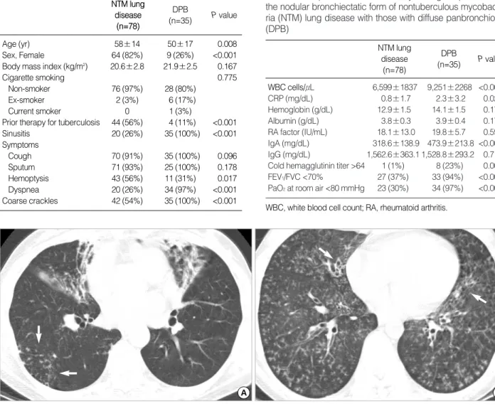
Fig. 1.Typical radiographic presentations of the nodular bronchiectatic form of nontuberculous mycobacterial lung disease and diffuse panbronchiolitis. (A) A 57-yr-old woman with Mycobacterium avium lung disease. Axial image of chest CT shows tubular bronchiectasis associated with lung volume loss in both right middle lobe and lingular division of the left upper lobe. Also note clustered centilobular micronodules and branching linear opacities (tree-in-bud pattern) in the right lower lobe (arrows). (B) A 23-yr-old woman with diffuse pan- brochiolitis. Axial image of c hest CT shows innumerable centrilobular micronodules and branching linear opacities or bronchioloectasis in entire lung. Also note bronchiectasis in both right middle lobe and lingular division of the left upper lobe (arrows).
A B
of patients with NTM lung disease and in all patients with DPB (Table 3).
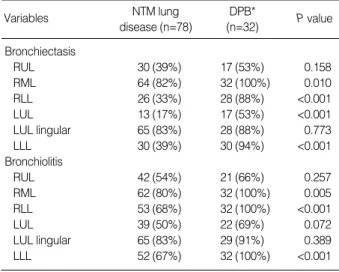
The most common HRCT findings were the presence of bronchiectasis and bronchiolitis, which were seen in all pati- ents with NTM lung disease and DPB. However, the involve- ment of bronchiectasis and bronchiolitis on chest HRCT was more extensive in patients with DPB. Bronchiectasis and bronchiolitis were usually observed in more lobes in patients with DPB than in those with NTM lung disease (P<0.001, P<0.001, respectively) (Table 3) (Fig. 1). In patients with DPB, the involvement of bronchiectasis and bronchiolitis in the right middle lobe and both lower lobes was also more common than in those with NTM lung disease (Table 4).
However, a cavity (or cavities) was more commonly found in patients with NTM lung disease (23% vs. 0%, P=0.003).
DISCUSSION
The purpose of this study was to identify clinicoradiograph- ic differences between the nodular bronchiectatic form of NTM lung disease and DPB. We found that patients with the nodular bronchiectatic form of NTM lung disease were of an older age, had a female predilection, and were more like- ly to have a history of tuberculous treatment, while patients with DPB had sinusitis more frequently. Hemoptysis was related to NTM lung disease, and exertional dyspnea and coarse crackles were related to DPB. In patients with DPB, marked obstructive abnormalities, hypoxemia, increased in- flammatory laboratory findings, and more lobes involved with bronchiectasis or bronchiolitis on HRCT were more
frequently observed.
The NTM lung disease has been differentiated into two distinct subtypes, the upper lobe cavitary form and the nodu- lar bronchiectatic form (1, 2). The nodular bronchiectatic form was reported in more than half of NTM lung disease cases and occurs predominantly in non-smoking, middle-aged or elderly women without previous or underlying lung dis- eases (1, 2). The radiographic findings show bilateral nodu- lar or reticulonodular changes, particularly in the middle lobe of the right lung and the lingular segment of the upper lobe of the left lung (1, 2). In the present study, more than 80%
of patients with nodular bronchiectatic form of NTM lung disease had involvement in the middle lobe of the right lung and the lingular segment of the left lung on chest HRCT scans.
Since the first comprehensive report of DPB was published in 1983 (15), there has been dissemination of knowledge about DPB. The diagnosis is based on the clinical and radiographic findings, pulmonary function test, and laboratory findings of patients as shown in the diagnostic criteria of the Ministry of Health and Welfare of Japan (10). Our study evaluated patients who fulfilled these diagnostic criteria. This is the reason nearly 100% show exertional dyspnea, crackles, and sinusitis in DPB. But, even in the nodular bronchiectatic form of NTM lung disease, exertional dyspnea, crackles, and sinusitis were observed in 26%, 54%, and 26% of patients, respectively. As the diagnostic criteria reflect chronic respi- ratory impairment, other laboratory findings suggest non- specific inflammation (16). Our study also demonstrated the increased value of WBC, CRP, and IgA in patients with DPB
NTM lung
disease DPB*
Variables P value
Chest radiography (n=78) (n=34)
Cavity (or cavities) 5 (6%) 0 0.320
Reticulonodular opacities 78 (100%) 34 (100%) 1.000
Distribution 0.216
Upper lung zone (s) 0 0
Middle and/or lower 43 (55%) 23 (68%) lung zone (s)
Both 35 (45%) 11 (32%)
Bilateral lung lesions 70 (90%) 34 (100%) 0.103
Chest HRCT (n=78) (n=32)
Cavity (or cavities) 18 (23%) 0 0.003
No. of involved lobes
Bronchiectasis 2.9±1.3 4.8±1.4 <0.001 Bronchiolitis 4.0±1.3 5.3±1.1 <0.001 Table 3.Comparison of chest radiography and HRCT findings between patients with the nodular bronchiectatic form of nontu- berculous mycobacteria (NTM) lung disease and those with diffuse panbronchiolitis (DPB)
*Chest radiography was not available in one patient and chest HRCT was not available in three patients with DPB.
HRCT, high-resolution computed tomography.
NTM lung disease (n=78)
DPB*
(n=32)
Variables P value
Bronchiectasis
RUL 30 (39%) 17 (53%) 0.158
RML 64 (82%) 32 (100%) 0.010
RLL 26 (33%) 28 (88%) <0.001
LUL 13 (17%) 17 (53%) <0.001
LUL lingular 65 (83%) 28 (88%) 0.773
LLL 30 (39%) 30 (94%) <0.001
Bronchiolitis
RUL 42 (54%) 21 (66%) 0.257
RML 62 (80%) 32 (100%) 0.005
RLL 53 (68%) 32 (100%) <0.001
LUL 39 (50%) 22 (69%) 0.072
LUL lingular 65 (83%) 29 (91%) 0.389
LLL 52 (67%) 32 (100%) <0.001
Table 4.Comparison of the distribution of bronchiectasis and bronchiolitis on chest HRCT between patients with the nodular bronchiectatic form of nontuberculous mycobacteria (NTM) lung disease and those with diffuse panbronchiolitis (DPB)
*Chest CT was not available in three patients with DPB.
HRCT, high-resolution computed tomography; RUL, right upper lobe;
RML, right middle lobe; RLL, right lower lobe; LUL, left upper lobe; LLL, left lower lobe.
and all of them were significantly higher than those with nodular bronchiectatic form of NTM lung disease. As IgA in immunoglobulin isotype is found predominantly in airway secretion and has a protective effect of the mucosal immune system against respiratory tract infection, increased IgA level in DPB patients might suggest that DPB is more character- ized by chronic airway inflammation (17).
A previous study reported that bilateral bronchiectasis, especially combined with multiple small nodules and branch- ing centrilobular nodular structures on chest HRCT, was asso- ciated with NTM infections, followed by DPB (7). Our study demonstrated that both groups had similar findings on the chest radiography films, and bronchiectasis and bronchioli- tis on chest HRCT without significant differences. However, cavitary lesions were observed only in the nodular bronchiec- tatic form of NTM lung disease, and the number of involved lobes, especially bilateral lower lobes including the right mid- dle lobe, was greater in DPB patients.
The importance of discriminating between the nodular bronchiectatic form of NTM lung disease and DPB is because of the different treatments for these diseases. In DPB, since the introduction of erythromycin therapy by Kudoh in the mid-1980s, the prognosis of DPB has changed from a fatal to a curable disease (10, 13). Presently, low-dose and long- term erythromycin monotherapy is established as the most efficient treatment, and the clinicians start erythromycin as soon as the patient is diagnosed (10). When erythromycin is found ineffective, the newer macrolides, such as clarithromycin or azithromycin are considered as the second choice.
These macrolides are also important in the treatment of NTM lung disease; especially MAC lung disease and M. absces- sus lung disease. In addition to M. kansasii, MAC and M.
abscessus are the most commonly encountered pathogens and more than half of patients with MAC or M. abscessus present with the nodular bronchiectatic form on chest CT scans. Intro- duction of the newer macrolides resulted in a major therapeu- tic advance in the treatment of MAC lung disease (1, 2). When a macrolide was administered alone, however, the organism was more likely to acquire macrolide-resistance which was strongly associated with a poor treatment outcome (14). Thus, combination therapy including macrolides is recommended for patients with MAC lung disease. In M. abscessus lung dis- ease, macrolides are the only oral antibiotics this organism is susceptible to, and macrolide monotherapy without other intravenous antibiotics, such as cefoxitin, amikacin and imi- penem cannot achieve negative sputum conversion (1, 2).
Hence, in NTM lung disease, if macrolide monotherapy is prescribed as it is in DPB, it can lead to the acquisition of macrolide-resistance of the NTM infection and even treat- ment failure. In addition, patients with DPB are recommend- ed for immediate treatment. While, as the course of NTM pulmonary infections of the nodular bronchiectatic form can be indolent, clinicians must determine when to start antibi- otic treatment (1, 2).
In conclusion, this study demonstrates that some clinical and radiographic findings are helpful in discriminating bet- ween the nodular bronchiectatic form of NTM lung disease and DPB. However, there is considerable overlap in the clin- ical and radiographic appearances of nodular bronchiectatic form of NTM lung disease and DPB. Still, there is no criti- cal discriminating clinical or radiographic characteristics between them that is as specific as the microbiologic docu- mentation. The correct diagnosis, including aggressive micro- biologic evaluation, should be made for the appropriate man- agement of patients presenting with bilateral bronchiectasis and bronchiolitis.
REFERENCES
1. Wallace RJ Jr, Cook JL, Glassroth J, Griffith DE, Olivier KN, Gordin F. American Thoracic Society statement: diagnosis and treatment of disease caused by nontuberculous mycobacteria. Am J Respir Crit Care Med 1997; 156: S1-25.
2. Griffith DE, Aksamit T, Brown-Elliott BA, Catanzaro A, Daley C, Gordin F, Holland SM, Horsburgh R, Huitt G, Iademarco MF, Ise- man M, Olivier K, Ruoss S, von Reyn CF, Wallace RJ Jr, Winthrop K. An official ATS/IDSA statement: diagnosis, treatment, and pre- vention of nontuberculous mycobacterial diseases. Am J Respir Crit Care Med 2007; 175: 367-416.
3. Koh WJ, Kwon OJ, Lee KS. Diagnosis and treatment of nontuber- culous mycobacterial pulmonary diseases: a Korean perspective. J Korean Med Sci 2005; 20: 913-25.
4. Swensen SJ, Hartman TE, Williams DE. Computed tomographic diagnosis of Mycobacterium avium-intracellulare complex in patients with bronchiectasis. Chest 1994; 105: 49-52.
5. Primack SL, Logan PM, Hartman TE, Lee KS, Muller NL. Pul- monary tuberculosis and Mycobacterium avium-intracellulare: a comparison of CT findings. Radiology 1995; 194: 413-7.
6. Jeong YJ, Lee KS, Koh WJ, Han J, Kim TS, Kwon OJ. Nontubercu- lous mycobacterial pulmonary infection in immunocompetent patients:
comparison of thin-section CT and histopathologic findings. Radiol- ogy 2004; 231: 880-6.
7. Koh WJ, Lee KS, Kwon OJ, Jeong YJ, Kwak SH, Kim TS. Bilater- al bronchiectasis and bronchiolitis at thin-section CT: diagnostic implications in nontuberculous mycobacterial pulmonary infection.
Radiology 2005; 235: 282-8.
8. Chung MJ, Lee KS, Koh WJ, Lee JH, Kim TS, Kwon OJ, Kim S.
Thin-section CT findings of nontuberculous mycobacterial pulmonary diseases: comparison between Mycobacterium avium-intracellulare complex and Mycobacterium abscessus infection. J Korean Med Sci 2005; 20: 777-83.
9. Chung MJ, Lee KS, Koh WJ, Kim TS, Kang EY, Kim SM, Kwon OJ, Kim S. Drug-sensitive tuberculosis, multidrug-resistant tuber- culosis, and nontuberculous mycobacterial pulmonary disease in non-AIDS adults: comparisons of thin-section CT findings. Eur Radi- ol 2006; 16: 1934-41.
10. Azuma A, Kudoh S. Diffuse panbronchiolitis in East Asia. Respirol-
ogy 2006; 11: 249-61.
11. Akira M, Kitatani F, Lee YS, Kita N, Yamamoto S, Higashihara T, Morimoto S, Ikezoe J, Kozuka T. Diffuse panbronchiolitis: evalua- tion with high-resolution CT. Radiology 1988; 168: 433-8.
12. Nishimura K, Kitaichi M, Izumi T, Itoh H. Diffuse panbronchioli- tis: correlation of high-resolution CT and pathologic findings. Radi- ology 1992; 184: 779-85.
13. Poletti V, Casoni G, Chilosi M, Zompatori M. Diffuse panbronchi- olitis. Eur Respir J 2006; 28: 862-71.
14. Griffith DE, Brown-Elliott BA, Langsjoen B, Zhang Y, Pan X, Girard W, Nelson K, Caccitolo J, Alvarez J, Shepherd S, Wilson R, Graviss EA, Wallace RJ Jr. Clinical and molecular analysis of macrolide
resistance in Mycobacterium avium complex lung disease. Am J Respir Crit Care Med 2006; 174: 928-34.
15. Homma H, Yamanaka A, Tanimoto S, Tamura M, Chijimatsu Y, Kira S, Izumi T. Diffuse panbronchiolitis. A disease of the transi- tional zone of the lung. Chest 1983; 83: 63-9.
16. Tsang KW, Ooi CG, Ip MS, Lam WK, Ngan H, Chan EY, Hawkins B, Ho CS, Amitani R, Tanaka E, Itoh H. Clinical profiles of Chinese patients with diffuse panbronchiolitis. Thorax 1998; 53: 274-80.
17. Kilian M, Mestecky J, Russell MW. Defense mechanisms involving Fc-dependent functions of immunoglobulin A and their subversion by bacterial immunoglobulin A proteases. Microbiol Rev 1988; 52:
296-303.