INTRODUCTION
Retinoblastoma is the most common intraocular tumor of childhood and accounts for 11% of all cancers that occur during the first year of life (1). Because treatments for retino- blastoma cure over 90% of patients, organ and vision preser- vation and the minimization of late treatment side-effects are important secondary treatment goals. Retinoblastoma had been treated by external beam radiotherapy (EBR), and for many years this was the accepted treatment standard (2- 7). However, greater knowledge of radiation induced mor- bidities and of secondary tumor risks after radiation therapy (8, 9) have encouraged the use of primary chemotherapy plus conservative focal therapy over the past decade.
Despite the recent trend toward chemoreduction (10-13), radiotherapy remains an excellent means of preserving vision in children (aged >1 yr) with retinoblastoma, because the tumor is radiosensitive and routinely responds to radiother- apy. Moreover, technologic advances made in the radiation oncology enable more precise targeting for tumor, avoiding healthy tissues, and the risks of secondary nonocular cancer reduced. Brachytherapy (14-16) has been used for selected cases in expert hospitals, and stereotactic conformal radiothera-
py (17-19), and proton therapy (20, 21) could also be consid- ered components in the modern radiotherapy armamentari- um for retinoblastoma. However, available clinical data for stereotactic conformal therapy and proton therapy is limited.
The Korea Cancer Center Hospital (KCCH) has consider- able experience of treating retinoblastoma in Korea, and thus, we retrospectively reviewed our experiences of treating reti- noblastoma patients with EBR, as initial treatment, to deter- mine its long-term effects on subsequent tumor control, and its associated complication rates and prognostic factors. In addition, we reviewed EBR and brachytherapy clinical data in the hope of providing guidelines regarding indications for radiation therapy in patients with retinoblastoma.
MATERIALS AND METHODS Patients and tumor characteristics
The medical records of all patients diagnosed as reti- noblastoma who received EBR as an initial treatment at the KCCH between July 1987 and June 1998, were reviewed.
A total of 36 eyes in 29 patients with intraocular retino-
546
Sang Yul Choi1, Mi-Sook Kim2, SungYul Yoo2, ChulKoo Cho2, YoungHoon Ji2, KumBae Kim2, YoungSeok Seo2, Kyung Duk Park3, JunAh Lee3, and Tai-Won Lee4
Departments of Ophthalmology1, Radiation Oncology2, and Pediatrics3, Korea Institute of Radiological &
Medical Sciences, and Lee’s Eye institute4, Seoul, Korea
Address for Correspondence Mi-Sook Kim, M.D.
Department of Radiation Oncology, Korea Cancer Center Hospital, Korean Institute of Radiological &
Medical Sciences, 75 Nowon-gil, Nowon-gu, Seoul 139-706, Korea
Tel : +82.2-970-1264, Fax : +82.2-970-2412 E-mail : [email protected]
Long Term Follow-up Results of External Beam Radiotherapy as Primary Treatment for Retinoblastoma
The authors reviewed their experiences of external beam radiotherapy (EBR) as an initial treatment in retinoblastoma patients to determine its long-term effect on subsequent tumor control and complications. A total of 32 eyes in 25 patients that underwent EBR for retinoblastoma were reviewed retrospectively. The patients con- sisted of 21 boys and 4 girls of median age at treatment of 7.1 months. Radiation doses ranged from 35 to 59.4 Gy. The 10-yr ocular and patient survivals were 75.4%
and 92.3%, respectively. Nine of the 32 eyes progressed; 7 of these were enucle- ated and 2 were salvaged by focal treatment. According to the Reese-Ellsworth clas- sification, 4 of 5 eyes of Group II, 13 of 16 Group III eyes, 2 of 4 Group IV eyes, and 5 of 7 Group V eyes were retained, and of the 32 eyes, 13 had visual acuity better than 20/200. Eleven patients experienced a radiation-induced complication. No patient developed a second malignancy during follow-up. Despite the limited number of patients enrolled, EBR may provide a mean of preserving eyeball and vision for some advanced lesions.
Key Words : Retinoblastoma; External Beam Radiotherapy; Complication
Received : 7 April 2009 Accepted : 18 August 2009
ⓒ 2010 The Korean Academy of Medical Sciences.
This is an Open Access article distributed under the terms of the Creative Commons Attribution Non-Commercial License (http://creativecommons.org/licenses/by-nc/3.0) which permits unrestricted non-commercial use, distribution, and reproduction in any medium, provided the original work is properly cited.
blastoma underwent EBR as an initial treatment. Of these, 4 patients were excluded due to a short follow-up duration (<3 yr). Patient details and data concerning tumor features, treatment parameters, and complications were collected by chart review.
The characteristics of patients and tumors are described in Table 1. Briefly, the study subjects were 21 boys and 4 girls of median age at treatment commencement of 7.1 months (range 7 weeks to 65 months). Twenty-one presented with bilateral involvement and 4 with unilateral disease. The most frequent presenting finding was leukocoria in 16 patients (64%) followed by strabismus in 6 (24%). For the 21 patients with bilateral involvement, 28 eyes received external beam radiation, and 14 eyes were enucleated due to advanced dis- ease without visual potential before EBR. Of the 32 eyes treat- ed, 0 were of Reese-Ellsworth (RE) Group I, 5 were Group II, 16 were Group III, 4 were Group IV, and 7 were Group V (5 in RE Group Va and 2 in RE Group Vb). All of two
eyes in Group Vb had a localized vitreous seeding pattern.
Treatment and follow-up
Treatment simulation was done for with patients under sedation while wearing a thermoplast head mask immobiliza- tion device. All patients were treated in the supine position using a linear accelerator at a photon energy of 6 MV with compensating bolus as needed. Twenty patients (27 eyes) were treated using opposed lateral fields alone (mainly patients with bilateral disease), while five patients (5 eyes) were treated using anterior and lateral wedged pair fields with no attempt to shield the lens. The most frequently used field size (exclud- ing half beam blocking) was 4×4 cm, but field sizes ranged from 3×3.5 cm to 6×4 cm. Treatment doses ranged from 35 to 59.4 Gy (median 41.6 Gy) in fractions of 1.6 to 2.0 Gy.
Chemotherapy was usually administered for high stage con- tralateral tumors that had been enucleated to control micro-
Pt, patient; RE, reese-ellsworth classification; OS, oculus survival; Od, oculus dexter; Os, ocular sinister; Bil, bilateral; LP, light perception; LR, local recurrence; Un, unilateral; RD, retinal detachment; VH, vitreous hemorrhage; HM, hand motion.
Irradiated
eye RE Dose
(Gy) Clinical course Visual acuity OS (mon) Complications
Pt
1 Od (Bil) 2a 37.4 LP (+) 80
Os (Bil) 3a 50.0 20/250 80
2 Od (Bil) 3a 38.0 Phthisis→enucleation Enucleation 74 Cataract, phthisis
Os (Bil) 4a 38.0 LR Enucleation 35
3 Od (Bil) 2a 43.0 LP (+) 116
Os (Bil) 3a 43.0 20/20 116
4 Os (Bil) 2a 46.0 20/30 140 Cataract
Od (Bil) 3a 44.0 LR Enucleation 16
5 Od (Bil) 3a 48.0 20/1000 150
Os (Bil) 3a 49.0 20/15 150
6 Od (Bil) 5a 50.0 LR Enucleation 8
Os (Bil) 5b 50.0 20/25 104
7 Od (Bil) 5a 59.4 20/1000 153
Os (Bil) 5a 50.0 LR Enucleation 10
8 Os (Un) 2b 35.0 LR Enucleation 13 RD, VH, facial asymmetry
9 Os (Bil) 4a 38.0 LR Enucleation 34 Cataract
10 Os (Bil) 3a 38.0 20/25 127 Cataract
11 Os (Un) 3a 38.0 20/500 117
12 Os (Bil) 3a 38.0 20/25 131 RD
13 Os (Bil) 3a 41.0 LR→laser 20/60 64
14 Os (Bil) 3a 41.0 20/25 96
15 Od (Bil) 4a 41.6 LR→cryotherapy 20/1000 148 Cataract, RD, VH
16 Os (Bil) 2a 41.6 20/100 105
17 Od (Bil) 3a 41.6 20/150 136 Cataract
18 Os (Bil) 5b 45.0 20/25 71
19 Od (Bil) 4a 46.0 20/20 78
20 Od (Un) 3a 46.0 LR Enucleation 37
21 Od (Bil) 3a 46.8 HM (+) 85 Cataract
22 Od (Bil) 5a 48.0 20/500 150 Cataract
23 Os (Bil) 3a 48.0 20/100 55
24 Os (Bil) 3a 48.0 20/50 132
25 Os (Un) 5a 56.0 LP (+) 78 Cataract, RD,
microophthalmia Table 1. Demographic data for retinoblastoma patients
scopic tumors. Twenty-two patients received cyclophospha- mide plus vincristine with or without doxorubicin at various doses and cycle numbers.
Additional cryotherapy or laser therapy was administered after EBR when tumor progression was detected. Enucleation was performed in cases with definite tumor progression after additional treatment or due to a severe complication.
Evaluations and statistics
Evaluations were performed at each follow-up to determine tumor sizes and visual acuities, and to detect new lesions and complications, such as, retinopathy, cataract, neovascular glau- coma, and midfacial hypoplasia. In addition, we analyzed prog- nostic factors, such as, gender, age, and the RE classification according to ocular survival using the log-rank test. In addi- tion, the relationship between RE classification and visual acuity was analyzed using the t-test. The Kaplan-Meier me- thod was used to estimate overall and ocular survivals. Over- all survival was calculated from EBR commencement to final follow-up or death, whereas ocular survival was calculated from EBR commencement to time of enucleation or final fol- low-up with an intact eye.
RESULTS
Median follow up was 150 months (range 55-249 months), and the 10-yr ocular and overall survival rates were 75.4%
and 92.3%, respectively (Fig. 1). Two patients died after in- volvement of the central nervous system.
Nine of the 32 eyes developed new lesions or reactivation of previous lesions. Of these, 7 eyes were enucleated and 2 were salvaged by cryotherapy and laser treatment (Table 1).
One additional enucleation was performed due to phthisis bulbi (patient No. 2 in Table 1). Therefore, ocular preserva- tion was achieved in 24 of the 32 (75.4%) eyes. According to RE classification, 4 of 5 eyes were retained in Group II,
13 of 16 in Group III, 2 of 4 in Group IV, and 5 of 7 in Group V. Vision was preserved in 24 (75%) out of 32 treated eyes.
Of the preserved 24 eyes, 9 (37.5%) had a visual acuity bet- ter than 20/40; 5 (20.8%) had an acuity worse than 20/40 but better than 20/200, and 10 (41.7%) had vision worse than 20/200. All the patients with visual acuity less than 20/200 presented with the involvement of the posterior pole, and showed macular degeneration following treatment.
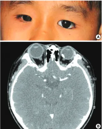
Records showed that 11 patients experienced a radiation- induced complication; 6 had cataracts alone, 1 had retinal detachment alone and 4 had one more complications (Table 1). Nine cataracts were recorded and removed when neces- sary, and vision restoration or improvement was achieved in all cases. The median time to cataract development was 5 yr and 9 months (range 13 to 125 months). Serous retinal detach- ment was detected in 3 eyes; 2 were transient and reattached after 3 months and 6 months respectively. One patient devel- oped vitreous hemorrhage and underwent enucleation. Midfa- cial hypoplasia occurred to some degree in all patients. Typi- cally, deforming hypoplasia was recorded in 2 patients. Fig.
2 shows typical hypoplasia in a treated eye by CT and pho- tography. No patient developed a second malignancy during follow-up.
An age of less than 13 months was found to be a signifi- cantly favorable factor of ocular survival by univariate analy- sis, but this was not confirmed by multivariate analysis. No
Fig. 1. Overall and ocular survival.
Survival
1.0
0.8
0.6
0.4
0.2
0.00 2 4 6 8 10 12 14 16 18 20 22
Years from radiotherapy
Overall survival: 92.3%
Ocular survival: 75.4%
Fig. 2. Midfacial hypoplasia and microophthalmia. (A) Midfacial hypoplasia is noted on the right. (B) CT scan shows smaller eye- ball and orbit on the left.
A
B
other prognostic factor was identified during this study (Table 2), and in particular, no significant relationship was found between RE classification and visual acuity.
DISCUSSION
EBR has a valuable role in the treatment of retinoblastoma, but radiation-induced secondary tumors jeopardize the role played by EBR in retinoblastoma. Large-scale cohort studies performed to quantify cancer risks in retinoblastoma treated by radiotherapy (9) have found that radiotherapy contribut- ed significantly to the risks of developing brain, nasal cavi- ty, and eye and orbit cancers. Notably, the risk of cancer of the nasal cavity increased by 1,364-fold in hereditary retino- blastoma treated by EBR. External beam radiation is usual-
ly favored for the treatment of bilateral retinoblastoma, which is almost always hereditary, and thus, all possible efforts should be made to reduce the risk of secondary malignancies after radiotherapy.
Various EBR techniques have been developed to reduce radi- ation dose to the lens. A review of EBR technology revealed that the lateral field is usually used because it requires lower lens doses (2-7). In most cases, radiation-induced cataract is not an obstacle to the vision preservation. However, reduc- tions in doses administered to the orbital cavity, optic nerve, or cranial bone appear to be more important. Recently, more meticulous techniques, such as, intensity modulated radia- tion therapy (IMRT) and stereotactic hypofractionated radi- ation therapy, have been introduced, which administer lower doses of radiation to critical organs. Reisner et al. (7) compared several EBR techniques, that is, electron beam, the lateral 2 field and anterior-lateral 2 field techniques, and IMRT, and found that IMRT had an advantage over the other techniques, because it allowed greater dose reductions to the orbit and lacrimal gland, while maintaining therapeutic doses to the ora serrata retinae and vitreous. Recently, Sahgal et al. (18) reported that stereotactic fractionated radiation therapy for localized tumor masses can achieve markedly lower doses to surrounding critical normal tissues than conventional radia- tion therapy. Furthermore, proton therapy (20, 21) is also likely to reduce cranial bone radiation dose due to the radia- tion quality of the Bragg peak. However, few clinical trials have been performed and clinical data is scarce.
Brachytherapy (14-16) was introduced in the 1920’s to treat ocular tumors and reduce the exposure to normal tis- sue around tumors, and has been further developed in terms of new radioisotopes, implant designs, and techniques of place- ment. Table 3 details the ocular survival and cataract inci- dence rates of conventional radiation therapy techniques and
LC, local control; NA, not assessed; MV, megavoltage; MeV, megavoltage electron.
Series Year No. of
eyes Method Dose (Gy)/
Fraction No. or dose
Ocular survival rate (%)
Incidences of cataract
(%) Method of
radiation
EBR Schipper (3) 1983 54 Oblique 6 MV photon enhanced 45/15 fraction 81 33
dynamic wedge
Hernandez (24) 1996 34 Ant & lateral 4 MeV photon field, 34.5-49.5/1.5-2.0 Gy 73 41 no lens shielding
Blach (25) 1996 113 lat collimated 6 MV photon field 42-46/21-23 fraction 84 (LC) 22 67 Ant lens-sparing electron beam 38-50/15-20 fraction 38 (LC)
Phillips (2) 2003 47 single and two field technique 30-50/(about 2 Gy) 72 23
Present series 32 Lat or ant & lateral 6 MeV photon field 35-55/(1.6-2.0 Gy) 74 28
Brachy- Shields (14) 2001 208 I-125(178), 106-Ru(7) Co-60(17), 40 to tumor apex 79 31
therapy I-192(6)
Merchant (22) 2004 25 I-125 44 (35-47.6) to tumor apex 60 (crude) NA
Shields (16) 2006 84 I-125 40 to tumor apex +2 mm 95 43
Schueler (15) 2006 175 106-Ru 50 or 70 to tumor apex 87 17
Abouzeid (23) 2008 41 106-Ru 50 to tumor apex 76 (crude) 10
Table 3. Review of retinoblastoma treated by EBR or plaque brachytherapy Prognostic factors No. of
cases
Ocular
survival (%) P value Gender
Male 25 75.3 0.7
Female 7 71.4
Age (mon)
<13 17 94.1 0.008
≥13 15 52.5
RE
2-3 21 80.0 0.2
4-5 11 63.6
RT dose (Gy)
<41 8 50.0 0.08
≥41 24 83.3
Table 2. Prognostic factors by univariate analysis
RE, Reese-Ellsworth classification; RT, radiotherapy.
brachytherapy; it is worth noting that the radiation doses, fraction numbers, and beam delivery techniques used at dif- ferent institutes over the last 20 yr has varied considerably.
The literature review revealed that ocular survival ranged from 38% to 84% (median 73%) for EBR, but from 60%
to 95% (median 79%) for brachytherapy. Furthermore, radi- ation-induced cataract was found to be the most common complication of radiation therapy; the incidence of cataract ranged from 22% to 41% (median 28%) for EBR and from 10% to 43% (median 17%) for brachytherapy. However, it should be noted that because retinoblastoma is rarely encoun- tered in infants, skilled treatment is required to obtain good outcomes after EBR and brachytherapy.
In the present study, we analyzed patient and ocular sur- vivals, and long-term treatment toxicities. Our results, in terms of ocular survival and the incidence of cataract, com- pare well with those of other EBR series (22-25). Long-term follow-up findings revealed that late radiation effects were milder than expected, and that favorable visual outcome had been maintained. Furthermore, ocular survival analysis by RE classification, despite the limitations imposed by small patient numbers, showed that involved eyes with an RE clas- sification of 4 or 5 achieved 64% ocular survival, which indi- cates that radiation therapy should be considered before enu- cleation as primary or secondly treatment for even advanced retinoblastoma.
In conclusion, our long-term results show that EBR has an important role to play in the avoidance of enucleation in retinoblastoma with small lesions or advanced lesions with vitreous seeding. Furthermore, non-conventional radiation therapies as brachytherapy, stereotactic radiotherapy, IMRT, and proton therapy, are likely to reduce complication rates.
Additional research is required to establish new indications for the various radiation therapy techniques available for the treatment of retinoblastoma.
REFERENCES
1. John Y, Malcolm S, Steven R, Jonathan L, Greta B. Retinoblastoma.
Cancer incidence and survival among children and adolescents.
National Cancer Institute, 1999.
2. Phillips C, Sexton M, Wheeler G, McKenzie J. Retinoblastoma:
review of 30 years’ experience with external beam radiotherapy.
Australas Radiol 2003; 47: 226-30.
3. Schipper J. An accurate and simple method for megavoltage radia- tion therapy of retinoblastoma. Radiother Oncol 1983; 1: 31-41.
4. McCormick B, Ellsworth R, Abramson D, Haik B, Tome M, Gra- bowski E, LoSasso T. Radiation therapy for retinoblastoma: com- parison of results with lens-sparing versus lateral beam techniques.
Int J Radiat Oncol Biol Phys 1988; 15: 567-74.
5. Weiss DR, Cassady JR, Petersen R. Retinblastoma: a modification in radiation therapy technique. Radiology 1975; 114: 705-8.
6. Krasin MJ, Crawford BT, Zhu Y, Evans ES, Sontag MR, Kun LE,
Merchant TE. Intensity-modulated radiation therapy for children with intraocular retinoblastoma: potential sparing of the bony orbit.
Clin Oncol (R Coll Radiol) 2004; 16: 215-22.
7. Reisner ML, Viegas CM, Grazziotin RZ, Santos Batista DV, Carneiro TM, Mendonca de Araujo CM, Marchiori E. Retinoblastoma--com- parative analysis of external radiotherapy techniques, including an IMRT technique. Int J Radiat Oncol Biol Phys 2007; 67: 933-41.
8. Pagani JJ, Bassett LW, Winter J, Gold RH, Brawer M. Osteogenic sarcoma after retinoblastoma radiotherapy. AJR Am J Roentgenol 1979; 133: 699-702.
9. Kleinerman RA, Tucker MA, Tarone RE, Abramson DH, Seddon JM, Stovall M, Li FP, Fraumeni JF Jr. Risk of new cancers after radio- therapy in long-term survivors of retinoblastoma: an extended fol- low-up. J Clin Oncol 2005; 23: 2272-9.
10. Sohajda Z, Damjanovich J, Bardi E, Szegedi I, Berta A, Kiss C. Com- bined local treatment and chemotherapy in the management of bilat- eral retinoblastomas in Hungary. J Pediatr Hematol Oncol 2006;
28: 399-401.
11. Rouic LL, Aerts I, Levy-Gabriel C, Dendale R, Sastre X, Esteve M, Asselain B, Bours D, Doz F, Desjardins L. Conservative treatments of intraocular retinoblastoma. Ophthalmology 2008; 115: 1405-10.
12. Antoneli CB, Ribeiro KC, Steinhorst F, Novaes PE, Chojniak MM, Malogolowkin M. Treatment of retinoblastoma patients with chemore- duction plus local therapy: experience of the AC Camargo Hospi- tal, Brazil. J Pediatr Hematol Oncol 2006; 28: 342-5.
13. Kim H, Lee JW, Kang HJ, Park HJ, Kim YY, Shin HY, Yu YS, Kim IH, Ahn HS. Clinical results of chemotherapy based treatment in retinoblastoma patients: a single center experience. Cancer Res Treat 2008; 40: 164-71.
14. Shields CL, Shields JA, Cater J, Othmane I, Singh AD, Micaily B.
Plaque radiotherapy for retinoblastoma: long-term tumor control and treatment complications in 208 tumors. Ophthalmology 2001;
108: 2116-21.
15. Schueler AO, Fluhs D, Anastassiou G, Jurklies C, Neuhauser M, Schilling H, Bornfeld N, Sauerwein W. Beta-ray brachytherapy with 106Ru plaques for retinoblastoma. Int J Radiat Oncol Biol Phys 2006;
65: 1212-21.
16. Shields CL, Mashayekhi A, Sun H, Uysal Y, Friere J, Komarnicky L, Shields JA. Iodine 125 plaque radiotherapy as salvage treatment for retinoblastoma recurrence after chemoreduction in 84 tumors.
Ophthalmology 2006; 113: 2087-92.
17. Munier FL, Verwey J, Pica A, Balmer A, Zografos L, Abouzeid H, Timmerman B, Goitein G, Moeckli R. New developments in exter- nal beam radiotherapy for retinoblastoma: from lens to normal tis- sue-sparing techniques. Clin Exprimet Ophthalmol 2008; 36: 78-89.
18. Sahgal A, Millar BA, Michaels H, Jaywant S, Chan HS, Heon E, Gallie B, Laperriere N. Focal stereotactic external beam radiother- apy as a vision-sparing method for the treatment of peripapillary and perimacular retinoblastoma: preliminary results. Clin Oncol (R Coll Radiol) 2006; 18: 628-34.
19. Cormack RA, Kooy HM, Bellerive MR, Loeffler JS, Petersen RA, Tarbell NJ. A stereotactic radiation therapy device for retinoblas- toma using a noncircular collimator and intensity filter. Med Phys 1998; 25: 1438-42.
20. Krengli M, Hug EB, Adams JA, Smith AR, Tarbell NJ, Munzenrid- er JE. Proton radiation therapy for retinoblastoma: comparison of various intraocular tumor locations and beam arrangements. Int J Radiat Oncol Biol Phys 2005; 61: 583-93.
21. Lee CT, Bilton SD, Famiglietti RM, Riley BA, Mahajan A, Chang EL, Maor MH, Woo SY, Cox JD, Smith AR. Treatment planning with protons for pediatric retinoblastoma, medulloblastoma, and pelvic sarcoma: how do protons compare with other conformal tech- niques? Int J Radiat Oncol Biol Phys 2005; 63: 362-72.
22. Merchant TE, Gould CJ, Wilson MW, Hilton NE, Rodriguez-Galin- do C, Haik BG. Episcleral plaque brachytherapy for retinoblastoma.
Pediatr Blood Cancer 2004; 43: 134-9.
23. Abouzeid H, Moeckli R, Gaillard MC, Beck-Popovic M, Pica A, Zografos L, Balmer A, Pampallona S, Munier FL. (106)Ruthenium brachytherapy for retinoblastoma. Int J Radiat Oncol Biol Phys 2008; 71: 821-8.
24. Hernandez JC, Brady LW, Shields JA, Shields CL, DePotter P, Karlsson UL, Markoe AM, Amendola BE, Singh A. External beam radiation for retinoblastoma: results, patterns of failure, and a pro- posal for treatment guidelines. Int J Radiat Oncol Biol Phys 1996;
35: 125-32.
25. Blach LE, McCormick B, Abramson DH. External beam radiation therapy and retinoblastoma: long-term results in the comparison of two techniques. Int J Radiat Oncol Biol Phys 1996; 35: 45-51.