INTRODUCTION
Prostate cancer (PCa) is the second most common malig
nancy and the fifth leading cause of cancer death in men with a total of 1.09 million new PCa cases and 307,500 diseaserelated deaths in 2012 [1]. In Malaysia, PCa is the fifth most common cancer among Malaysian men [2]. The incidence of PCa increases with age, and the disease is most often diagnosed after the age of 65 years. Hence, 60% of cases are detected at a late stage (stages 3 and 4), with only
Role of co-expression of estrogen receptor beta and Ki67 in prostate adenocarcinoma
Nornazirah Azizan1, Firdaus Hayati2, Nur Maya Sabrina Tizen3, Wirda Indah Farouk3, Noraidah Masir3
Departments of 1Pathobiology and Medical Diagnostic, 2Surgery, Faculty of Medicine and Health Sciences, Universiti Malaysia Sabah, Sabah, 3Department of Pathology, Faculty of Medicine, Universiti Kebangsaan Malaysia, Kuala Lumpur, Malaysia
Purpose: To evaluate the expression of estrogen receptor (ER)-beta and Ki67 in prostate cancer and study their relationship.
Materials and Methods: We analyzed 101 cases of prostate adenocarcinoma diagnosed from January 2011 to June 2015 in 100 patients. Immunohistochemical staining of ER-beta and Ki67 was analyzed according to Gleason score categorized into prognostic groups of 1 to 5. Double-immunofluorescent staining of ER-beta and Ki67 was performed in a total of 20 cases to study the co- expression and the relationship between these markers within the same tumor.
Results: A total of 53 of 101 cases (52.5%) were positive for ER-beta expression. There was a positive correlation whereby a high percentage of ER-beta expression was seen in the higher prognostic groups (groups 4 and 5; p=0.007). High Ki67 expression was observed in the higher prognostic group, whereas low Ki67 or negative expression was found in the lower prognostic group (p<0.001). The majority of cases evaluated with double-immunofluorescent staining (14/20) showed co-expression of ER-beta and Ki67 at the individual cell level.
Conclusions: ER-beta and Ki67 are independent tumor markers in high prognostic groups. Hence, co-expression of ER-beta and Ki67 indicates a more aggressive tumor with a poorer prognosis.
Keywords: Estrogen receptor beta; Ki-67 antigen; Neoplasm grading; Prostate neoplasms
This is an Open Access article distributed under the terms of the Creative Commons Attribution Non-Commercial License (http://creativecommons.org/licenses/by-nc/4.0) which permits unrestricted non-commercial use, distribution, and reproduction in any medium, provided the original work is properly cited.
Received: 20 February, 2018 • Accepted: 3 May, 2018 Corresponding Author: Nornazirah Azizan
Department of Pathobiology and Medical Diagnostic, Faculty of Medicine and Health Sciences, Universiti Malaysia Sabah, Jalan UMS, 88450 Kota Kinabalu, Sabah, Malaysia
TEL: +60-125460459, FAX: +60-88321377/+60-88321373, E-mail: [email protected] ORCID: http://orcid.org/0000-0002-5831-3734
ⓒ The Korean Urological Association
16% at stage 1 and 24% at stage 2 [2].
Estrogen receptor (ER)beta is the predominant ER in normal prostate and is preferentially expressed in epithelial cells [3]. The role of ERbeta in the pathogenesis or prognosis of PCa is unclear, however. It has been suggested that it controls proliferation and prevents hyperplasia in rodent prostate because ERbeta knockout mice show prostatic hyperplasia with aging and high epithelial proliferation with a low apoptosis index [4]. In the absence of ERbeta, the epithelium does not fully differentiate and a large fraction
www.icurology.org
Investig Clin Urol 2018;59:232-237.
https://doi.org/10.4111/icu.2018.59.4.232 pISSN 2466-0493 • eISSN 2466-054X
of epithelial cells retain their capacity to proliferate [4].
Few studies have examined the variety of pattern of ER expression in prostate tumor. One previous study revealed that ERbeta expression is reduced in highergrade prostatic adenocarcinoma compared with low and intermediategrade prostatic adenocarcinoma [5]. It has also been shown that ERbeta is highly expressed in normal human prostate and that there is a progressive loss of expression in prostatic hyperplasia and to a greater extent in invasive cancer [6].
Evidence also exists of increased ER staining intensity in malignant prostate [7]. It has been postulated that ERbeta switches its role during cancer progression. In the early phases of PCa progression, ERbeta presents as a tumor
suppressing agent and then becomes a tumor promoter as cancer progresses. It may also have a role in the process of prostatic hyperplasia and malignancy [8,9]. The expression of this biomarker may thus be useful as a predictive marker in tumor staging and prognosis and may also shed new light on targeted treatment in prostate adenocarcinoma.
Ki67 is a protein found in proliferating cells. Ki67 recognizes a proliferationspecific nuclear antigen. Normal prostate cells proliferate slowly with low Ki67 expression. In prostate tumors, high Ki67 expression is associated with a higher Gleason score and more aggressive cancer [1012]. Data from previous reports (on individual markers) suggest that ERbeta is absent or downregulated in highgrade prostate adenocarcinoma and that Ki67 is highly expressed in high
grade prostate adenocarcinoma. However, no studies have looked at the coexpression or relationship between ERbeta and cell proliferation within the same tumor. In the present study, we investigated the expression of ERbeta and Ki67 and the relationship with the proliferation rate in PCa of various Gleason scores.
MATERIALS AND METHODS
This study was conducted in Universiti Kebangsaan Malaysia Medical Center, Malaysia. Ethical approval was obtained from the local ethics committee (reference number FF2014146). A total of 101 paraffinembedded blocks from cases of prostate adenocarcinoma of different Gleason grades from 100 patients diagnosed from January 2011 to June 2015 were retrieved from the department archive along with their corresponding H&Estained slides. The PCa tissues were obtained from transrectal biopsy and transurethral resection of the prostate. Representative areas of prostate adenocarcinoma were marked on each H&E
stained slide and the corresponding areas on paraffin blocks were cored for construction of tissue microarrays (TMAs)
as well as for full tissue sections. A total of 46 cases were constructed for TMAs and 55 cases were used for full tissue sections. Representative 3µm paraffin sections were made for immunohistochemical staining for ERbeta and Ki67.
Doubleimmunofluorescent staining was performed on a total of 20 cases, including 14 cases of prostate adeno
carcinoma showing strong positivity for ERbeta with a high Ki67 proliferation rate, 4 cases showing weak positivity for ERbeta with a high Ki67 proliferation rate, and 2 cases showing weak positivity for ERbeta with a low Ki67 proliferation rate.
Mouse monoclonal antibody against Ki67 was used at a dilution of 1:100 with normal tonsil tissue as the positive control. Rabbit monoclonal antibody against ERbeta was used at a dilution of 1:300 with human ovarian serous cystadenocarcinoma as the positive control.
Immunohistochemistry staining was performed by using the protocol from EnVisionTM FLEX+ (mouse, high pH; Dako, Santa Clara, CA, USA). Primary antibodies were diluted to optimal concentration using Dako REALTM Antibody Diluent (Dako). Tissue blocks were sectioned and an initial deparaffinization and pretreatment step was performed in the Dako PTLink using the EnVisionTM FLEX Target Retrieval Solution (Dako), high pH, followed by cooling at room temperature for 20 minutes and rinsing under running tap water for 3 minutes. The slides were subsequently incubated with EnVisionTM FLEX peroxidase
blocking reagent for 5 minutes followed by the washing step.
Slides were then incubated with primary antibody at room temperature followed by incubation with EnVisionTM FLEX/
HRP (Dako) for 20 minutes. Sections were then incubated with 1X DABcontaining substrate working solution for 10 minutes and then counterstained with hematoxylin.
Doubleimmunofluorescent staining was performed at optimal concentrations of primary and secondary antibodies.
After deparaffinization and pretreatment, the slides were incubated for 45 minutes at room temperature with the primary antibody mixture, washed with TBS for 10 minutes, and incubated in the dark moist chamber with a mixture of 2 isotypespecific secondary antibodies conjugated to Alexa Fluor 594® (red) and Alexa Fluor 488® (green) for 45 minutes. The slides were then washed with TBS for 30 minutes, followed by a counterstaining step using DAPI II (Abbott Molecular Inc., Des Plaines, IL, USA). Human ovarian cystadenocarcinoma tissue was used as a positive control.
Sections stained for ERbeta were scored qualitatively and those stained for Ki67 were scored quantitatively at ×40 magnification. ERbeta was interpreted as positive when the
cells showed nuclear immunoreactivity in 10% or more of the tumor cells (Fig. 1). Negative staining was interpreted if no staining was seen or when less than 10% of tumor cells showed positive expression regardless of staining intensity.
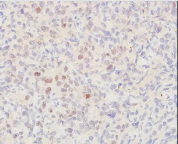
Results for Ki67 were interpreted as negative if there was a lack of nuclear immunoreactivity (no staining at all), low in cases with positive nuclear staining of less than 10% (Fig. 2) of the cells, and high in cases with positive nuclear staining of 10% or more of the cells (Fig. 3).
For doubleimmunofluorescent staining, ERbeta and Ki67 expression was detected in the nuclei of tumor cells.
The nuclei that were positive for ERbeta were stained red and those positive for Ki67 were stained green. Nuclei with ERbeta and Ki67 coexpression appeared yellowish.
The slides were reviewed by two observers to determine
the Gleason score and interpret the immunohistochemical and doubleimmunofluorescent staining. The cases were categorized and analyzed according to prognostic Gleason grade grouping of group 1 to group 5 (group 1, Gleason score of less than or equal to 6; group 2, Gleason score of 3+4;
group 3, Gleason score of 4+3; group 4, Gleason score of 8;
and group 5, Gleason scores of 9 and 10).
Statistical analysis was performed using IBM SPSS ver.
22.0 software (IBM Co., Armonk, NY, USA). Chisquare and Fisher’s exact test were used for comparative analysis. The pvalues of <0.05 were considered statistically significant.
RESULTS
A total of 53 of 101 cases (52.5%) were positive for ER
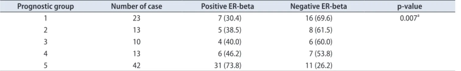
beta expression at variable levels of intensity. ERbeta was observed in 7 of 23 cases (30.4%) from group 1, 5 of 13 cases (38.5%) from group 2, 4 of 10 cases (40.0%) from group 3, 6 of 13 cases (46.2%) from group 4, and 31 of 42 cases (73.8%) from group 5 (Table 1). Fig. 4 shows the number of positive cases according to prognostic group. Statistical analysis with the Pearson chisquare test showed a correlation or association between ERbeta expression and high prognostic group in which a high percentage of ERbeta positivity was found in the high prognostic group (p=0.007; Table 1).
Of 101 cases, 98 cases (97.0%) were positive for Ki67 and 3 cases (3.0%) were negative for Ki67. As shown in Table 2, among the proliferating cells, 50 of 98 cases showed a low Ki67 proliferation rate (less than 10%) and 48 of 98 cases showed a high Ki67 proliferation rate (10% or more).
The Ki67 proliferation rate was evaluated and correlated with prognostic Gleason group as shown in Table 2. A high Fig. 1. Strong positive staining pattern for estrogen receptor beta (im-
munohistochemical staining, ×20).
Fig. 2. Low Ki67 staining pattern. Strong positive staining pattern for estrogen receptor beta (immunohistochemical staining, ×10).
Fig. 3. High Ki67 staining pattern. Strong positive staining pattern for estrogen receptor beta (immunohistochemical staining, ×20).
Ki67 proliferation rate was seen in the higher prognostic groups (groups 4 and 5), whereas a low Ki67 proliferation rate or negative staining was found in the lower prognostic
group (Fig. 5). This was statistically significant (p<0.001, Fisher’s exact test; Table 2). The correlation between the Ki67 proliferation rate and ERbeta expression showed that 77.1% of cases with a high Ki67 rate were positive for ER
Table 1. ER-beta expression in relation to prognostic Gleason group
Prognostic group Number of case Positive ER-beta Negative ER-beta p-value
1 23 7 (30.4) 16 (69.6) 0.007a
2 13 5 (38.5) 8 (61.5)
3 10 4 (40.0) 6 (60.0)
4 13 6 (46.2) 7 (53.8)
5 42 31 (73.8) 11 (26.2)
Values are presented as number only or number (%).
ER, estrogen receptor.
a:Pearson chi-square test.
Table 2. Ki67 proliferation rate in relation to prognostic Gleason group
Prognostic group Number of case Ki67
p-value
High Low
1 23 1 (4.3) 19 (82.6) <0.001a
2 13 2 (15.4) 11 (84.6)
3 10 1 (10.0) 9 (90.0)
4 13 8 (61.5) 5 (38.5)
5 42 36 (85.7) 6 (14.3)
Values are presented as number only or number (%).
a:Fisher exact test.
1 80 70 60 50 40 30 20 10
Percentageofcases
Prognostic Gleason group 0
2 3 4 5
Negative ER-beta Positive ER-beta
Fig. 4. Pattern of estrogen receptor (ER)-beta expression in relation to prognostic group.
1 100
90 80 70 60 50 40 30 20 10
Percentageofcases
Prognostic Gleason group 0
2 3 4 5
Negative Ki67 Low Ki67 High Ki67
Fig. 5. Pattern of Ki67 proliferation rate in relation to prognostic group.
Table 3. Correlation between Ki67 proliferation rate and ER-beta expression
Ki67 Number of case ER-beta expression
p-value
Positive Negative
Negative 3 0 (0.0) 3 (100.0) <0.001a
Low 50 16 (32.0) 34 (68.0)
High 48 37 (77.1) 11 (22.9)
Values are presented as number only or number (%).
ER, estrogen receptor.
a:Fisher exact test.
beta expression, whereas 68.0% of cases with a low Ki67 proliferation rate and 100% of cases with negative Ki67 proliferation were negative for ERbeta expression (Table 3). Statistical analysis using Fisher’s exact test showed a significant correlation between ERbeta expression and a high proliferation rate (p<0.001).
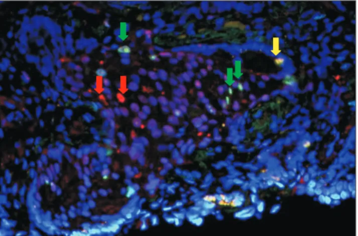
A total of 20 cases from various prognostic groups were evaluated by doubleimmunofluorescent staining for ERbeta and Ki67. Among the cases evaluated, 14 of 20 cases showed positive coexpression of ERbeta and Ki67 within the same tumor (Fig. 6). In a few cases that coexpressed both markers, some of the proliferating cells were, however, negative for ERbeta (Fig. 2). In 6 of 20 cases, the tumor cells did not co
express ERbeta and Ki67, showing that the two markers are mutually exclusive (Fig. 6).
DISCUSSION
We found that ERbeta was mainly expressed in cases with a high Gleason score (scores of 9 and above). Our findings showed a gradual increment in the number of positive cases with a Gleason score of 4 to 8 and an exponen
tial increase in positivity for cases with Gleason scores of 9 and 10. This is in contrast with the findings of Asgari and Morakabati [5] in which there was reduced expression of ERbeta in highergrade prostatic adenocarcinoma compared with low and intermediategrade cancer. We have no obvious explanation for the inverse expression of ERbeta and Gleason score, but we can postulate that inadequate antibody specificity, ineffective antigenbearing tissue retrieval, or the presence of unknown isoforms of ER protein may have affected the quality of immunohistochemistry [5].
Besides, it was previously described that ERbeta, as detected by PPG5/10 antibody, may have a role in the process of de
differentiation of prostate adenocarcinoma, with a higher level present in less differentiated tumor [13]. Our findings support the potential of ERbeta as a cancerpromoting agent as proposed by Christoforou et al. [8] in 2014.
It was previously shown that the expression of ERbeta is more intense in PCa than in benign prostatic hyperplasia [14]. Although we did not compare the staining intensity between benign and malignant prostate tissue, our own observation showed that the intensity of ERbeta staining is not restricted to the Gleason score.
The proliferation rate was high in cases with a Gleason score of 8 and above. Among those with positive Ki67 staining, the cases in prognostic groups 4 and 5 (Gleason scores of 8, 9, and 10) showed a higher percentage of proliferation rate than did the lower prognostic Gleason group. These findings agree with previous studies by Cowen et al. [11] and Sulik et al. [12] that reported that Ki67 levels were significantly higher in tumors with a high Gleason score. A study by Fixemer et al. [15] concluded that ER
beta is expressed in the secretory epithelium of the prostate and possibly reflects androgendependent cancer cells and might have a chemopreventive effect. However, our study focused on prostate adenocarcinoma alone, not normal and premalignant tissue.
The correlation between the Ki67 proliferation rate and ERbeta expression showed that those cases with high Ki67 proliferation were positive for ERbeta expression and those cases that were negative for Ki67 were negative for ER
beta expression. These findings indicate that proliferating prostate adenocarcinoma cells are expressing ERbeta. This observation was confirmed with doubleimmunofluorescent staining in which the majority of cases (14/20) showed co
expression of the markers at the individual cell level.
However, in a smaller number of cases, coexpression was lacking, showing that ERbeta is expressed in individual cells independent of the proliferation marker. Whether the latter group (ERbeta positive, Ki67 negative) had a better prognosis remains to be determined.
Limitations
The use of a control group is important to avoid observational bias during the interpretation of data.
However, financial constraints were the main culprit leading to the exclusion of a control group in this study.
In the future, we will try to improve on this issue with a subsequent study or a continuation of this study.
Fig. 6. Double-immunofluorescent staining (×40) showing cells with positive co-expression for estrogen receptor (ER)-beta and Ki67 stained yellow (yellow arrow) while some proliferating cells not ex- pressing ER-beta are stained green (green arrows) and some ER-beta positive cells that are not proliferating are stained red (red arrows).
CONCLUSIONS
Positive ERbeta expression and a high Ki67 proliferation rate was associated with a high prognostic group. ERbeta and Ki67 are independent markers within tumor cells;
hence, coexpression of ERbeta and Ki67 indicates a more aggressive tumor with a poorer prognosis and possibly cells that will respond to targeted therapy.
CONFLICTS OF INTEREST
The authors have nothing to disclose.
REFERENCES
1. Wong MC, Goggins WB, Wang HH, Fung FD, Leung C, Wong SY, et al. Global incidence and mortality for prostate cancer:
analysis of temporal patterns and trends in 36 countries. Eur Urol 2016;70:862-74.
2. Ministry of Health. Summary of Malaysian national cancer registry report 2007-2011. Putrajaya: Ministry of Health;
2017;1-19.
3. Hartman J, Ström A, Gustafsson JÅ. Current concepts and significance of estrogen receptor β in prostate cancer. Steroids 2012;77:1262-6.
4. Imamov O, Morani A, Shim GJ, Omoto Y, Thulin-Andersson C, Warner M, et al. Estrogen receptor beta regulates epithelial cellular differentiation in the mouse ventral prostate. Proc Natl Acad Sci U S A 2004;101:9375-80.
5. Asgari M, Morakabati A. Estrogen receptor beta expression in prostate adenocarcinoma. Diagn Pathol 2011;6:61.
6. Horvath LG, Henshall SM, Lee CS, Head DR, Quinn DI, Makela S, et al. Frequent loss of estrogen receptor-beta expres- sion in prostate cancer. Cancer Res 2001;61:5331-5.
7. Dunsmuir WD, Gillett CE, Meyer LC, Young MP, Corbishley C, Eeles RA, et al. Molecular markers for predicting prostate cancer stage and survival. BJU Int 2000;86:869-78.
8. Christoforou P, Christopoulos PF, Koutsilieris M. The role of estrogen receptor β in prostate cancer. Mol Med 2014;20:427- 34.
9. Al-Maghrabi JA, Hassan TM, Abdel-Meguid TA, Mosli HA.
Expression of estrogen alpha and beta receptors in prostate cancer and hyperplasia: immunohistochemical analysis. Afr J Urol 2010;16:79-87.
10. Muñoz E, Gómez F, Paz JI, Casado I, Silva JM, Corcuera MT, et al. Ki-67 immunolabeling in pre-malignant lesions and car- cinoma of the prostate. Histological correlation and prognostic evaluation. Eur J Histochem 2003;47:123-8.
11. Cowen D, Troncoso P, Khoo VS, Zagars GK, von Eschenbach AC, Meistrich ML, et al. Ki-67 staining is an independent cor- relate of biochemical failure in prostate cancer treated with radiotherapy. Clin Cancer Res 2002;8:1148-54.
12. Sulik M, Maruszak K, Puchalska J, Misiukiewicz-Poć M. Ex- pression of Ki-67 as a proliferation marker in prostate cancer.
Pol Ann Med 2011;18:12-9.
13. Torlakovic E, Lilleby W, Torlakovic G, Fosså SD, Chibbar R.
Prostate carcinoma expression of estrogen receptor-beta as de- tected by PPG5/10 antibody has positive association with pri- mary Gleason grade and Gleason score. Hum Pathol 2002;33:
646-51.
14. Royuela M, de Miguel MP, Bethencourt FR, Sánchez-Chapado M, Fraile B, Arenas MI, et al. Estrogen receptors alpha and beta in the normal, hyperplastic and carcinomatous human prostate. J Endocrinol 2001;168:447-54.
15. Fixemer T, Remberger K, Bonkhoff H. Differential expression of the estrogen receptor beta (ERbeta) in human prostate tis- sue, premalignant changes, and in primary, metastatic, and recurrent prostatic adenocarcinoma. Prostate 2003;54:79-87.