Received on April 21, 2010. Revised on April 29, 2010. Accepted on May 7, 2010.
CC This is an open access article distributed under the terms of the Creative Commons Attribution Non-Commercial License (http://creativecommons.org/licenses/by-nc/3.0) which permits unrestricted non-commercial use, distribu- tion, and reproduction in any medium, provided the original work is properly cited.
*Corresponding Author. Tel: 82-43-261-2826; Fax: 82-43-268-2732; E-mail: cklee@chungbuk.ac.kr Keywords: Aspirin, Ibuprofen, Antigen processing, Dendritic cell
Cyclooxygenase Inhibitors, Aspirin and Ibuprofen, Inhibit MHC-restricted Antigen Presentation in Dendritic Cells
Hyun-Jin Kim1, Young-Hee Lee1, Sun-A Im1, Kyungjae Kim2 and Chong-Kil Lee1*
1College of Pharmacy, Chungbuk National University, Cheongju 361-763, Korea, 2College of Pharmacy, SahmYook University, Seoul 139-742, Korea
Background: Nonsteroidal anti-inflammatory drugs (NSAIDs) are widely used to relieve pain, reduce fever and inhibit inflammation. NSAIDs function mainly through inhibition of cyclooxygenase (COX). Growing evidence suggests that NSAIDs also have immunomodulatory effects on T and B cells. Here we examined the effects of NSAIDs on the anti- gen presenting function of dendritic cells (DCs). Methods:
DCs were cultured in the presence of aspirin or ibuprofen, and then allowed to phagocytose biodegradable micro- spheres containing ovalbumin (OVA). After washing and fix- ing, the efficacy of OVA peptide presentation by DCs was evaluated using OVA-specific CD8 and CD4 T cells. Results:
Aspirin and ibuprofen at high concentrations inhibited both MHC class I and class II-restricted presentation of OVA in DCs. In addition, the DCs generated in the presence of low concentrations of the drugs exhibit a profoundly suppressed capability to present MHC-restricted antigens. Aspirin and ibuprofen did not inhibit the phagocytic activity of DCs, the expression level of total MHC molecules and co-stimulatory molecules on DCs. Ibuprofen rather increased the expression level of total MHC molecules and co-stimulatory molecules on DCs. Conclusion: These results demonstrate that aspirin and ibuprofen inhibit the intracellular processing event of the phagocytosed antigen, and further suggest that prolonged administration of NSAIDs in high doses may impair the capa- bility of DCs to present antigens in asiociation with MHC molecules.
[Immune Network 2010;10(3):92-98]
INTRODUCTION
Nonsteroidal anti-inflammatory drugs (NSAIDs) inhibit cyclo- oxygenase (COX), the rate-limiting enzyme for the synthesis of prostaglandins (1,2). Two COX isoforms have been identi- fied in eukaryotic cells, COX-1 and COX-2. COX-1 is con- stitutively expressed in most cells, while COX-2 is inducibly expressed in a more limited array of cells at inflammatory sites (3,4). Thus, the ability of NSAIDs to inhibit COX-2 activ- ity may explain their therapeutic effects as anti-inflammatory drugs (2-5). Most of the NSAIDs that are currently in clinical use inhibit both COX-1 and COX-2, although some inhibit one isosome to a greater extent than the other (6).
There is growing evidence showing that NSAIDs may have immunomodulatory activities not apparently related to the inhibition of prostaglandin synthesis. NSAIDs were shown to inhibit T cell proliferation, expression of activa- tion-related molecules such as CD25 and CD71, and the production of cytokines such as IL-2, IFN-γ and TNF-α (7-9). Aspirin and salicylates have been shown to inhibit NF-κB activation, which is indispensible in the tran- scription of pro-inflammatory cytokines (10,11). Ibuprofen, indomethacin and fenoprofen have also been shown to in- hibit PMA-induced cytokine synthesis in human peripheral blood lymphocytes (12). It has also been demonstrated that aspirin, ibuprofen, tylenol and naproxen suppress antibody production in human peripheral blood mononuclear cells (13). In support of the immunomodulatory activity of
NSAIDs on lymphocytes, it has been shown that COX-1 is constitutively expressed on T cells, whereas the expression of COX-2 is inducibly up-regulated in T cells upon stim- ulation (9,14).
In the present study, we examined the effects of aspirin and ibuprofen on the MHC-restricted presentation of the exogenous antigen, ovalbumin (OVA), in DCs, which are the most important accessory cells for the activation of naïve T cells and the generation of primary T cell responses (15). We found that the drugs inhibit MHC-restricted exogenous anti- gen presentation, especially when the DCs are exposed to the drugs during early the stage of differentiation. Since T cells can only recognize antigens presented on MHC molecules, the inhibition of MHC-restricted antigen processing pathways by the drugs may have profound implications in explaining the immunomodulatory effects of the drugs such as the in- hibition of immune activation and the production of pro-in- flammatory cytokines by T cells.
MATERIALS AND METHODS Cells and cell lines
T cell hybridomas, B3Z86/90.14 (B3Z) and DOBW, were kindly provided by Dr. Nilabh Shastri (University of Califor- nia, Berkeley, CA) and by Dr. Clifford V. Harding (Case Western Reserve University, Cleveland, OH), respectively (16,17). The DC cell line (DC2.4) was obtained from the Dana-Farber Cancer Institute, Boston, MA, USA (18).
Generation of bone marrow-derived DCs (BM-DCs) DCs were generated from total BM cells as described pre- viously (19). Briefly, BM cells obtained from femurs of BALB/c mmice were cultured in a 6-well plates (5×10⁶/well) in a culture medium supplemented with 200 U/ml rmGM-CSF.
At days 3 and 4 from the initiation of the culture, nonadherent cells were discarded by replacing the culture medium with fresh medium containing the cytokines after gentle shaking.
DCs were harvested by gentle pipetting at day 6.
Preparation of OVA-microspheres
Microspheres containing OVA were prepared using a sol- vent-evaporation method, as described previously (20), using OVA dissolved in 3% polyvinyl alcohol (4 mg/ml) and poly (DL-lactide-co-glycolide) (PLGA; lactide:glycolide=50:50;
Sigma-Aldrich, St. Louis, MO, USA) dissolved in a mixture of acetone and ethanol (9:1) (5%). The concentration of OVA
was determined by micro-bicinchoninic acid assay kit (Pierce, Rockford, IL) according to the manufacturer’s instructions af- ter lysing the microspheres in a lysis buffer containing 0.1%
SDS and 0.1 N NaOH. For phagocytosis assays, micro- pheres containing both OVA and fluorescein isothiocyanate (FITC) were prepared by adding FITC (final, 5 mg/ml) to a mixture of acetone and ethanol (9:1) together with PLGA (final, 5%).
MHC class I-restricted presentation assay
LacZ T cell activation assays were used to assess the amounts of cross-presented OVA peptides, as previously described (20). Briefly, DCs were cultured in the presence of different concentrations of aspirin (Sigma-Aldich) or ibuprofen (Sigma- Aldich) for 18 h in 96-well plates (1×10⁵/well), and then add- ed with OVA-microspheres (50μg/ml as OVA). After 2 h in- cubation at 37oC, the plate was washed twice with 300μl/
well of pre-warmed PBS, and then fixed with 100μl/well of ice-cold 1.0% paraformaldehyde for 5 min at room tem- perature. The plate was washed 3 times with 300μl/well of PBS, and B3Z cells were added (2×10⁵/well). After incubat- ing for 4 h at 37oC, lacZ activity was measured either by col- orimetric analysis after incubating freeze-thaw lysed cells with β-galactosidase substrate, chlorophenol red β-D-galactopyr- anoside (Calbiochem, Darmstadt, Germany), as described previously (20).
MHC class II-restricted presentation assay
BM-DCs were cultured in the presence of different concen- trations of aspirin or ibuprofen for 18 h in 96-well plates (1
×105/well), and then added with OVA-microspheres (50 μ g/ml as OVA). After 2 h incubation at 37oC, unphagocytized OVA-microspheres were removed by suction, and then fixed with ice-cold 1.0% paraformaldehyde for 5 min at room temperature. The plate was then washed twice with 300 μl/
well of pre-warmed media, and added with DOBW cells (1
×105/well). After 24 h incubation at 37oC, the plate was cen- trifuged at 1,800 rpm, and the culture supernatant was col- lected and assayed for IL-2 content using an IL-2 ELISA kit (BD Biosciences, San Jose, CA).
Phagocytosis assay
DCs were cultured in the presence of different concentrations of aspirin or ibuprofen for 18 h in 6-well plates (2×10⁶ cells/well), and then added with microspheres (average diam- eter, 300 nm) containing both ovalbumin (OVA) and fluo-
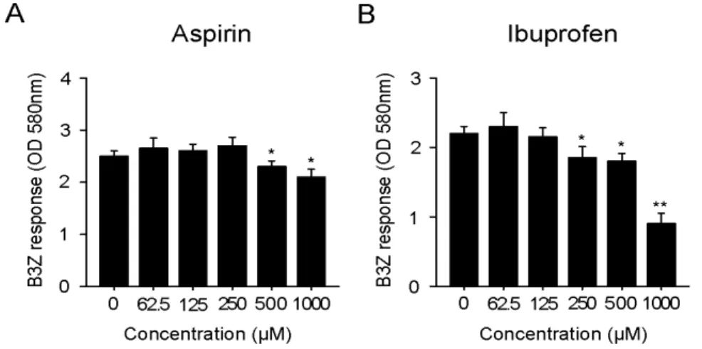
Figure 1. Effects of aspirin (A) and ibu- profen (B) on the cross-presentation of exogenous OVA. DC2.4 cells were incu- bated with the indicated amounts of the drugs for 18 h, and then added with OVA-microspheres. After 2 h incubation, the cells were washed, fixed, and the amounts of OVA peptides presented on MHC class I molecules were assessed using B3Z cells. The amount of β-galactosidase expressed in B3Z cells was determined by an enzymatic assay using chlorophenolred β-D-galactopyranoside as a substrate. *p
<0.05, **p<0.01 compared with un- treated control.
rescein isothiocyanate (FITC). After 2 h, unphagocytozed mi- crospheres were removed by washing with pre-warmed PBS.
The plate was chilled on ice for 20 min, then the cells were harvested by treating with Cell stripper solution (Cellgro Mediatech, Herndon, VA) as suggested in the manufacturer’s instruction, fixed in 1% paraformaldehyde in PBS, and flow cytometric analysis was performed on a FACS Calibur flow cytometer (Becton Dickinson).
Phenotypic analysis
DCs were cultured in the presence of different concentrations of aspirin or ibuprofen for 18 h in 6-well plates (2×10⁶ cells/well). The plate was then chilled on ice for 20 min, and the cells were harvested by treating with Cell stripper solution (Cellgro Mediatech). The cells were stained with monoclonal antibodies recognizing murine cell surface molecules after blocking of FcR-binding anti-CD16/CD32 monoclonal anti- body (clone 2.4G2), and flow cytometric analysis was per- formed on a FACS Caliver (Becton-Dickinson). The mono- clonal antibodies, anti-H-2Kb (clone AF6-88.5), anti-I-Ab (clone AF6-120.1), anti-CD40 (clone 3/23), anti-CD54 (clone 3E2), anti-B7-1 (clone 16-10A1), anti-B7-2 (clone GL1), and isotype-matched control antibodies were purchased from BD Biosciences.
RESULTS
Aspirin and ibuprofen block the MHC-restricted presentation of exogenous OVA
To examine the effects of the aspirin and ibuprofen on the cross-presentation, DC2.4 cells were cultured in the presence
of the drugs for 18 h, and then allowed to phagocytose OVA-microspheres for 2 h. The DC2.4 cells were then washed, fixed with paraformaldehyde, and the amount of OVA pep- tide-class I MHC complexes was measured using a T cell hy- bridoma, B3Z, which recognizes OVA peptide (SIINFEKL)- H-2Kb complex and expresses β-galactosidase (16). As shown in Fig. 1, aspirin inhibited MHC class I-restricted OVA pre- sentation at concentrations of 500μM or above. The inhibitory activity of ibuprofen on MHC class I-restricted OVA pre- sentation was observed at concentrations of 250μM or above.
The effects of the drugs on the MHC class II-restricted pre- sentation of exogenous OVA were examined in DCs gen- erated from bone marrow cells (BM-DCs) with GM-CSF.
BM-DCs were treated with different concentrations of the drugs for 18 h, and then allowed to phagocytize OVA-micro- spheres for 2 h. The DCs were then washed, fixed with paraf- ormaldehyde, and then the amount of OVA peptide-class II MHC complexes was measured using OVA-specific CD4 T cell hybridoma, DOBW cells. As shown in Fig. 2, aspirin inhibited MHC class II-restricted OVA presentation at concentration of 250μM or above. The inhibitory activity of ibuprofen on MHC class II-restricted OVA presentation was observed at concentrations of 125μM or above.
DCs developed in the presence of aspirin or ibuprofen are suppressed in the antigen presenting function
To examine the effects of aspirin and ibuprofen on the ac- quisition of antigen presenting function, the drugs were add- ed to DC differentiation-inducing cultures at different time points, days 0, 3 and 5 after the initiation of the culture.
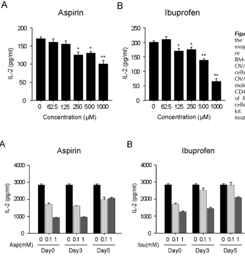
Figure 2. Effects of aspirin and ibuprofen on the class II MHC-restricted presentation of exogenous OVA. Indicated amounts of aspirin or ibuprofen were added to cultures of BM-DCs for 18 h, and then added with OVA-microspheres. After 2 h incubation, the cells were washed, fixed, and the amounts of OVA peptides presented on MHC class II molecules were assessed using OVA-specific CD4 T cell hybridoma, DOBW. The amounts of IL-2 produced from OVA-specific CD4 T cells were assayed by a commercial IL-2 ELISA kit. *p<0.05, **p<0.01 compared with un- treated control.
Figure 3. DCs differentiated in the presence of aspirin or ibuprofen are suppressed in MHC class II-restricted antigen presentation capability. Indi- cated amounts of aspirin or ibu- profen were added to mouse bone marrow cells together with GM-CSF from the initiation of the DC diffe- rentiation-inducing culture (day0), at 3 days (Day3), or at 5 days (day5) after the initiation of the cultures.
BM-DCs were harvested on day 6 from the initiation of the culture, and the ability to present exogenous antigen in association with MHC class II molecules was ascertained as in Fig. 2.
BM-DCs were harvested on day 6 from the initiation of the culture, and the ability to present exogenous antigen in asso- ciation with MHC class II molecules was examined as in Fig.
2. As shown in Fig. 3A, DCs generated from mouse bone marrow cells in the presence of aspirin from the initiation of the culture (Day0) or 3 days after the initiation of the cul- ture (Day3) were profoundly suppressed in MHC class II-re- stricted OVA presenting capability. This inhibitory effect of aspirin appeared to be dose dependent. When developing DCs were exposed to aspirin at a later time point (Day5, 5 days after the initiation of culture), the suppressive activ- ity of aspirin on antigen presenting capability was weaker compared to the earlier exposures (Day0 and Day3).
Likewise, DCs generated in the presence of ibuprofen from earlier time points of differentiation (Day0 and Day5) were significantly suppressed in MHC class II-restricted antigen presenting capability. We also found that DCs generated in
the presence of aspirin or ibuprofen were suppressed in the MHC class I-restricted exogenous antigen presentation, and the suppressive activities of the drugs were more prominent when developing DCs were exposed to the drugs at earlier time points (data not shown).
Aspirin and ibuprofen do not inhibit the phagocytic activity of DCs
To examine whether the suppressed capacity of aspirin or ibuprofen-treated DCs to present OVA peptides in association with MHC molecules was due to decreased phagocytic activ- ity, DC2.4 cells were cultured with the drugs (1 mg/ml) for 18 h, and then added with microspheres containing both OVA and FITC. After 2 h incubation, unphagocytized microspheres were washed, and the cells were harvested by gentle pipet- ting after cooling on ice. Flow cytometric analysis of the har- vested cells showed that neither of the drugs suppressed the
Figure 4. Effects of aspirin and ibuprofen on the phagocytic activity.
DC2.4 cells were cultured with aspirin or ibuprofen for 18 h, and then added with microspheres containing both OVA and FITC. After 2 h incubation, unphagocytized microspheres were washed, and the cells were harvested by gentle pipetting, and then analyzed by flow cytometry. Shaded histograms represent the phagocytic activity of DCs cultured in the presence of aspirin or ibuprofen, and thick line histograms represent the phagocytic activity of DCs cultured in the absence of the drugs. DCs that were not incubated with FITC-labeled microspheres are shown as thin line histograms (left).
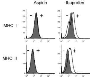
Figure 5. Effects of aspirin and ibuprofen on the expression of MHC molecules. DC2.4 cells were cultured with aspirin or ibuprofen for 18 h, and then the cells were harvested by gentle pipetting. The expression levels of class I and class II MHC molecules were assessed using anti-H-2Kb and anti-I-Ab monoclonal antibodies. Shaded histograms represent the expression levels of H-2Kb and I-Ab molecules in DC2.4 cells cultured in the absence of the drugs. Thick line histograms represent the expression levels of H-2Kb and I-Ab molecules in DC2.4 cells cultured in the presence of the drugs.
phagocytic activity of DC2.4 cells (Fig. 4).
Ibuprofen, but not aspirin, slightly increases the expression of MHC molecules and co-stimulatory molecules
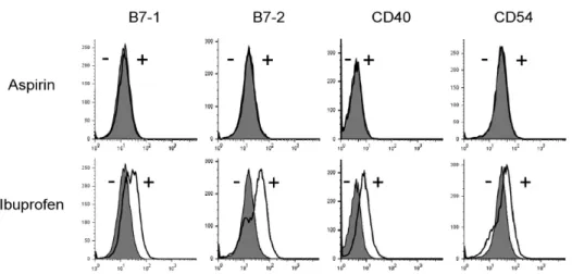
To examine whether the suppressed capacity of aspirin or ibuprofen-treated DCs to present OVA peptides in association with MHC molecules was due to the inhibition of expression of MHC molecules on the cell surface, DC2.4 cells were cul- tured with the drugs (1 mg/ml) 18 h, and then the expression levels of MHC class I and class II molecules were determined by anti-H-2Kb and anti-I-Ab monoclonal antibodies. As shown in Fig. 5, aspirin did not affect the expression of MHC class I or class II molecules. Ibuprofen, which inhibited MHC-re- stricted exogenous OVA presentation, even slightly increased the expression of MHC class I and class II molecules. Flow cytometric analysis for major co-stimulatory molecules such as B7-1, B7-2, and CD40 also showed that aspirin did not affect the expression of these co-stimulatory molecules (Fig.
6). Again, ibuprofen, which inhibited MHC-restricted exoge- nous OVA presentation, even slightly increased the ex- pression of co-stimulatory molecules such as B7-1, B7-2, and
CD40 (Fig. 6).
DISCUSSION
The present study demonstrates that DCs exposed to aspirin or ibuprofen for 18 h are suppressed in MHC-restricted exog- enous antigen presenting capability. Aspirin and ibuprofen significantly inhibited MHC class I-restricted exogenous anti- gen presenting capability of DCs at doses of 500μM or above and of 250μM or above, respectively. The MHC class II-re- stricted exogenous antigen presenting capability of DCs was inhibited at lower concentration of the drugs-250μM or above for aspirin and 125μM or above for ibuprofen. When aspirin is used as an anti-inflammatory drug in doses of 1,200 mg/day, the in vivo therapeutic levels are in between 100∼
2,000μM (21). Administration of ibuprofen in doses of 400 mg three times/day translates to up to 120μM in the plasma (22). Taken together, our results suggest that aspirin and ibu- profen suppress antigen presenting function of DCs at clin- ically relevant concentrations.
Because NSAIDs are usually used for prolonged time peri- ods in the treatment of chronic inflammatory diseases, we
Figure 6. Effects of aspirin and ibuprofen on the expression of co-stimulatory molecules. DC2.4 cells were cultured with aspirin or ibuprofen for 18 h, and then the cells were harvested by gentle pipetting. The expression levels of co-stimulatory molecules were assessed using monoclonal antibodies recognizing mouse cell surface molecules. Shaded histograms represent the expression levels of co-stimulatory molecules in DC2.4 cells cultured in the absence of the drugs. Thick line histograms represent the expression levels of co-stimulatory molecules in DC2.4 cells cultured in the presence of the drugs.
were curious to investigate whether the DCs exposed to aspirin or ibuprofen during development could acquire normal func- tion in terms of antigen processing and presentation. Our re- sults show that DCs generated from mouse bone marrow cells in the presence of aspirin (100μM) or ibuprofen (100μM) from the early stage of differentiation (Day 0 or 3 after the differentiation initiation culture) were profoundly suppressed in MHC-restricted exogenous antigen presenting capability.
These results suggest that aspirin and ibuprofen, when used in prolonged and higher doses, exert suppressive activity on adaptive immunity in addition to the well-known mechanism of COX inhibition.
DCs play a key role in the initiation of primary immune responses (23-25). DCs can acquire and process antigens in the periphery, and migrate to secondary lymphoid tissues where they prime the primary T cell responses. The capa- bility of DCs to activate even naïve T cells in a primary re- sponse has been explained by their ability to express high levels of MHC class II molecules and co-stimulatory mole- cules. The present study shows that aspirin and ibuprofen suppress MHC class II-restricted antigen presentation more strongly than MHC class I-restricted antigen presentation in DCs. The effects of the expression of co-stimulatory mole- cules appeared to be different between the two drugs.
Aspirin did not affect the expression of co-stimulatory mole- cules in DC2.4 cells, whereas ibuprofen slightly increased the
expression of co-stimulatory molecules. Ibuprofen also slight- ly increased the expression of MHC class I and class II mole- cules in DC2.4 cells. Although ibuprofen slightly increased the expression of co-stimulatory molecules as well as MHC molecules, the DCs treated with ibuprofen were suppressed in activating OVA-specific CD4 T cells compared to normal DCs. Because T cells can only recognize antigens presented in the form of short peptide-MHC complexes presented by professional antigen presenting cells (22-25), inhibition of the MHC-restricted antigen presenting function of DCs by ibupro- fen might have had significant effects in activating T cells.
In all of the experiments described in the present study, DCs were exposed to the drugs for 18 h, and the cells were allowed to phagocytose OVA-microspheres for 2 h. The DCs were washed to remove unphagocytosed OVA-microspheres, fixed with paraformaldehyde, and then washed thoroughly again to remove paraformaldehyde before functional assays with OVA-specific CD4 or CD8 T cells. Thus, the activation of OVA-specific T cells must be due to enhanced expression of OVA peptides on DCs, and not due to the carryover of the drugs to T cell cultures.
In summary, we have demonstrated that aspirin and ibu- profen at high concentrations inhibit both MHC class I and class II-restricted exogenous antigen presentation. In addition, we showed that the DCs generated in the presence of these drugs are profoundly suppressed in MHC-restricted antigen
presentation capability. These results suggest that aspirin and ibuprofen may exert their anti-inflammatory activity by the in- hibition of the antigen presenting function of DCs in addition to the well-known mechanism of COX inhibition, especially when these drugs are used in prolonged durations.
ACKNOWLEDGEMENTS
This work was supported by the research grant of the Chungbuk National University in 2008.
CONFLICTS OF INTEREST
The authors declare no financial or commercial conflicts of interest.
REFERENCES
1. Vane JR: Inhibition of prostaglandin synthesis as a mecha- nism of action for aspirin-like drugs. Nat New Biol 231;
232-235, 1971
2. Smith WL, DeWitt DL, Garavito RM: Cyclooxygenases:
structural, cellular, and molecular biology. Annu Rev Biochem 69;145-182, 2000
3. Smith WL, Dewitt DL: Prostaglandin endoperoxide H syn- thases-1 and -2. Adv Immunol 62;167-215, 1996
4. Griswold DE, Adams JL: Constitutive cyclooxygenase (COX-1) and inducible cyclooxygenase (COX-2): rationale for selective inhibition and progress to date. Med Res Rev 16;181-206, 1996
5. DeWitt DL: Cox-2-selective inhibitors: the new super aspirins. Mol Pharmacol 55;625-631, 1999
6. Cryer B, Feldman M: Cyclooxygenase-1 and cyclooxy- genase-2 selectivity of widely used nonsteroidal anti-in- flammatory drugs. Am J Med 104;413-421, 1998
7. Kazmi SM, Plante RK, Visconti V, Taylor GR, Zhou L, Lau CY: Suppression of NF kappa B activation and NF kappa B-dependent gene expression by tepoxalin, a dual inhibitor of cyclooxygenase and 5-lipoxygenase. J Cell Biochem 57;299-310, 1995
8. Hall VC, Wolf RE: Effects of tenidap and nonsteroidal anti- inflammatory drugs on the response of cultured human T cells to interleukin 2 in rheumatoid arthritis. J Rheumatol 24;1467-1470, 1997
9. Iñiguez MA, Punzón C, Fresno M: Induction of cyclo- oxygenase-2 on activated T lymphocytes: regulation of T cell activation by cyclooxygenase-2 inhibitors. J Immunol 163;111-119, 1999
10. Kopp E, Ghosh S: Inhibition of NF-kappa B by sodium sali- cylate and aspirin. Science 265;956-959, 1994
11. Yin MJ, Yamamoto Y, Gaynor RB: The anti-inflammatory agents aspirin and salicylate inhibit the activity of I(kappa)B kinase-beta. Nature 396;77-80, 1998
12. Jiang C, Ting AT, Seed B: PPAR-gamma agonists inhibit production of monocyte inflammatory cytokines. Nature 391;82-86, 1998
13. Bancos S, Bernard MP, Topham DJ, Phipps RP: Ibuprofen and other widely used non-steroidal anti-inflammatory drugs inhibit antibody production in human cells. Cell Immunol 258;18-28, 2009
14. Paccani SR, Boncristiano M, Ulivieri C, D'Elios MM, Del PG, Baldari CT: Nonsteroidal anti-inflammatory drugs suppress T-cell activation by inhibiting p38 MAPK induction. J Biol Chem 277;1509-1513, 2002
15. Banchereau J, Briere F, Caux C, Davoust J, Lebecque S, Liu YJ, Pulendran B, Palucka K: Immunobiology of den- dritic cells. Annu Rev Immunol 18;767-811, 2000 16. Karttunen J, Sanderson S, Shastri N: Detection of rare anti-
gen-presenting cells by the lacZ T-cell activation assay sug- gests an expression cloning strategy for T-cell antigens.
Proc Natl Acad Sci U S A 89;6020-6024, 1992
17. Harding CV, Collins DS, Kanagawa O, Unanue ER: Lipo- some-encapsulated antigens engender lysosomal process- ing for class II MHC presentation and cytosolic processing for class I presentation. J Immunol 147;2860-2863, 1991 18. Shen Z, Reznikoff G, Dranoff G, Rock KL: Cloned dendritic
cells can present exogenous antigens on both MHC class I and class II molecules. J Immunol 158;2723-2730, 1997 19. Lee JK, Lee MK, Yun YP, Kim Y, Kim JS, Kim YS, Kim
K, Han SS, Lee CK: Acemannan purified from Aloe vera induces phenotypic and functional maturation of immature dendritic cells. Int Immunopharmacol 1;1275-1284, 2001 20. Lee YH, Lee YR, Im SA, Park SI, Kim KH, Gerelchuluun
T, Song S, Kim K, Lee CK: Calcineurin inhibitors block MHC-restricted antigen presentation in vivo. J Immunol 179;5711-5716, 2007
21. Borthwick GM, Johnson AS, Partington M, Burn J, Wilson R, Arthur HM: Therapeutic levels of aspirin and salicylate directly inhibit a model of angiogenesis through a Cox-in- dependent mechanism. FASEB J 20;2009-2016, 2006 22. Blain H, Boileau C, Lapicque F, Nédélec E, Loeuille D,
Guillaume C, Gaucher A, Jeandel C, Netter P, Jouzeau JY:
Limitation of the in vitro whole blood assay for predicting the COX selectivity of NSAIDs in clinical use. Br J Clin Pharmacol 53;255-265, 2002
23. Heath WR, Carbone FR: Cross-presentation, dendritic cells, tolerance and immunity. Annu Rev Immunol 19;47-64, 2001 24. Huang AY, Golumbek P, Ahmadzadeh M, Jaffee E, Pardoll
D, Levitsky H: Role of bone marrow-derived cells in pre- senting MHC class I-restricted tumor antigens. Science 264;961-965, 1994
25. Sigal LJ, Crotty S, Andino R, Rock KL: Cytotoxic T-cell im- munity to virus-infected non-haematopoietic cells requires presentation of exogenous antigen. Nature 398;77-80, 1999