- 223 -
Imaging Science in Dentistry 2018; 48: 223-6 https://doi.org/10.5624/isd.2018.48.3.223
Intraosseous cervical pneumatocysts are rare, asymp- tomatic benign lesions incidentally found on radiographs.
These lesions commonly involve the iliac or sacral bone and are rarely visible on the cervical spine.1 Vertebral pneumatocysts are most commonly seen in the location of spondylosis in adults.2 Awareness of these characteristic features can help radiologists distinguish pneumatocysts from osseous lesions.1 The etiology of pneumatocysts is unknown, but they have been claimed to be caused by the accumulation of nitrogen gas.3 Intraosseous gas collection is a phenomenon of the bone that occurs in various con- ditions, including osteomyelitis, trauma, neoplasm, and osteonecrosis.4,5 The most common cause of intraosseous gas collection in the vertebral bodies is the presence of intraosseous pneumatocysts.6 The appearance of pneuma- tocysts in the cervical spine is uncommon, and 21 cases have been reported in the literature.7 Recent studies have
proven that cervical lesions are more common2 than re- ported; therefore, meticulous radiographic evaluation will allow such lesions to be identified. Cone-beam computed tomography(CBCT) is predominantly used to diagnose dental abnormalities, as well as aiding in the evaluation of structures such as the temporomandibular joint, paranasal sinus, upper cervical vertebrae, the base of the skull, and the pharyngeal airspace. It is more likely that dentists will encounter pneumatocyst-like lesions in the cervical spine while interpreting diagnostic radiographs.8 Medium and large fields of view are the image volumes that enable di- agnosticians to identify incidental findings of the cervical spine on CBCT.8
This report presents a case with 8 lytic lesions in the cervical vertebrae(C2-C5) diagnosed incidentally when a patient presented for dental implant treatment planning.
Case Report
A 60-year-old female patient was referred for a CBCT scan to the Oral and Maxillofacial Imaging Center of the University of Texas School of Dentistry at Houston for an
Multiple intraosseous cervical pneumatocysts: A case report of a rare incidental finding on cone-beam computed tomography
Aniket B. Jadhav1,*, Sangeetha Gajendran Sarah1, Robert Cederberg2, Aditya Wagh3, Sudarat Kiat-amnuay2
1Department of Diagnostic and Biomedical Sciences, The University of Texas School of Dentistry at Houston, Houston, TX, USA
2Department of General Practice and Dental Public Health, The University of Texas School of Dentistry at Houston, Houston, TX, USA
3University of Pittsburgh School of Dental Medicine, Pittsburgh, PA, USA
ABSTRACT
This report presents a case of cervical pneumatocysts as an incidental finding on cone-beam computed tomography.
Pneumatocysts are gas-containing lesions of unknown etiology. They usually present in the ilium or sacrum, adjacent to the sacroiliac joint. In the literature, 21 case reports have described cervical pneumatocysts. Cervical pneumatocysts should be differentiated from other lesions, such as osteomyelitis, osteonecrosis, and neoplasms, as well as post-traumatic and post-surgical cases. Computed tomography, cone-beam computed tomography, and magnetic resonance imaging are appropriate tools to diagnose cervical pneumatocysts.(Imaging Sci Dent 2018; 48:
223-6)
KEY WORDS: Cone-Beam Computed Tomography; Bone Cysts; Intraosseous Pneumatocyst; Cervical Vertebrae
Copyright ⓒ 2018 by Korean Academy of Oral and Maxillofacial Radiology
This is an Open Access article distributed under the terms of the Creative Commons Attribution Non-Commercial License(http://creativecommons.org/licenses/by-nc/3.0) which permits unrestricted non-commercial use, distribution, and reproduction in any medium, provided the original work is properly cited.
Imaging Science in Dentistry·pISSN 2233-7822 eISSN 2233-7830 Received May 8, 2018; Revised July 20, 2018; Accepted August 2, 2018
*Correspondence to : Dr. Aniket B. Jadhav
Department of Diagnostic and Biomedical Sciences, The University of Texas School of Dentistry at Houston, 7500 Cambridge St Suite # 5371, Houston, TX 77054, USA Tel) 1-713-486-4134, Fax) 1-713-486-4416, E-mail) [email protected]
Multiple intraosseous cervical pneumatocysts: A case report of a rare incidental finding on cone-beam computed tomography
- 224 -
evaluation of the anterior maxilla for dental implant treat- ment planning. The patient’s medical history was positive for a benign brain tumor and visual impairment. The stan- dard Imaging Center protocol was requested for CBCT scanning and interpretation, which consisted of a medi- um field of view(9cm ×15cm, 200-μm voxels) at 90 kVp and 10mA. The scan was acquired on a Kodak 9500 CBCT unit(Carestream Health, Rochester, NY, USA) and the scan was retrieved on an InVivo-5.0 DICOM viewer (Anatomage Inc., San Jose, CA, USA). The CBCT vol- ume revealed multiple radiolucencies with sclerotic mar- gins on C2-C5 that measured approximately 2-10mm (Figs. 1-6). The radiolucencies were primarily located on the lamina, adjacent to the facet joints and close to the endplates. No destruction of the adjacent bone or exten- sion into the intervertebral space was evident. The pixel intensity values ranged from -750 to -1000, indicat-
ing gas density. Eight lytic lesions were revealed on the CBCT scan. Cystic-appearing radiolucencies on the cer- vical vertebrae can be associated with a wide spectrum of conditions, and lytic metastasis, subchondral cysts, osteo- arthritis, and other analogous conditions were considered in the differential diagnosis. The benign-appearing radio- graphic appearance and range of pixel intensity values were pathognomonic of intravertebral gas formation and suggestive of cervical pneumatocysts.
Discussion
Pneumatization of bone commonly occurs in the cra- niofacial bones irrespective of age, gender, and race. In- traosseous lesions are diagnosed in conditions such as osteomyelitis, trauma, irradiated neoplasms, intraosseous ganglia, osteonecrosis, methyl methacrylate prosthesis,
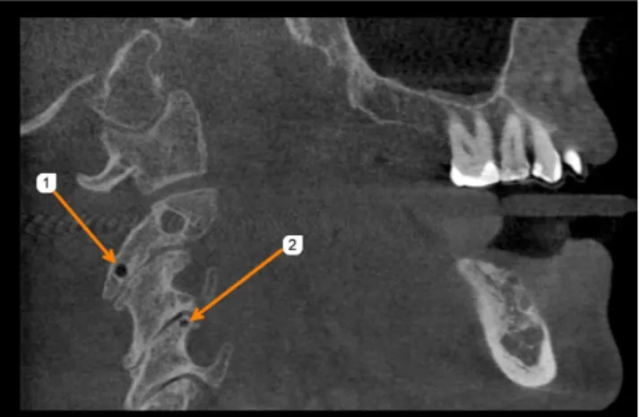
Fig. 1. Sagittal cone-beam computed tomographic image shows cystic lesions(#1 and #2) with sclerotic margins on C-2 and C-4.
Fig. 2. Sagittal cone-beam computed tomographic image shows a large cystic lesion(#3) with sclerotic margins on the right lateral mass of C-3.
Fig. 3. Sagittal cone-beam computed tomographic image shows a small cystic lesion(#4) with sclerotic margin on the left lateral mass of C-3.
Fig. 4. Sagittal cone-beam computed tomographic image shows a small cystic lesion(#5) with sclerotic margins on C-4.
- 225 -
Aniket B. Jadhav et al
and postoperative conditions.9
Cervical pneumatocysts are very rare; so far, 21 cases have been reported in the literature.8 On computed to- mography, pneumatocysts appear as lytic low-density le- sions of variable dimension. These lytic lesions show at- tenuation values of -950 to -580 Hounsfield units(HU), which is pathognomonic of pneumatocysts.10 Intraosseous pneumatocysts are benign lesions and require no specific treatment, although growing lesions may need to be mon- itored if they pose a risk of fracture.10
Ramirez et al.11 first described an intraosseous pneu- matocyst with an unknown etiology in 1984; this benign lesion consisted predominantly of nitrogen gas. Although they are more common in the sacroiliac joint, intraosse- ous pneumatocysts have occasionally been reported to be present in the vertebrae, humeral head, clavicle, cer- vical rib, scapula, acetabulum, and pubis.5 Many authors
have reported that pneumatocysts did not progress in size.
However, Nakayama et al.13 stated that pneumatocysts in the cervical spine and lumbar spine suddenly progressed to fluid-filled cysts 6-16 months after the initial presenta- tion. In the case reported by Yamamoto et al.,5 the pneu- matocysts resolved spontaneously and appeared as flu- id-filled cysts on follow-up imaging.
Kitagawa et al.4 reported a case of growing cervical pneumatocysts, while the study conducted by Matsukubo et al.14 reviewed 500 neck computed tomography exams and found multiple lytic lesions on the cervical spine, with most occurrences in the C4-C7 segment. The pres- ent report describes the incidental finding of multiple intraosseous cervical pneumatocysts involving the C2- C5 segment in a 60-year-old female patient referred for CBCT imaging for dental implant treatment planning.
Intraosseous pneumatocysts are gas-filled cystic cavities, and the exact cause of the development of these cavities is unknown. Karasick et al.14 suggested that the source of gas might be from the intervertebral disc through the vertebral endplate, although the gas inside pneumatocysts is mostly composed of nitrogen. Some authors have pro- posed that negative pressure within the lesion causes the release of nitrogen from the surrounding soft tissues or articular joints.7,13 In the degenerated spine, gas often ac- cumulates in the intervertebral discs, which is known as the vacuum phenomenon. This phenomenon results from the extension of gas through the defect from the interver- tebral disc into the vertebral body, and it causes intraver- tebral pneumatocysts.7 The spontaneous progression of vertebral intraosseous pneumatocysts could result in flu- id-filled cysts.7
In the case reported by Kitagawa et al.,4 direct commu- nication was observed between the intervertebral disc and pneumatocysts on C6. In our case, no such direct com- munication was observed. The lesions described in this report showed variable dimensions, with largest being approximately 10mm.4 In our case, the pneumatocysts were primarily seen in the lamina of the cervical vertebra and were adjacent to the facet joints, with minimal degen- erative changes on osseous portions of the cervical spine.
Since CBCT utilizes pixel intensity values, which closely resemble HU values, all the lesions in our case showed gas attenuation, with pixel intensity values ranging from -1000 to -750. Osteolytic lesions, such as those seen in metastatic lesions, osteomyelitis, and osteonecrosis, should be differentiated from pneumatocysts by their ra- diographic appearance and gas attenuation characteristics through computed tomography and CBCT.
Fig. 5. Sagittal cone-beam computed tomographic image shows small cystic lesions(#6 and #7) with sclerotic margins on articular processes of C-3 and C-4.
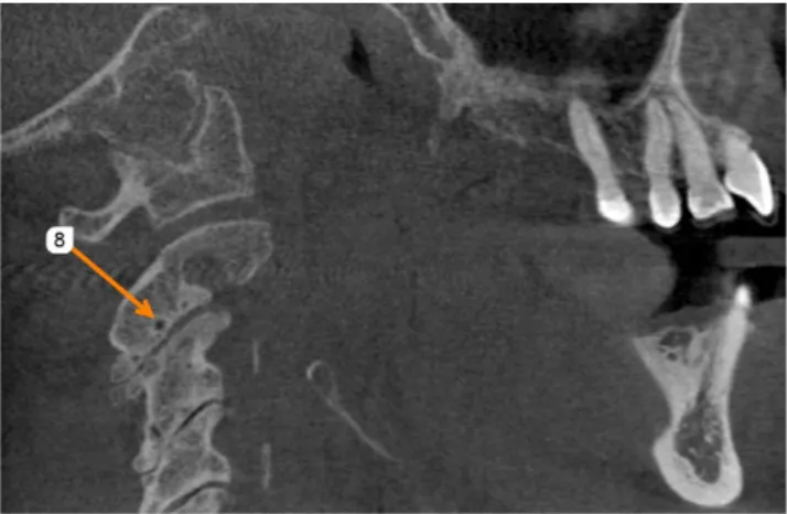
Fig. 6. Sagittal cone-beam computed tomographic image shows a small cystic lesion(#8) with sclerotic margin on C-2.
Multiple intraosseous cervical pneumatocysts: A case report of a rare incidental finding on cone-beam computed tomography
- 226 -
In conclusion, CBCT has revolutionized dentistry over the last decade, with multiple applications that range from diagnosis to treatment planning. More incidental findings are emerging as CBCT technology advances. It is there- fore imperative to report such instances to educate our peers in the dental world and, at the same time, to under- stand the liability associated with CBCT scan findings during the routine practice of dentistry. Our case report presented innocuous benign lesions seen on an CBCT exam. Although these lesions did not warrant further medical treatment, follow-up imaging may still be need- ed to assess their expansion potential. Vertebral pneuma- tocysts are uncommon, but predominantly occur in the cervical spine in patients with degenerative disc disease.
They may appear as possible metastatic lesions on plain radiographs, but CBCT, in combination with further con- sultations with specialists, can confirm the nature of these lesions.
Disclosure: This case report was submitted and pre- sented at the annual American Academy of Oral and Max- illofacial Academy meeting, and the abstract has been published in the journal Oral Surgery, Oral Medicine, Oral Pathology, and Oral Radiology.
References
1. Sen D, Satija L, Saxena S, Rastogi V, Singh M. Intraosseous pneumatocyst of the cervical vertebra. Med J Armed Forces India 2015; 71: 380-3.
2. Matsukubo Y, Kashiwagi N, Uemura M, Tatsumi S, Takahashi H, Hyodo T, et al. Intravertebral pneumatocysts of the cervical
spine. Neuroradiology 2013; 55: 1341-4.
3. Coşar M, Eser O, Aslan A, Korkmaz S, Boyaci G, Değirmenci B, et al. Vertebral body pneumatocyst in the cervical spine and review of the literature. Turk Neurosurg 2008; 18: 197-9.
4. Kitagawa T, Fujiwara A, Tamai K, Kobayashi N, Saiki K, Omata S, et al. Enlarging vertebral body pneumatocysts in the cervical spine. AJNR Am J Neuroradiol 2003; 24: 1707-10.
5. Yamamoto T, Yoshiya S, Kurosaka M, Nagira K, Takabatake M, Hamamoto H, et al. Natural course of an intraosseous pneumatocyst of the cervical spine. AJR Am J Roentgenol 2002; 179: 667-9.
6. Arslan G, Ceken K, Cubuk M, Ozkaynak C, Luleci E. Verte- bral pneumatocysts. Acta Radiol 2001; 42: 20-3.
7. Sunder S, Prasad R. Vertebral body pneumatocyst in the cer- vical spine - a case report. Apollo Med 2008; 5: 121-4.
8. Husain MA, Tetradis S, Mallya SM. Intraosseous pneumato- cysts of the cervical spine: a report of four cases and review of literature. Oral Surg Oral Med Oral Pathol Oral Radiol 2015;
119: e49-54.
9. Laufer L, Schulman H, Hertzanu Y. Vertebral pneumatocyst.
A case report. Spine(Phila Pa 1976) 1996; 21: 389-91.
10. Renshaw H, Patel A, Boctor DS, Hakmi MA. ‘Abnormal’
cervical imaging?: Cervical pneumatocysts - a case report of a cervical spine pneumatocyst. J Orthop 2015; 12(Suppl 1):
S83-5.
11. Ramirez H Jr, Blatt ES, Cable HF, McComb BL, Zornoza J, Hibri NS. Intraosseous pneumatocyst of the ilium. Findings on radiographs and CT scans. Radiology 1984; 150: 503-5.
12. Oehler E, Valour F, Pascart T. Intraosseous pneumatocysts: a case based review. Clin Rheumatol 2013; 32: 129-34.
13. Nakayama T, Ehara S, Hama H. Spontaneous progression of vertebral intraosseous pneumatocysts to fluid-filled cysts.
Skeletal Radiol 2001; 30: 523-6.
14. Karasick D, Eason MA. Vertebral pneumatocyst mimicking susceptibility artifact on MR imaging. AJR Am J Roentgenol 1998; 170: 221.