https://doi.org/10.5624/isd.2017.47.4.261
Introduction
The maxillary sinus, the largest paranasal sinus, is a pyramid-shaped structure. The base of this pyramid is in the lateral wall of the nasal cavity pointing towards the zygomatic process of the maxilla.1 After tooth loss, the alveolar process of the posterior maxilla is resorbed and, as a result, the vertical bone dimension is reduced. Many
older individuals do not have enough bone volume to sup- port an implant between the sinus floor and alveolar crest, and thus, implant surgery can be complicated.2
Today, the sinus lift procedure is a standard procedure for the treatment of atrophic posterior maxilla before the insertion of implants. It is one of the most reliable proce- dures in pre-prosthetic surgery.3 Perforation of the sinus membrane is one of the complications of this surgery.
Some sinus anatomical variations increase the risk of si- nus membrane perforation. One of these variations is the existence of a maxillary sinus septum.4,5
Maxillary sinus septa have been studied more than other structures inside the maxillary sinus. Several studies2,5-13
Frequency of different maxillary sinus septal patterns found on cone-beam computed tomography and predicting the associated risk of sinus membrane perforation during sinus lifting
Ali Khalighi Sigaroudi1, Zahra Dalili Kajan2,*, Shabnam Rastgar3, Hamid Neshandar Asli4
1Department of Maxillofacial Surgery, School of Dentistry, Guilan University of Medical Sciences, Rasht, Iran
2Dental Sciences Research Center, Department of Maxillofacial Radiology, School of Dentistry, Guilan University of Medical Sciences, Rasht, Iran
3Private Dental Office, Guilan University of Medical Sciences, Rasht, Iran
4Dental Sciences Research Center, Department of Prosthodontics, School of Dentistry, Guilan University of Medical Sciences, Rasht, Iran
AbstrAct
Purpose: Analyzing different patterns of maxillary sinus septa in cone-beam computed tomography(CBCT) images and predicting maxillary sinus membrane perforations.
Materials and Methods: In this cross-sectional study, CBCT images of 222 patients ranging from 20 to 81 years old were evaluated. One hundred fifty-two patients(93 females and 59 males) who had maxillary sinus septa in axial views were included in this study. Cross-sectional images were used to determine classifications of sinus septa and the risk of membrane perforation using a method modified from Al-Faraje et al. Variables of sex, age, and dental status were considered. Chi-squared and Kruskal-Wallis tests were used for data analysis(P<.05).
results: In this study, 265 maxillary sinus septal patterns were found. The mean age of the patients was 44.1±14.7 years old. The Class I and VII-div II patterns had the greatest and least prevalence, respectively. Furthermore, there was a significant relationship between the location of septa and the frequency of membrane perforation risk(P<.05).
In this study, the relationship of different patterns of septa with dental status did not differ significantly(P>0.05).
conclusion: A higher prevalence of moderate risk of membrane perforation in the molar region relative to the premolar region was observed. Furthermore, maxillary sinus septa occur most frequently in the molar region, demonstrating the importance of paying attention to this region during sinus lift surgery. This study did not show any relationship between tooth loss and the presence of septa.(Imaging Sci Dent 2017; 47: 261-7)
Key words: Cone-Beam Computed Tomography; Oral Surgical Procedures; Maxillary Sinus
Copyright ⓒ 2017 by Korean Academy of Oral and Maxillofacial Radiology
This is an Open Access article distributed under the terms of the Creative Commons Attribution Non-Commercial License(http://creativecommons.org/licenses/by-nc/3.0) which permits unrestricted non-commercial use, distribution, and reproduction in any medium, provided the original work is properly cited.
Imaging Science in Dentistry·pISSN 2233-7822 eISSN 2233-7830 Received May 3, 2017; Revised October 28, 2017; Accepted October 31, 2017
*Correspondence to : Prof. Zahra Dalili Kajan
Department of Maxillofacial Radiology, School of Dentistry, End of Professor Samii Blvd. Rasht, Iran
Tel) 98-13-3336-3622, Fax) 98-13-3336-3621, E-mail) [email protected]
have evaluated different aspects of these septa, including their prevalence, location, height, and morphology.
Al-Faraje et al.1 classified the morphology of septa into 6 different patterns and described specific clinical consid- erations for each one. Wen et al.8 presented a classifica- tion system for septa based on the location, number, ori- entation, and size of the antral septa and treatment consid- erations for each type to avoid complications during sinus lift surgery. These studies demonstrated the importance of detailed knowledge of maxillary sinus septa morphology to arrange a precise treatment plan and to avoid complica- tions during surgery.
In a retrospective cohort study, the presence of antral septa and their interference in Schneiderian membrane el- evation in 45 patients undergoing sinus lifts was assessed.
Interfering septa were present in 28.8% of the cases hav- ing antral septa. Membrane perforation was reported in 11.5% of cases.14 Thus, pre-operative radiographic exam- inations using panoramic radiographs or cone-beam com- puted tomography(CBCT) could be helpful in successful surgical planning and to overcome such complications.
Therefore, the aim of this study was to determine the frequency of the different patterns of the maxillary sinus septa in an Iranian population and to predict the risk of si- nus membrane perforation based on different scores.
Materials and Methods
In this statistical cross-sectional study, we evaluated cone-beam computed tomography(CBCT) images of 222 patients who were more than 20 years old and were re- ferred to a maxillofacial radiology clinic in northern Iran between September 2014 and March 2016 for radiograph- ic examination of the maxilla or paranasal sinuses. An ex- pert maxillofacial radiologist with more than 15 years of experience reported the presence of maxillary sinus septa in 152 CBCT images. Thus, 152 CBCT images were in- cluded in the second step of this study. This study was approved by the Ethics Committee of Guilan University of Medical Sciences: IR.GUMS.REC.1394.514. All pro- cedures followed were in accordance with the ethics stan- dards of the responsible committee on human experimen- tation(Guilan University of Medical Sciences) and with the Helsinki Declaration of 1975, as revised in 2008. In- formed consent to be included in the study was obtained from all patients.
Motion artifacts, the presence of maxillary sinus le- sions, the destruction of maxillary sinus walls, fractures of the maxillary sinus walls, and a past medical history of
maxillary sinus surgery were exclusion criteria.
The dental status on each side of the posterior portion of the maxilla was divided into 2 groups: adentate(less than 2 teeth in the premolar and molar region) and den- tate(2 or more teeth in the premolar and molar region).
Demographic data, including age and sex, were extracted from the information registered in CBCT software before performing the procedure.
CBCT images were taken using a New Tom VG(QR Srl Company, Verona, Italy) by selecting the “zoom” mode.
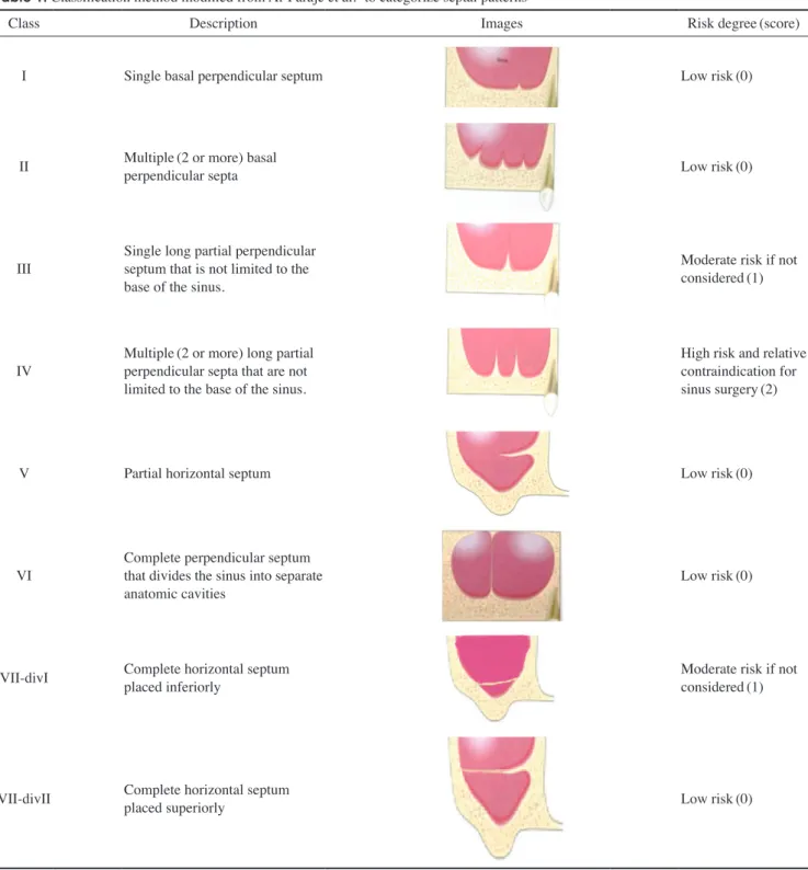
An expert maxillofacial radiologist and a final-year den- tal student evaluated the right and left maxillary sinuses in axial views with a 1-mm thickness and sequential slice interval for the presence of sinus septa. They reported the CBCT findings in agreement with each other. They then determined the classification and location of these septa based on mesiodistal cross-sectional and axial directions, and then totally reconstructed the panoramic views. To do so, they reconstructed mesiodistal cross-sectional images with a 1-mm thickness and distance of sequential slice with a 60-mm image width on premolar molar regions ad- jacent to the maxillary sinuses that had internal septa. To reconstruct cross-sectional images in the mesiodistal di- rection, the reconstruction line perpendicular to the alve- olar bone in the premolar-molar area was drawn and then the software automatically reconstructed them from the buccal to the palatal side. They determined the location of the septa and then classified the sinus septa as well as the degree and score of the sinus membrane perforation risk according to a classification method modified from that of Al-Faraje et al.1 (Table 1).
Before starting this study, 2 maxillofacial surgeons with more than 10 years of experience in implant surgery pro- vided input on the classification of Al-Faraje et al.1 (Fig.
1). Their comments were expertized on the CBCT find- ings by a maxillofacial radiologist as the observer of the CBCT images in this research. They established a mod- ification of the Al-Faraje et al.1 classification method in agreement with each other. Finally, based on the risk de- scriptions in Al-Faraje et al., classifications, and the sur- geons’ experiences; the risk of the membrane perforation was scored from 0(low) to 2(high risk).
Cross-sectional images were further evaluated to mea- sure the height of the maxillary sinus septa. The septa that had 3mm or more height were considered to be ‘long’.
Fifty images were reevaluated by the observers 2 weeks later. The intraobserver reliability was found to be 95%.
Moreover, the locations of the septa were determined according to their presence in premolar or/and molar re-
gions. Figures 1 and 2 reveal 2 different kinds of septal classifications.
All data were analyzed with SPSS version 22(IBM Corporation, Armonk, NY, USA). A chi-squared test was used to compare the quantitative variables according to different patterns of maxillary sinus septa. The mean ages of the patients with different patterns of maxillary sinus septa were compared with a Kruskal-Wallis test. P<.05
was considered significant.
results
In this study, CBCT images of 222 patients were eval- uated. Among them, 152 of the patients(68.4%), 93 fe- males(61.2%) and 59 males(38.8%), showed at least one pattern of septa in one of the maxillary sinuses. A total of
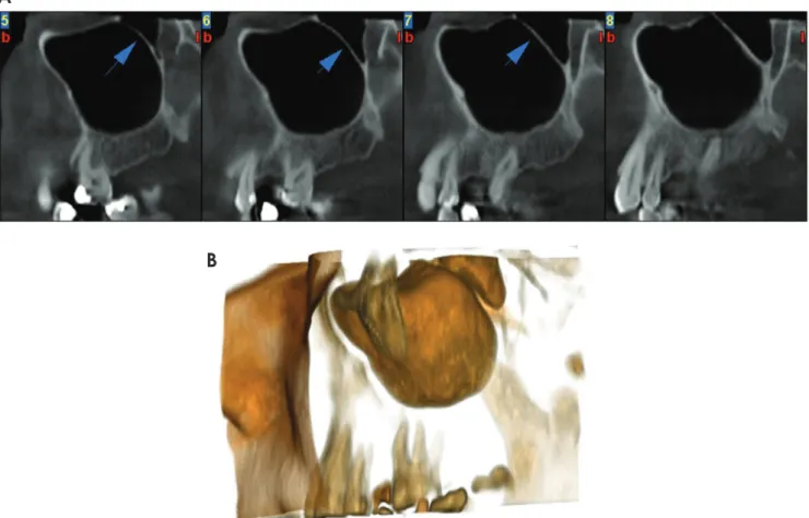
Fig. 1. Class IV sinus septal pattern: 2 long partial perpendicular septa(arrows) that are not limited to the base of the sinus in cross-section- al(A) and three-dimensional(B) images.
A
B
Fig. 2. Cross-sectional images of the class II sinus septal pattern: multiple basal perpendicular septa(arrows).
42.1% of 152 patients had internal septa only in one max- illary septum, and 57.9% had internal septa in both maxil- lary sinuses. Meanwhile, if all the patients(with and with- out septa) were considered, the prevalence of sinus septa in the female and male populations were 29% and 35.2%, respectively. In total, 240 maxillary sinuses had internal septa. Twenty-five maxillary sinuses showed more than one pattern of maxillary sinus septa in one sinus. There-
fore, there were 265 instances of the varying pattern of maxillary sinus septa taken together. The ages of the pa- tients ranged from 20 to 81 years old. The mean age was 44.1±14.7 years, and the median age was 43.5 years.
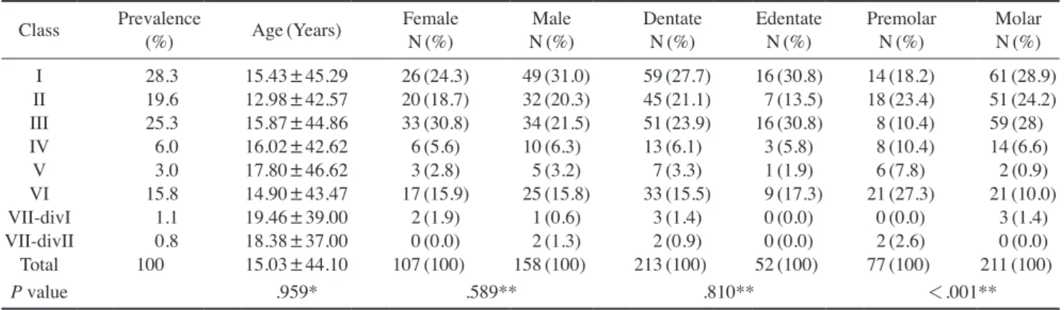
Class I and Class III patterns of maxillary sinus septa had the highest prevalence, which was 28.3%(75) and 25.3%(67), respectively. The lowest prevalence values belonged to Class VII-divI and Class VII-divII, which
Table 1. Classification method modified from Al-Faraje et al.1 to categorize septal patterns
Class Description Images Risk degree(score)
I Single basal perpendicular septum Low risk(0)
II Multiple(2 or more) basal
perpendicular septa Low risk(0)
III Single long partial perpendicular septum that is not limited to the base of the sinus.
Moderate risk if not considered(1)
IV Multiple(2 or more) long partial perpendicular septa that are not limited to the base of the sinus.
High risk and relative contraindication for sinus surgery(2)
V Partial horizontal septum Low risk(0)
VI Complete perpendicular septum that divides the sinus into separate
anatomic cavities Low risk(0)
VII-divI Complete horizontal septum
placed inferiorly Moderate risk if not
considered(1)
VII-divII Complete horizontal septum
placed superiorly Low risk(0)
were 1.1%(3) and 0.8%(2), respectively(Table 2).
Table 2 also shows the mean age and distribution of different patterns of maxillary sinus septa based on sex, dental status, and location. The Class I pattern had the highest frequency in females(31%) and in the dentate group(27.7%). The Class III pattern was more frequent in males(30.8%) and was concurrent with the Class I pattern in the edentate group(30.8%). There was no significant difference in the mean age of the patients with different patterns of maxillary sinus septa.
In this study, there was no significant difference among different patterns of maxillary sinus septa according to sex(P value=.589) or dental status(P=.810)(Table 2).
The most common septal patterns were Class VI in the premolar region(27.3%) and Class I in the molar region (28.9%). Overall, the distribution of sinus septa varied significantly by location(P=.001).
The mean age of the patients with different score rang- es for the risk of sinus membrane perforation are shown in Table 3. Age was not found to be significantly related to the degree of risk of sinus membrane perforation(P=
.193).
The prevalence values of the scores for membrane per- foration risk were 68%(low risk), 26%(moderate risk),
and 6%(high risk). There was also no significant differ- ence between genders(P=.216) or by dental status(P=
.686) in the membrane perforation risk level. Overall, the risk of membrane perforation was higher in molar than premolar regions. However, the risk scores have signif- icant differences between premolar and molar tooth re- gions.(P=.004)(Table 3).
discussion
In this study, the prevalence of the presence of maxil- lary sinus septa was 68.4% in the patients’ CBCT images studied and 54.05% of the maxillary sinuses evaluated.
Van Zyl and van Heerden10 reported a prevalence of 69%
in CT scans of 200 patients with one or more sinus septa and of 56% in the maxillary sinuses evaluated. Rancitelli et al.9 reported 38.1% as the prevalence of maxillary sinus septa in 228 maxillary sinuses in CBCT images.
Nevertheless, in several other studies, the prevalence of maxillary sinus septa in CT scans11-13,15 was reported to be 24%-37%. Gosau et al.16 examined 65 cadaver heads and reported an incidence of sinus septa of 27%. Ella et al.17 studied male cadavers and CT scans of male patients.
In their study, 39% of the maxillary sinuses had sinus
Table 2. The mean age and prevalence of different patterns of maxillary sinus septa based on age, sex, dental group, and location Class Prevalence
(%) Age(Years) Female
N(%) Male
N(%) Dentate
N(%) Edentate
N(%) Premolar
N(%) Molar
N(%) I 28.3 15.43±45.29 26(24.3) 49(31.0) 59(27.7) 16(30.8) 14(18.2) 61(28.9) II 19.6 12.98±42.57 20(18.7) 32(20.3) 45(21.1) 7(13.5) 18(23.4) 51(24.2) III 25.3 15.87±44.86 33(30.8) 34(21.5) 51(23.9) 16(30.8) 8(10.4) 59(28) IV 6.0 16.02±42.62 6(5.6) 10(6.3) 13(6.1) 3(5.8) 8(10.4) 14(6.6)
V 3.0 17.80±46.62 3(2.8) 5(3.2) 7(3.3) 1(1.9) 6(7.8) 2(0.9) VI 15.8 14.90±43.47 17(15.9) 25(15.8) 33(15.5) 9(17.3) 21(27.3) 21(10.0) VII-divI 1.1 19.46±39.00 2(1.9) 1(0.6) 3(1.4) 0(0.0) 0(0.0) 3(1.4) VII-divII 0.8 18.38±37.00 0(0.0) 2(1.3) 2(0.9) 0(0.0) 2(2.6) 0(0.0)
Total 100 15.03±44.10 107(100) 158(100) 213(100) 52(100) 77(100) 211(100)
P value .959* .589** .810** <.001**
*Kruskal-Wallis, **Chi-squared test
Table 3. The prevalence of risk for membrane perforation Risk score Age(Years) Female
N(%) Male
N(%) Dentate
N(%) Edentate
N(%) Premolar
N(%) Molar
N(%)
Low 43.96±14.67 113(71.5) 67(62.6) 147(69.0) 33(63.5) 61(79.2) 136(64.5)
Moderate 44.84±15.92 35(22.2) 34(31.8) 53(24.9) 16(30.8) 8(10.4) 61(28.9)
High 42.62±16.02 10(6.3) 6(5.6) 13(6.1) 3(5.8) 8(10.4) 14(6.6)
Total 44.10±15.03 158(100) 107(100) 213(100) 52(100) 77(100) 211(100)
P value .193* .216** .686** .004**
*Kruskal-Wallis, **Chi-squared test
septa. In other cadaver studies, Rosano et al.18 and Nai- toh et al.19 found the incidence of sinus septa was 37%
and 33.3%, respectively. Their studies had limited sample sizes: 30 and 15 human skulls, respectively. This variabil- ity might be related to the differences in the populations examined, sample sizes, methods of evaluation and in- clusion criteria, minimum septa height, and consideration of the septa as sinus septa. Malkinson et al.14 evaluated the sinus septa during sinus lift surgery. They reported the septa existed in 40% of the cases. Krennmair et al.5 reported a prevalence of 27.7% in 65 clinically examined maxilla and 16% in 200 CT-examined maxillary sinuses.
Shen et al.20 evaluated a large sample size with CT scans obtained from 423 patients. Thirty percent of the patients had sinus septa. However, they used 2.5-mm thick CT images, while the present study used 1-mm thick CBCT images, which could explain the reason for the difference in reported results. Neugebauer et al.21 evaluated CBCT scans of 1,029 patients, and they found septa in 47% of the patients and 33.2% of the sinuses. Koymen et al.22 as- sessed 205 dental CT scans of maxilla. Septa were found in 35.4% of the maxillary sinuses.
In this study, the sinus pattern with the highest preva- lence was the Class I classification. This finding means that this pattern was short enough to not interfere with si- nus lift surgery and a lateral window design. Additionally, the Class VI pattern of septa(long and multiple perpen- dicular septa) had the highest membrane perforation risk and a low prevalence(6%). If this type of septum existed in the sinus, it would interfere with sinus lift surgery.
Thus, the surgeons would have to design 2 separate win- dows.
On the other hand, the risk of sinus membrane perfora- tion was low in 68%; however, in 32%, the estimated risk was medium to high.
We found there was a significant difference between the frequencies of septa presence in molar areas compared to premolar areas. Class III, IV, and VII-div I were consid- ered medium- to high-risk groups relative to the others, and these patterns were in molar rather than premolar ar- eas.According to the results of this study, 26.7% of ex- isting sinus septa were located in the premolar region, and 73.3% were in the molar region. This finding, that is, the higher prevalence of sinus septa in the molar re- gion, was similar to the findings of Malec et al.(66%),11 Rancitelli(70.4%),9 Neugebauer et al.(79.6%),21 Shen et al.(81.44%),22 Park et al.(77.4%),12 Kim et al.(74.5%),13 Lee et al.(77.7%),2 and Velasquez-Plata et al.(76%).15
The molar area in these studies included the middle and posterior regions. However, the molar region must be in- vestigated precisely to find maxillary sinus septa that in- terfere with sinus lift surgery.
There was no significant difference between dentate and edentate groups regarding the presence of different patterns of sinus septa. The prevalence of sinus septa in the dentate group(80.4%) was higher than in the edentate group(19.6%). This finding was similar to those of Shen et al.20 and Velásquez-Plata et al.15 The percentage of si- nus septa in the atrophic alveolar ridge(21.28%) was less than the total non-atrophic and partially atrophic ridge (43.94).20 In contrast, the prevalence of sinus septa in edentulous(atrophic) patients was more than in the den- tate group in Lee et al.,2 Pommer et al.,23 and Krennmair et al.5
In our study, the mean age of patients with maxillary si- nus septa was 44.1±14.7 years old. This finding differed from those of Malec et al.11 (65±15 years in males and 65±15 years in females).
The findings of this study regarding the degree of mem- brane perforation risk and its relationship to the other variables of sex, dental status, and location of sinus sep- ta were not comparable with other studies because other studies not consider similar variables.
This study suggests that maxillofacial surgeons must consider the type of maxillary sinus septal pattern with an accurate examination of radiographs before applying implants and performing sinus lift surgery. Additionally, considering the location of the surgery and the related membrane perforation risk can be helpful to design of a treatment plan with less complications during and after surgery.
In conclusion, according to this study, the molar region is the most frequent region for the presence of maxillary sinus septa. There was a significant difference between the location of sinus septa and the frequency of mem- brane perforation risk. We found a higher prevalence of low perforation risk in the molar region. Additionally, this study found that there was no relationship between dental status and the presence of sinus septa. Sex, age, and den- tal status were not factors related to sinus septal patterns.
Acknowledgments
We would like to thank Fatemeh Salamat(Department of Biostatistics, Guilan University of Medical Sciences, Rasht, Iran) for her help with analysis of the data.
references
1. Al-Faraje L. Surgical complications in oral implantology: eti- ology, prevention, and management. Hanover Park: Quintes- sence; 2011. p. 153-60.
2. Lee WJ, Lee SJ, Kim HS. Analysis of location and prevalence of maxillary sinus septa. J Periodontal Implant Sci 2010; 40:
56-60.
3. Nkenke E, Stelzle F. Clinical outcomes of sinus floor augmen- tation for implant placement using autogenous bone or bone substitutes: a systematic review. Clin Oral Implants Res 2009;
20 Suppl 4: 124-33.
4. van den Bergh JP, ten Bruggenkate CM, Disch FJ, Tuinzing DB. Anatomical aspects of sinus floor elevations. Clin Oral Implants Res 2000; 11: 256-65.
5. Krennmair G, Ulm CW, Lugmayr H, Solar P. The incidence, location, and height of maxillary sinus septa in the edentulous and dentate maxilla. J Oral Maxillofac Surg 1999; 57: 667-72.
6. Underwood AS. An inquiry into the anatomy and pathology of the maxillary sinus. J Anat Physiol 1910; 44: 354-69.
7. Maestre-Ferrín L, Galán-Gil S, Rubio-Serrano M, Peñarro- cha-Diago M, Peñarrocha-Oltra D. Maxillary sinus septa: a systematic review. Med Oral Patol Oral Cir Bucal 2010; 15:
e383-6.
8. Wen SC, Chan HL, Wang HL. Classification and management of antral septa for maxillary sinus augmentation. Int J Peri- odontics Restorative Dent 2013; 33: 509-17.
9. Rancitelli D, Borgonovo AE, Cicciù M, Re D, Rizza F, Frigo AC, et al. Maxillary sinus septa and anatomic correlation with the Schneiderian membrane. J Craniofac Surg 2015; 26: 1394- 10. van Zyl AW, van Heerden WF. A retrospective analysis of 8.
maxillary sinus septa on reformatted computerised tomogra- phy scans. Clin Oral Implants Res 2009; 20: 1398-401.
11. Malec M, Smektala T, Tutak M, Trybek G, Sporniak-Tutak K.
Maxillary sinus septa prevalence and morphology-computed tomography based analysis. Int J Morphol 2015; 33: 144-8.
12. Park YB, Jeon HS, Shim JS, Lee KW, Moon HS. Analysis of the anatomy of the maxillary sinus septum using 3-dimension- al computed tomography. J Oral Maxillofac Surg 2011; 69:
1070-8.
13. Kim MJ, Jung UW, Kim CS, Kim KD, Choi SH, Kim CK, et al. Maxillary sinus septa: prevalence, height, location, and morphology. A reformatted computed tomography scan analy- sis. J Periodontol 2006; 77: 903-8.
14. Malkinson S, Irinakis T. The influence of interfering septa on the incidence of Schneiderian membrane perforations during maxillary sinus elevation surgery: a retrospective study of 52 consecutive lateral window procedures. Oral Surg 2009; 2:
19-25.
15. Velásquez-Plata D, Hovey LR, Peach CC, Alder ME. Maxil- lary sinus septa: a 3-dimensional computerized tomographic scan analysis. Int J Oral Maxillofac Implants 2002; 17: 854- 16. Gosau M, Rink D, Driemel O, Draenert FG. Maxillary sinus 60.
anatomy: a cadaveric study with clinical implications. Anat Rec(Hoboken) 2009; 292: 352-4.
17. Ella B, Noble Rda C, Lauverjat Y, Sédarat C, Zwetyenga N, Siberchicot F, et al. Septa within the sinus: effect on elevation of the sinus floor. Br J Oral Maxillofac Surg 2008; 46: 464-7.
18. Rosano G, Taschieri S, Gaudy JF, Lesmes D, Del Fabbro M.
Maxillary sinus septa: a cadaveric study. J Oral Maxillofac Surg 2010; 68: 1360-4.
19. Naitoh M, Suenaga Y, Kondo S, Gotoh K, Ariji E. Assessment of maxillary sinus septa using cone-beam computed tomog- raphy: etiological consideration. Clin Implant Dent Relat Res 2009; 11 Suppl 1: e52-8.
20. Shen EC, Fu E, Chiu TJ, Chang V, Chiang CY, Tu HP. Prev- alence and location of maxillary sinus septa in the Taiwanese population and relationship to the absence of molars. Clin Oral Implants Res 2012; 23: 741-5.
21. Neugebauer J, Ritter L, Mischkowski RA, Dreiseidler T, Scherer P, Ketterle M, et al. Evaluation of maxillary sinus anatomy by cone-beam CT prior to sinus floor elevation. Int J Oral Maxillofac Implants 2010; 25: 258-65.
22. Koymen R, Gocmen-Mas N, Karacayli U, Ortakoglu K, Ozen T, Yazici AC. Anatomic evaluation of maxillary sinus septa:
surgery and radiology. Clin Anat 2009; 22: 563-70.
23. Pommer B, Ulm C, Lorenzoni M, Palmer R, Watzek G, Zech- ner W. Prevalence, location and morphology of maxillary sinus septa: systematic review and meta-analysis. J Clin Peri- odontol 2012; 39: 769-73.