86
86 THE EWHA MEDICAL JOURNALTHE EWHA MEDICAL JOURNAL
Endotracheal Intubation Using McGrath Videolaryngoscope in Klippel-Feil Syndrome
Ji Seon Chae, Jae Hee Woo, Chi Hyo Kim, Eun Hee Chun, Hee Jung Baik, Min Hee Choi
Department of Anesthesiology and Pain Medicine, Ewha Womans University College of Medicine, Seoul, Korea
Introduction
When a patient has congenital or acquired anatomical defect in head and neck regions, it might be difficult to obtain glottic exposure for endotracheal intubation. Klippel-Feil syndrome is a clinical triad with low posterior hairline, short neck, and lim- ited range of motion in neck [1]. These patients require much attention during anesthesia because of the high probability of neurological damage from cervical spine instability during endo- tracheal intubation and body posture change [2-4].
The algorithm for anticipated difficult tracheal intubation includes supraglottic airway device, awake endotracheal intuba- tion and awake fiberoptic intubation [5]. Traditionally, awake fiberoptic intubation has been suggested as a standard approach.
However, it is associated with possible complications, includ- ing unpleasant experience, hypertension, tachycardia, epistaxis,
over-sedation with resultant hypoxemia, and airway loss [6].
Awake endotracheal intubation with direct laryngoscopy es- sentially requires careful skills with extensive experiences and patient’s cooperation.
Recently, videolaryngoscopes have gained popularity in manag- ing airway in patients with limited neck extension, teeth problem or abnormal airway anatomy. Videolaryngoscopes offer glottic ex- posure, which is superior to that obtained with direct laryngoscopy in patients with anticipated difficult airway as well as in general population [7]. Videolaryngoscopes are designed similar to con- ventional laryngoscopes, enabling clinicians to use them success- fully without special training. Also, videolaryngoscopes can provide an exposure of glottic opening in patients requiring manual in- line stabilization [8]. We report a case of successful endotracheal intubation using a videolaryngoscope in a patient with congenital anatomic defects in the cervical spine from Klippel-Feil syndrome.
Case Report
Ewha Med J 2018;41(4):86-89
https://doi.org/10.12771/emj.2018.41.4.86 eISSN 2234-2591
Patients with Klippel-Feil syndrome require much attention during anesthesia be- cause of congenital abnormalities in head and neck regions and the high probability of neurological damage from cervical spine instability during endotracheal intuba- tion. We report a case of successful endotracheal intubation using a videolaryngo- scope in a patient with Klippel-Feil syndrome who experienced difficult transnasal intubation. (Ewha Med J 2018;41(4):86-89)
Received August 16, 2018 Revised September 27, 2018 Accepted September 28, 2018 Corresponding author Jae Hee Woo
Department of Anesthesiology and Pain Medicine, Ewha Womans University College of Medicine, 1071 Anyangcheon-ro, Yangcheon- gu, Seoul 07985, Korea
Tel: 82-2-2650-5285, Fax: 82-2-2655-2924 E-mail: [email protected]
Key Words
Klippel-Feil syndrome; Laryngoscopes;
Intubation
This is an Open Access article distributed under the terms of the Creative Commons Attribution Non-Commercial License (http://creativecommons.org/licenses/by-nc/4.0) which permits unrestricted non-commercial use, distribution, and reproduction in any medium, provided the original work is properly cited.
87
THE EWHA MEDICAL JOURNAL Klippel-Feil Syndrome and Videolaryngoscope
Case
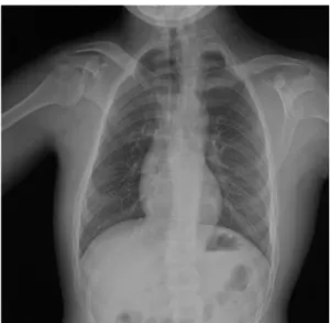
A 22-year-old male patient (147 cm, 45 kg), previously diagnosed to have grade 2 mental retardation, was hospital- ized for tooth extraction under general anesthesia. The patient had a history of surgical repair of ventricular septal defect and patent ductus arteriosus at 2 years of age and a C1–2 fusion for congenital upper spinal deformity and cervical instability at 4 years of age in other hospitals. Six years ago, in 2009, the patient underwent tooth extraction under general anesthesia in our hospital. The patient had facial asymmetry, with maximum mouth opening of 2 cm and thyromental distance of 4 cm, and was confirmed as Mallampati class 4. Chest radiograph revealed no pulmonary abnormality, but higher right scapula and scoliosis with Cobb’s angle of approximately 15o were confirmed (Fig.
1).
To evaluate the cervical spine, flexion and extension radio- graphs were taken. Non-segmentation of C3–6 and segmental instability between C2 and C3 were confirmed by limitations in neck extension and flexion as well as by traces of C1–2 fusion (Fig. 2). Neurosurgical consultation advised to avoid excessive neck extension, flexion, and rotation because of cervical insta- bility and severe limitations in cervical range of motion.
The patient underwent tooth extraction under general anes- thesia in 2009, and the anesthesia record revealed that trans- nasal endotracheal intubation had been tried for 30 minutes and finally failed. Then orotracheal intubation was successfully done by direct laryngoscopy with Schucman blade. It was recorded
that the nasal cavity of the patient was asymmetric and severely bowed. Considering the whole circumstances of this patient, awake orotracheal intubation was planned for the current opera- tion. Because orotracheal intubation was successfully done in previous operation, it was decided to lightly sedate the patient with dexmedetomidine infusion for visual confirmation of glottic exposure with videolaryngoscope. Only when glottic exposure with videolaryngoscope was confirmed, anesthetic agents would be given to the patient for endotracheal intubation.
On the patient’s arrival at the operation room, standard monitors, including electrocardiogram, non-invasive blood pres-
A B Fig. 2. Cervical spine radiographs. Flexion
(A) and extension view (B).
L-PA
Fig. 1. Chest radiograph.
88 THE EWHA MEDICAL JOURNAL Chae JS, et al
sure, and pulse oximetry, were used throughout the surgery.
Midazolam 1 mg was given intravenously and 10% lidocaine were sprayed on the patient’s nasal and oral cavity. Then, dexmedetomidine infusion of 1 μg/kg/hr was initiated. After 10 minutes, the patients showed a sluggish response to light glabellar tap or loud auditory stimulus, indicating that he was moderately sedated. The patient’s neck was placed in neutral position and Cormack-Lehane grade 2 was confirmed using the McGrath (Aircraft Medical, Edinburgh, UK) videolaryngoscope (Fig. 3). Thiopental sodium 250 mg, rocuronium 30 mg, and fentanyl 50 μg were immediately given for induction of general anesthesia, and then endotracheal intubation was performed with a RAE (Ring-Adair-Elwin) tube of internal diameter 6.5 without any difficulty using McGrath videolaryngoscope. Blood pressure and pulse rate were maintained within 30% of the baseline value without a decrease in oxygen saturation through- out the procedure. Surgery was performed for 20 minutes. After 70 minutes of anesthesia, the patient was extubated successfully in the operating room after intravenous injection of glycopyr- rolate 0.4 mg and pyridostigmine 10 mg for reversal of muscle relaxation. At the first day postoperatively, the patient was dis- charged without any complication.
Discussion
Thorough evaluation before anesthesia, including meticulous
review of previous anesthesia records, and thoughtful preparation based on planned anticipation are essential parts of anesthetic management of patient with congenital or acquired anatomical defect in head and neck region.
Awake transnasal endotracheal intubation using fiberoptic bronchoscope has been reportedly chosen for airway manage- ment of patients with Klippel-Feil syndrome, who present with short neck, small mandible and limited range of motion in neck [9]. For our patient, previous attempts of nasal intubation had failed, and then orotracheal intubation by direct laryngoscopy with Shucman blade was successfully done. Considering previous experiences, transnasal intubation could not be a choice for this patient, and orotracheal intubation was only an option for this patient.
Nevertheless, immediate induction of general anesthesia, in- cluding full relaxation with neuromuscular blocking agent, was not a prudent approach for this patient, because unanticipated difficulty in airway maintenance could be met. Furthermore, even a successful previous anesthetic experiences do not guaran- tee to secure the airway in patients with Klippel-Feil syndrome, because cervical fusion becomes progressively worse over time [10]. Therefore, we planned to try an airway evaluation with videolaryngoscope under sedation with dexmedetomidine infu- sion. Fortunately, this exploratory videolaryngoscopy successfully produced glottic exposure of Cormack-Lehane grade 2. Then, orotracheal intubation was immediately done under the general anesthesia, and the neck of this patient was maintained in neu- tral position throughout the whole procedures. As patient coop- eration was not expected because of mental retardation, sedation during exploratory videolaryngoscopy was required even more.
Dexmedetomidine was chosen because it caused less respiratory depression than other sedative-hypnotics.
Videolaryngoscope was employed because it was shown to al- low glottic exposure without stretching the neck in patient with various airway conditions [8]. Video fluoroscopic investigations demonstrated that movement in upper cervical spines was sig- nificantly reduced during endotracheal intubation when videolar- yngoscopes were employed, compared to direct laryngoscopes even in patients simulating in-line stabilization of cervical spine [11,12]. Also, videolaryngoscopes were shown to improve glot- tic exposure and reduce the need for neck extension in patients with restricted neck motion [13,14]. Based on those beneficial effects, we chose videolaryngoscope as the first option for air- Fig. 3. Confirmation of glottic exposure with a videolaryngoscope
before anesthesia induction.
89
THE EWHA MEDICAL JOURNAL Klippel-Feil Syndrome and Videolaryngoscope
way management for patients with limited cervical spine mo- tion.
In the pre-anesthesia airway evaluation, any difficulty in en- dotracheal intubation should be anticipated in consideration of the distance of mouth opening (gap between anterior teeth in maxilla and mandible), the degree of anterior movement of the mandible, the degree of head and neck flexion, the upper lip bite test, the distance between thoracic cartilage and chin, Mal- lampati classification, and mandibular length [15]. The degree of a patient’s cooperation and previous history of endotracheal intubation also should be considered. A medical record of dif- ficult endotracheal intubation should be preserved to inform the physician at the next anesthesia, as well as the patient that the patient has a difficult airway to manage.
In conclusion, pre-anesthesia airway evaluation and consid- eration of appropriate plans are essential for anesthetic man- agement in a patient with Klippel-Feil syndrome. Anesthesia induction after assessment of glottic exposure using a vide- olaryngoscope under sedation can be a useful option in patients with anticipated difficult airway, requiring in-line stabilization of cervical spine.
References
1. Samartzis DD, Herman J, Lubicky JP, Shen FH. Classification of congenitally fused cervical patterns in Klippel-Feil patients: epi- demiology and role in the development of cervical spine-related symptoms. Spine (Phila Pa 1976) 2006;31:E798-E804.
2. Harley EH, Collins MD. Neurologic sequelae secondary to atlan- toaxial instability in Down syndrome. Implications in otolaryn- gologic surgery. Arch Otolaryngol Head Neck Surg 1994;120:159- 165.
3. Lennarson PJ, Smith DW, Sawin PD, Todd MM, Sato Y, Traynelis VC. Cervical spinal motion during intubation: efficacy of stabili- zation maneuvers in the setting of complete segmental instabil- ity. J Neurosurg 2001;94(2 Suppl):265-270.
4. Nagib MG, Maxwell RE, Chou SN. Identification and manage-
ment of high-risk patients with Klippel-Feil syndrome. J Neuro- surg 1984;61:523-530.
5. Law JA, Broemling N, Cooper RM, Drolet P, Duggan LV, Gries- dale DE, et al. The difficult airway with recommendations for management: part 2. The anticipated difficult airway. Can J An- aesth 2013;60:1119-1138.
6. Cho SY, Woo JH, Kim YJ, Chun EH, Han JI, Kim DY, et al. Airway management in patients with deep neck infections: a retrospec- tive analysis. Medicine (Baltimore) 2016;95:e4125.
7. Stroumpoulis K, Pagoulatou A, Violari M, Ikonomou I, Kalantzi N, Kastrinaki K, et al. Videolaryngoscopy in the management of the difficult airway: a comparison with the Macintosh blade. Eur J Anaesthesiol 2009;26:218-222.
8. Niforopoulou P, Pantazopoulos I, Demestiha T, Koudouna E, Xanthos T. Video-laryngoscopes in the adult airway manage- ment: a topical review of the literature. Acta Anaesthesiol Scand 2010;54:1050-1061.
9. Hase Y, Kamekura N, Fujisawa T, Fukushima K. Repeated an- esthetic management for a patient with Klippel-Feil syndrome.
Anesth Prog 2014;61:103-106.
10. Thompson E, Haan E, Sheffield L. Autosomal dominant Klippel- Feil anomaly with cleft palate. Clin Dysmorphol 1998;7:11-15.
11. Maruyama K, Yamada T, Kawakami R, Hara K. Randomized cross-over comparison of cervical-spine motion with the Air- Way Scope or Macintosh laryngoscope with in-line stabilization:
a video-fluoroscopic study. Br J Anaesth 2008;101:563-567.
12. Maruyama K, Yamada T, Kawakami R, Kamata T, Yokochi M, Hara K. Upper cervical spine movement during intubation:
fluoroscopic comparison of the AirWay Scope, McCoy laryngo- scope, and Macintosh laryngoscope. Br J Anaesth 2008;100:120- 124.
13. Malik MA, Maharaj CH, Harte BH, Laffey JG. Comparison of Macintosh, Truview EVO2, Glidescope, and Airwayscope laryn- goscope use in patients with cervical spine immobilization. Br J Anaesth 2008;101:723-730.
14. Komatsu R, Kamata K, Hoshi I, Sessler DI, Ozaki M. Airway scope and gum elastic bougie with Macintosh laryngoscope for tracheal intubation in patients with simulated restricted neck mobility. Br J Anaesth 2008;101:863-869.
15. Khan ZH, Maleki A, Makarem J, Mohammadi M, Khan RH, Zandieh A. A comparison of the upper lip bite test with hyomen- tal/thyrosternal distances and mandible length in predicting difficulty in intubation: a prospective study. Indian J Anaesth 2011;55:43-46.