Transl Clin Pharmacol
TCP
2018;26(3):103-110https://doi.org/10.12793/tcp.2018.26.3.103
Copyright © 2018 Yoon-Keun Kim
It is identical to the Creative Commons Attribution Non-Commercial License (http://creativecommons.org/licenses/by-nc/3.0/).
This paper meets the requirement of KS X ISO 9706, ISO 9706-1994 and ANSI/NISO Z.39.48-1992 (Permanence of Paper).
Introduction
Interest in extracellular vesicles (EVs) has dramatically in- creased over the course of three decades as their physiological importance and promising biomedical applications have been realized. Upon the initial discovery that eukaryotic cells release bi-layered vesicles into the extracellular environment in 1983, their function was not well understood and EVs were written off as the cellular equivalent of a garbage disposal.[1] However, extensive research conducted over the past several decades have revealed that EVs are expressed in prokaryotes, eukaryotes, and archaea at varying levels of environmental stress. This evi- dence suggests that EV secretion is an evolutionarily conserved, necessary function in cells across all domains of life.[2] While nomenclature confusion has previously been an issue in the field, the term extracellular vesicle is the general term used for
all lipid-bilayer encased spherical nanoparticles that transport protein, lipid and nucleic acid cargo from the cell into the extra- cellular milieu. The role of EVs in intercellular communication, long-distance transport of active biomolecules, receptor-medi- ated host cell entry, immune modulation, disease pathogenesis, and a variety of biomedical applications including bioengi- neered vaccine adjuvants and diagnostic biomarkers have be- come a research topic of great interest in the past decade.[3,4]
The functional significance of the commensal bacteria living in and on our bodies, or microbiota, has similarly undergone a reversal of favor in the scientific community since the turn of the century. While it has been well-established that communi- ties of bacteria naturally co-exist within our bodies, their rela- tionship with their human host was regarded as largely com- mensal or potentially pathogenic. Appreciation of the essential symbiotic role microbiota plays in health has dramatically increased since 2001 when Nobel Laureate Joshua Lederberg famously coined the term microbiome as it is currently defined:
the collection of genomes contributed by all microorganisms present in an ecosystem.[5] Since then, an explosion of research has revealed that the human microbiome has a multitude of es- sential functions in host health including immune modulation, nutrient metabolism, maintenance of the intestinal barrier, and
Reviewer
This article was invited and reviewed by the editors of TCP.
Microbe-derived extracellular vesicles as a smart drug delivery system
Jinho Yang
†, Eun Kyoung Kim
†, Andrea McDowell
†and Yoon-Keun Kim*
Institute of MD Healthcare Inc., Seoul 03923, Republic of Korea
*Correspondence: Y. K. Kim; Tel: +82-70-7812-8065, Fax: +82-2-2655-0768, E-mail: ykkim@mdhc.kr
†These authors contributed equally to this work.
The human microbiome is known to play an essential role in influencing host health. Extracellular vesicles (EVs) have also been reported to act on a variety of signaling pathways, distally transport cellular components such as proteins, lipids, and nucleic acid, and have immunomodulatory effects.
Here we shall review the current understanding of the intersectionality of the human microbiome and EVs in the emerging field of microbiota-derived EVs and their pharmacological potential. Mi- crobes secrete several classes of EVs: outer membrane vesicles (OMVs), membrane vesicles (MVs), and apoptotic bodies. EV biogenesis is unique to each cell and regulated by sophisticated signaling pathways. EVs are primarily composed of lipids, proteins, nucleic acids, and recent evidence sug- gests they may also carry metabolites. These components interact with host cells and control various cellular processes by transferring their constituents. The pharmacological potential of microbiome- derived EVs as vaccine candidates, biomarkers, and a smart drug delivery system is a promising area of future research. Therefore, it is necessary to elucidate in detail the mechanisms of microbi- ome-derived EV action in host health in a multi-disciplinary manner.
Keywords
Drug delivery system, Extracellular vesicles, Microbiome, Pharmabiotics pISSN: 2289-0882 eISSN: 2383-5427
TCP
Transl Clin PharmacolrEViEw
TCP
Transl Clin PharmacolMicrobe-derived extracellular vesicles
protection from pathogen colonization.[6-8]
As advancements in both EV and microbiome research have dramatically increased in the past decade, the intersectionality of the microbiome and EVs is emerging as a promising research avenue. Previously, only gram-negative bacteria were thought to produce EVs. However, it was recently determined that gram- positive bacteria also release ectosomes, a class of EVs that are packaged and released at the cellular membrane as cells grow.[9]
Furthermore, mounting evidence suggests that single-cell bac- teria as well as multicellular eukaryotic organisms undergo pro- grammed cell death, or apoptosis.[10] This introduces another layer of complexity to bacterial EV secretion as cells that un- dergo apoptosis release another class of EVs, apoptotic bodies, which carry cellular cargo from cells undergoing programmed cell death into the extracellular environment. Through further exploration of the emerging field of microbe-derived extracel- lular vesicles, researchers can better understand the complex in- terplay of host-microbiota interactions and develop innovative pharmacological health solutions using microbe-derived EVs as a smart drug delivery system.
Why microbiome-derived EVs?
EVs are released from all three domains of life including our own eukaryotic human cells, so why is special interest war- ranted to EVs originating from our commensal bacteria? Three decades ago the foundation of human genetics was based on the assumption that the vast, complex biological functions of the human body were carried out primarily by the genetic information contained in our own cells. Thus, launched the groundbreaking Human Genome Project in 1990 which sought to sequence and map the entire human genome in order to elucidate our genetic makeup. Early estimates of the number of genes contained in the human genome ranged from 50,000 upwards to 140,000. However, at the HGP’s completion in 2003 it was revealed that the human genome contained drastically fewer genes than previously estimated. At present, the human genome is estimated to encode approximately 20,500 genes, barely surpassing the genetic content of Caenorhabditis elegans, a 1 mm roundworm.[11,12]
While scientists were left scratching their heads over the unex- pectedly low number of protein-encoding genes in the human genome, interest in understanding the genetic contributions of the commensal microorganisms co-existing in and on our bodies, the microbiota, began to rise. The human microbiota was generally believed to outnumber human cells by a factor of 10, so logically the next step to uncover the missing piece of the genetic puzzle making up our bodies was to launch the Hu- man Microbiome Project (HMP) in 2007. The HMP conducted metagenomic sequencing, a culture independent methodology that selectively targets the conserved 16s ribosomal RNA region of bacteria, to identify up to the species level all the bacteria composing the microbial flora of healthy people at multiple body sites.[13] Whole genome shotgun sequencing and gene
mapping further elucidated the functional characteristics of core bacterial strains in our microbiota.
The HMP and other investigations around the world have laid the foundation for an ever-increasing understanding of our microbiome. Though it was commonly believed bacterial cells outnumber our own by a factor of 10, it now estimated to be a more modest factor of 1.1. Rather than raw cell count, the critical way microbiota outnumber their human counterparts is by their genetic contributions. It is estimated that on average 1,000 different bacterial species inhabit a healthy human host and that each species encodes roughly 2,000 genes.[14] Based on these currently accepted approximations, our microbiome yields approximately 2,000,000 genes, vastly outnumbering the 20,500 genes contributed by our own human genome. Thus, the human body can be viewed as a supraorganism comprised of trillions of both human and microbial cells shaped by 99% mi- crobial and 1% human genomic content.
The HGP and HMP have shown that the human genome com- prises less than 1% of the total genetic material present in our body, highlighting the importance of understanding how the composition and activity of the other 99% contributed by our microbiota impacts human health and disease. Imbalances in commensal microbial communities have been associated with a wide variety of diseases including obesity, diabetes, cancer, de- pression, anxiety, asthma, atopic dermatitis, nonalcoholic fatty liver disease (NAFLD), and Irritable Bowel Syndrome (IBS).[15- 17] As interest continues to shift from microbial composition to functionality, EVs derived from these bacterial communities have great potential as therapeutic and diagnostic tools to better understand the complex interactions between commensal mi- crobial communities and the host.
EV biogenesis
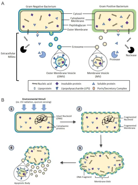
The biogenesis of EVs is a very tightly regulated process gov- erned by multiple signaling molecules and begins with receptor activation unique for each cell type.[18] Though eukaryotic EV biogenesis is well characterized, the mechanisms of bacterial EV biogenesis have only recently begun to be elucidated.[19] Here we will describe the current understanding of the biogenesis of ectosomes, also called shedding vesicles, in gram-negative and gram-positive bacteria as well as bacterial release of apoptotic bodies as summarized in Figure 1. Additionally, we will briefly discuss the well-defined mechanisms underlying eukaryotic exosome release that can offer insights into the process of EV biogenesis.
Gram-negative bacteria: Outer Membrane Vesicles (OMV) Gram-negative bacteria produce ectosomes, known as outer membrane vesicles (OMVs), that contain periplasmic con- stituents including proteins, lipoproteins, phospholipids, and lipopolysaccharide (LPS).[2] The first step in OMV formation is outward bulging of the outer membrane (OM). Links between the OM and the peptidoglycan are lost, either by movement
TCP
Transl Clin Pharmacol
Jinho Yang, et al.
of linking protein or by breaking the connection directly in various areas. This process can result in the incorporation of peptidoglycan fragments and parts of OM-peptidoglycan bridg- ing proteins in the OMV.[20] However, simply removing OM- peptidoglycan links cannot sufficiently explain the mechanism of OMV formation. Overexpressed and misfolded periplasmic proteins can cause budding and specific proteins are enriched/
excluded in OMVs, suggesting a more sophisticated biogenesis pathway. Gathering of periplasmic proteins as well as accumu- lation of curvature-inducing OM proteins could induce addi-
tional budding events.[20,21]
It is often proposed that OMVs are released from the bacte- rial cell when the bud grows to the point at which membrane curvature forces separation. Many membrane fission processes are energy-dependent, but the gram-negative bacterial envelope does not have a direct energy source such as ATP or NADPH, so it is not clear what would drive this fission. One possibil- ity is that energy is provided from the cytoplasm via an inner membrane conduit. Alternatively, the required energy could be stored by the folding of membrane-associated proteins and re-
Figure 1. Proposed bacterial extracellular vesicle secretion. Bacteria ubiquitously release extracellular vesicles (EVs) into the extracellular milieu roughly 10-300 nm in diameter. EVs are composed of a lipid bilayer containing soluble and insoluble proteins, nucleic acids, and lipids. The lipid bilayer encasing the vesicular contents confers protection from extracellular degradation by nucleases and proteases, allowing distal transfer of EV cargo. (A) Gram negative and positive bacteria secrete ectosomes, or outer membrane vesicles (OMV) and membrane vesicles (MV), respectively.
OMV biogenesis through budding of the outer membrane is well characterized, however the mechanism of MV biogenesis through the thick pepti- doglycan cell wall of gram positive bacteria is not yet fully understood. (B) Bacterial cells also undergo apoptosis, a conserved self-destruct mecha- nism instigated by environmental stimuli such as quorum sensing or UV damage. After the apoptotic pathway is initiated, nucleoid DNA is degraded and subsequently fragmented, followed by cell membrane fragmentation. Finally, blebbing of the cell membrane occurs, resulting in release of bac- terial apoptotic bodies containing bacterial cellular components.
A
B
TCP
Transl Clin PharmacolMicrobe-derived extracellular vesicles
leased by their conformational changes, as occurs for the mem- brane fusion step of many viruses.[20,22]
Gram-positive bacteria: Membrane Vesicles (MV)
Unlike gram-negative bacteria, gram-positive bacteria lack an outer membrane and have a much thicker peptidoglycan cell wall outside of the cell membrane, which led to the initial assumption that gram-positive bacteria could not release EVs.
However, increasing evidence has shown that many species of gram positive bacteria release EVs, or membrane vesicles.[9,23]
Currently, the specific mechanisms behind the release of EVs in gram-positive bacteria are not fully understood, but support for three exclusive hypotheses exists in scientific literature.
EVs may be forced through the cell wall by turgor pressure after release from the plasma membrane.[24] Alternatively, cell wall-modifying enzymes released with EVs may facilitate a loosening of the wall to enable EV release. Such a mechanism may be widespread, as EVs from the Gram-positive bacterium Staphylococcus aureus carry peptidoglycan-degrading enzymes, such as Sle1, that can manipulate the thick Gram-positive pep- tidoglycan cell wall.[9] Finally, EVs may be actively transited through channels. In other words, proteins channels or struc- tural cables may guide EVs to the extracellular environment.[24]
Microbial apoptotic bodies
Another development in the field of microbial membrane dy- namics is the recent attention given to prokaryotic programmed cell death (PCD), or apoptosis. It was originally presumed that possession of a genetically coded cellular self-destruct mecha- nism would confer no evolutionary advantage to single-celled organisms. However, recent evidence suggests that bacteria undergo apoptosis in an altruistic manner for the benefit of the
entire colony rather than the individual cell in order to respond to environmental stress, biofilm formation, and genetic trans- formation.[25] Once PCD signal pathways are activated, DNA damage and fragmentation are induced, leading to blebbing at the cell membrane, which results in cell fragmentation and the release of apoptotic bodies. These apoptotic bodies contain frag- ments of cellular DNA, RNA, proteins, and other cellular com- ponents.[22] While in-depth research of eukaryotic apoptosis has enabled a detailed understanding of the mechanisms of eukaryotic autolysis and subsequent release of apoptotic bodies, further research and studies must be conducted to understand the role of apoptotic body release from our microbiota in im- mune system modulation and intercellular signaling.
Eukaryotic cell-derived exosomes
Briefly, we will discuss the formation and release of exosomes, a class of EV released in all eukaryotic cells. Exosomes, while not present in microbiota, are a well-characterized EV class that offer insight into cellular mechanisms involved in EV biogenesis that may be conserved in other EV classes, such as ectosomes.
Exosomes biogenesis begins with the characteristic formation of intraluminal vesicles (ILVs) transported in multivesicular bodies (MVBs).[26] The best-described mechanism for forma- tion of MVBs and ILVs is carried out by the endosomal sorting complex required for transport (ESCRT). ESCRT machinery is formed at vesicle budding sites at the cell membrane through recruitment of four cytosolic protein complexes. The ESCRT-0 complex recognizes and sequesters ubiquitin-modified trans- membrane proteins in the endosomal membrane. The ESCRT-I and -II complexes are believed to be responsible for membrane budding containing the sorted cargo and ESCRT-III compo- nents drive vesicle scission.[27,28] ESCRT vesicular bodies are
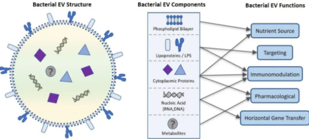
Figure 2. Proposed bacterial extracellular vesicle composition and related function. Bacterial EVs are enclosed in a phospholipid bilayer originating from the bacterial membrane that along with lipo- and cytosolic proteins can be used as a nutrient source by targeted cells. Cytoplasmic proteins contained within the EV also have roles in immunomodulation and potential pharmacological applications. Lipoproteins and LPS embedded in the phospholipid bilayer moderate site-specific targeting of the EV for intra- and intercellular communication and EV constituent delivery. Nucleic acids including sRNA, mRNA, tRNA, rRNA, and DNA are transported in bacterial EVs, further contributing to immunomodulation as well as horizontal gene transfer. Recent evidence that bacterial EVs are metabolically active suggest that bacterial EV may also contain metabolites, increasing the pharmacological potential of bacterial EVs.
TCP
Transl Clin Pharmacol
Jinho Yang, et al.
released from the cytosolic space by a fission event involving a ring of proteins at the bud-membrane interface. This protein ring complex constricts the neck of the bud by forming increas- ingly smaller rings and finally pinches the budding vesicle from the membrane to be released into the extracellular milieu.
[20,29] However, some evidence suggests that MVBs and ILVs can form in the absence of ESCRT.[30,31] Therefore, MVBs and their ILVs can be formed by both ESCRT-dependent and -inde- pendent mechanisms related to the cargo that is sorted within a given cell.[20]
Microbial EV components and functions
Despite their nanosized dimensions, microbial EVs contain a variety of functional components including microbe-derived lipids, luminal and membrane proteins, nucleic acid (DNA, siRNA, mRNA, tRNA, and sRNA), and possibly metabolites.
[3,20,24] After these components are released extracellularly within a spherical phospholipid bilayer, they have a variety of functional roles in host health as outlined in Figure 2. Here we will discuss these functions including drug delivery, targeting, immunomodulation, and gene transfer as they relate to the pharmacological potential of microbial EVs.
Role of lipids in microbial EVs
Drug delivery vehicle
The bilayered lipids of OMVs include mainly lipopolysaccha- ride (LPS) and outer membrane lipids.[32] A number of lipid vesicles called liposomes are incorporated in mammalian cells cultured in vitro with no observable cytotoxic effect.[33] Due to the variety of glycolipids and glycoproteins inserted into liposo- mal vesicles, uptake of vesicles of defined composition by cells offers a potential to modify cellular composition and to intro- duce biological activity of molecules into cells.[33]
EVs can be used to deliver a variety of cargo including cyclic nucleotides, enzymes, sugars, and anti-tumor drugs.[34-36]
Recently, nano or micro particles, such as virus-like particles and liposomal vesicles, are under investigation for their use as a vaccine delivery system.[37,38] Considering this evidence, EVs have notable advantages as pharmacological vehicles. Native OMVs have the immunogenic capacity to carry a wide spec- trum of endogenous antigens, and the natural self-adjuvanticity exerted by toll-like receptor (TLR) agonists, such as LPS.[39]
However, to use OMVs as vehicles, safety concerns about the inclusion of endotoxic LPS must be resolved due to the po- tential for excessive secretion of proinflammatory cytokines instigated by LPS.[40] Many studies have explored the potential use of vesicles as a drug delivery vehicle such as Kim et. al. who successfully modified OMVs to be utilized as multifunctional vaccine delivery vehicles.[39]
Specific targeting
Regardless of the exact lipid composition of EVs, the lipid
bilayer appears to be adapted to the EV’s target environment.
pH may differ among target tissues, and the vesicle needs to be rigid enough to maintain in these environments to func- tion optimally.[41] In addition, carbohydrate cell membrane determinants play a significant role in intercellular recognition processes. The carbohydrate recognition systems include the receptors for glycoprotein.[42] The existence of these receptors suggests that it should be possible to control the tissue distribu- tion and cellular uptake of phospholipid vesicles by attaching appropriate carbohydrate determinants to the vesicle surface.
[43,44] The modification of the vesicle surface with specific synthetic glycolipids has been shown that the tissue distribution and stability of these vesicles are significantly affected in mice.
[45,46] The surface properties of vesicles can be changed by size, surface charge, permeability, and the surface ligand groups.
[47] The physical state of lipids is important in determining the pathways by which lipid vesicles are incorporated into cultured cells. Thus, the method manipulating the lipid composition of vesicles have a potential to achieve some degree of targeting of vesicles and their contents to specific regions of the cell.[33]
Role of proteins in microbial EVs
Although lipids are accepted to play important roles in EVs functions, proteins are indispensable. Such proteins can play significant roles in regulatory processes, cellular responses, host- microbe interactions and targeting.[41] According to a variety of proteomic studies, a number of proteins are detected in mi- crobial Evs.[10,48] EV proteins are mainly located in either the membrane or luminal space and are understood to potentially function in immunity, targeting, and pathogenicity.
Membrane proteins
Outer membrane proteins mainly operate in flagellum assem- bly, pore formation, transport of specific substrates, and outer membrane stabilization. Additionally, various membrane pro- teins have been shown to be important for antibiotic resistance, proteolysis, and host-bacteria interaction.[32] For example, OprM is required for antibiotic efflux,[49] PonA is involved in β-lactam resistance,[50] and LasA is a protease virulence factor related to chronic lung infections such as cystic fibrosis.[51] In addition, EVs released by bacteria, such as Pseudomonas aeru- ginosa and Staphylococcus aureus, contain a variety of virulence factors that can stimulate the host immune system.[10,48]
Membrane proteins also play a critical role in targeting of the EV. The carbohydrate cell membrane recognition systems include the receptors for galactose-terminated glycoproteins in hepatocytes, 6-phosphomannosyl containing glycoproteins in fibroblasts, and mannose terminated glycoproteins in mac- rophages.[52-54] These membrane glycoprotein receptors are crucial for specific cell-targeting of bacterial EVs.
Luminal proteins
Proteins are not only localized in EV membranes, but also
TCP
Transl Clin PharmacolMicrobe-derived extracellular vesicles
inside the EV lumen. For example, α-hemolysin was shown to be localized in the lumen of EVs released by Staphylococcus aureus.[55] α-hemolysin is a clinically relevant toxin which kills many types of cells including keratinocytes[56] and has been reported to be associated with atopic dermatitis severity.[57,58]
In general, secreted soluble toxins are neutralized and lose their activity by engaging the host immune system.[21] EVs may of- fer additional protection to bacterial toxins by enveloping them in a cell membrane compared to soluble toxins. Furthermore, EV-associated α-hemolysin induces keratinocyte death more readily than soluble α-hemolysin. In addition, EV-associated α-hemolysin induces necrotic cell death by facilitating toxin entry into the cytoplasm of keratinocytes, while soluble α-hemolysin induces keratinocyte death via apoptosis. Thus, α-hemolysin delivered in the EV lumen enhances keratinocyte death and evasion of host immune defenses, illustrating the clinical significance of EVs in luminal protein delivery.[55]
Role of nucleic acid in microbial EVs
A diverse composition of genetic material is found in micro- bial Evs.[26] Most research has focused on the nucleic acid con- tents (DNA, RNA) of EVs in human and eukaryotic cell EVs. In contrast, much less is known regarding the properties and func- tions of the nucleic acid components of bacterial Evs.[59,60]
It has been well established that microbial EVs carry rRNA, tRNA, mRNA, and small RNA (sRNAs).[59,61] The majority of RNAs in EVs are shorter than 250 nucleotides in length and are therefore classified as sRNAs. It is interesting to note that many RNAs in EVs are noncoding, with prokaryotic RNAs in EVs fre- quently deriving from intergenic or nonprotein-coding regions.
[59,62] In addition, a large proportion of sRNAs in EVs are from uncharacterized intergenic regions and may also modulate gene expression in target cells.[59,63] Recent evidence suggests that bacterial OMVs also carry bacterial DNA mainly at the OMV surface, though their functional role and composition are not yet fully understood.[60] The presence of bacterial DNA in secreted EVs makes it possible to target bacteria-derived EVs systemically as potential biomarkers of disease through metage- nomic analysis of the conserved 16S rDNA region of bacteria.
[64]
Horizontal gene transfer (HGT)
The presence of RNA in EVs raises considerable interest in the use of microbial EVs as novel mediators of horizontal gene transfer (HGT). RNAs in EVs may attenuate or enhance the ex- pression of specific genes in the target cell.[59,65]
To affect gene expression in target cells directly, RNAs in EVs enter the cytoplasm by fusion of the EV with the target cell membrane. Once in the cytoplasm, microbial RNAs might interact with or be modified by target cell factors, such as the eukaryotic RNA-induced silencing complex (RISC) or RNases.
Complementarity between noncoding microbial sRNA in EVs and target cell RNAs might lead to the attenuation or enhance-
ment of gene expression by influencing transcription, transla- tion, or mRNA processing or stability. The mechanisms in- volved are likely to vary greatly depending on the specific RNA in EV and the presence of specific target cell factors. mRNA in EV might be translated to produce microbial proteins in target cells.[59]
Immunomodulatory effects
Host immune cells and other somatic cells express a variety of pattern-recognition receptors (PRRs) and other conserved pathogen-associated molecular patterns (PAMPs).[59] Pattern recognition receptors (PRRs) include Toll-like receptors (TLRs), nucleotide-binding oligomerization domain-like receptors (NLRs), retinoid acid inducible gene I (RIG-I)-like receptors (RLRs), and C-type lectin-binding domain receptors. It is rec- ognized that microbial nucleic acids, DNA and RNA, are sensed by either endosomal or cytoplasmic receptors in host cells.[66]
EVs from several bacterial species trigger PRR signaling to promote protective immunity, a property that has been har- nessed for vaccine development.[59] For instance, Staphylococ- cus aureus EV has shown potential as a vaccine candidate by inducing innate immunity dependent on TLR2 signaling in host cells.[67] Most research regarding bacterial EVs and im- mune modulation as described above has focused on immune regulation by LPS and proteins, which are constituents of bacte- rial EVs. However, currently there is no available literature that has verified the influence of nucleic acids contained in bacterial EVs on immunomodulation. Thus, further studies are needed to determine the role of nucleic acids associated with bacterial EVs in host immune modulation.
Metabolites in microbial EVs?
Recently, it has been demonstrated that EVs are metaboli- cally active and they present L-asparaginase activity in neural stem cells (NSCs). Although emerging evidence suggests that EVs can act as metabolic regulators, characterization of EVs in metabolic activity is a controversial issue.[68,69] In addition, little is known about the existence and function of metabolites in microbial EVs. Gut microbiota are known to produce a va- riety of metabolites such as N-acyl amides that can influence and regulate host physiology.[70] Thus, it is necessary to further investigate and characterize the presence and activity of me- tabolites in microbiota-derived EVs in order to harness their potential metabolic functions.
Conclusion and perspectives
In this review, we have elaborated on recent advancements in microbial EV research including a brief background on the intersectionality of EVs and the human microbiome, the biogenesis of bacterial EVs, the structure and function of their cargo, as well as future biomedical applications of microbiota- derived EVs. Further studies must be undertaken to thoroughly understand the mechanisms underlying the biogenesis of bacte-
TCP
Transl Clin Pharmacol
Jinho Yang, et al.
rial EVs, particularly MVs in gram-positive bacteria and bacte- rial apoptotic bodies. EV applications in medicine, especially as biomarkers and pharmabiotics, is an emerging field. Future biomedical applications of microbiota-derived EVs include bio- markers for disease diagnosis and prevention, drug delivery sys- tems for beneficial proteins and miRNAs, as well as adjuvants for vaccine development. As the underlying mechanisms of bac- terial EV biogenesis and subsequent physiological consequences to the human host become elucidated, their pharmacological potential can be fully realized.
Acknowledgements
This work was supported by the National Research Foundation of Korea(NRF) grant funded by the Korea government(MSIT) (NRF-20163A9B6901516 and NRF-2017M3A9F3047497).
Conflict of interest
- Authors: Nothing to declare - Reviewers: Nothing to declare - Editors: Nothing to declare
References
1. H Rashed M, Bayraktar E, K Helal G, Abd-Ellah MF, Amero P, Chavez- Reyes A, et al. Exosomes: From Garbage Bins to Promising Therapeutic Targets. Int J Mol Sci 2017;18: E538. doi: 10.3390/ijms18030538.
2. Deatherage BL, Cookson BT. Membrane vesicle release in bacteria, eu- karyotes, and archaea: a conserved yet underappreciated aspect of mi- crobial life. Infect Immun 2012;80:1948-1957. doi: 10.1128/IAI.06014-11.
3. Zaborowski MP, Balaj L, Breakefield XO, Lai CP. Extracellular vesicles:
Composition, biological relevance, and methods of study. Bioscience 2015;65:783-797.
4. McConnell MJ. Extracellular vesicles and immune modulation. Immunol Cell Biol 2018;96:681-682.
5. Lederberg J, Mccray AT. "Ome Sweet'Omics--A Genealogical Treasury of Words". The Scientist 2001:8.
6. Young VB. The role of the microbiome in human health and disease: an introduction for clinicians. BMJ 2017;356:j831. doi: 10.1136/bmj.j831.
7. Takiishi T, Fenero CIM, Câmara NOS. Intestinal barrier and gut microbio- ta: Shaping our immune responses throughout life. Tissue Barriers 2017;5:e1373208. doi: 10.1080/21688370.2017.1373208.
8. Rowland I, Gibson G, Heinken A, Scott K, Swann J, Thiele I, et al. Gut microbiota functions: metabolism of nutrients and other food components.
Eur J Nutr 2018;57:1-24. doi: 10.1007/s00394-017-1445-8.
9. Lee EY, Choi DY, Kim DK, Kim JW, Park JO, Kim S, et al. Gram-positive bac- teria produce membrane vesicles: proteomics-based characterization of Staphylococcus aureus-derived membrane vesicles. Proteomics 2009;9:5425- 5436. doi: 10.1002/pmic.200900338.
10. Peeters SH, de Jonge MI. For the greater good: Programmed cell death in bacterial communities. Microbiol Res 2018;207:161-169. doi: 10.1016/j.
micres.2017.11.016.
11. National Human Genome Research Institute. An Overview of the Human Genome Project. https://www.genome.gov/12011238/an-overview-of-the- human-genome-project/Accessed 03 Oct 2018
12. Hillier LW, Coulson A, Murray JI, Bao Z, Sulston JE, Waterston RH. Ge- nomics in C. elegans: so many genes, such a little worm. Genome Res 2005;15:1651-1660.
13. Lloyd-Price J, Mahurkar A, Rahnavard G, Crabtree J, Orvis J, Hall AB, et al. Strains, functions and dynamics in the expanded Human Microbiome Project. Nature 2017;550:61-66. doi: 10.1038/nature23889.
14. Gilbert JA, Blaser MJ, Caporaso JG, Jansson JK, Lynch SV, Knight R. Current understanding of the human microbiome. Nature Med 2018;24:392-400.
15. Wang B, Yao M, Lv L, Ling Z, Li L. The Human Microbiota in Health and Disease. Engineering 2017;3:71-82.
16. Carabotti M, Scirocco A, Maselli MA, Severi C. The gut-brain axis: interac- tions between enteric microbiota, central and enteric nervous systems.
Ann Gastroenterol 2015;28:203-209.
17. Huang YJ, Marsland BJ, Bunyavanich S, O'Mahony L, Leung DY, Muraro A, et al. The microbiome in allergic disease: Current understanding and future opportunities-2017 PRACTALL document of the American Acade- my of Allergy, Asthma & Immunology and the European Academy of Al- lergy and Clinical Immunology. J Allergy Clin Immunol 2017;139:1099- 1110. doi: 10.1016/j.jaci.2017.02.007.
18. Azmi AS, Bao B, Sarkar FH. Exosomes in cancer development, metasta- sis, and drug resistance: a comprehensive review. Cancer Metastasis Rev 2013;32:623-642. doi: 10.1007/s10555-013-9441-9.
19. Cocucci E, Meldolesi J. Ectosome and exosomes: shedding the confusion between extracellular vesicles. Trends Cell Biol 2015;6:364-372. doi:
10.1016/j.tcb.2015.01.004.
20. Kulp A, Kuehn MJ. Biological functions and biogenesis of secreted bacte- rial outer membrane vesicles. Annu Rev Microbiol 2010; 64:163-184. doi:
10.1146/annurev.micro.091208.073413.
21. McBroom AJ, Kuehn MJ. Release of outer membrane vesicles by Gram- negative bacteria is a novel envelope stress response. Mol Microbiol 2007; 63:545-558.
22. Akers JC, Gonda D, Kim R, Carter BS, Chen CC. Biogenesis of extracel- lular vesicles (EV): exosomes, microvesicles, retrovirus-like vesicles, and apoptotic bodies. J Neurooncol 2013;113:1-11. doi: 10.1007/s11060-013- 1084-8.
23. Rivera J, Cordero RJ, Nakouzi AS, Frases S, Nicola A, Casadevall A.
Bacillus anthracis produces membrane-derived vesicles containing bio- logically active toxins. Proc Natl Acad Sci U S A 2010;107:19002-19007.
doi: 10.1073/pnas.1008843107.
24. Brown L, Wolf JM, Prados-Rosales R, Casadevall A. Through the wall:
extracellular vesicles in Gram-positive bacteria, mycobacteria and fungi.
Nat Rev Microbiol 2015;13:620-630. doi: 10.1038/nrmicro3480.
25. Allocati N, Masulli M, Di Ilio C, De Laurenzi V. Die for the community: an overview of programmed cell death in bacteria. Cell Death Dis 2015;6:e1609.
doi: 10.1038/cddis.2014.570.
26. Abels ER, Breakefield XO. Introduction to Extracellular Vesicles: Biogen- esis, RNA cargo selection, Content, Release, and Uptake. Cell Mol Neu- robiol 2016;36:301-312. doi: 10.1007/s10571-016-0366-z.
27. Hanson PI, Cashikar A. Multivesicular body morphogenesis. Annu Rev Cell Dev Biol 2012;28:337-362. doi: 10.1146/annurev-cellbio-092910-154152.
28. Colombo M, Raposo G, Théry C. Biogenesis, secretion, and intercellular interactions of exosomes and other extracellular vesicles. Annu Rev Cell Dev Biol 2014;30:255-289. doi: 10.1146/annurev-cellbio-101512-122326.
29. Williams RL, Urbé S. The emerging shape of the ESCRT machinery. Nat Rev Mol Cell Biol 2007;8:355-368.
30. Ghossoub R, Lembo F, Rubio A, Gaillard CB, Bouchet J, Vitale N, et al.
Syntenin-ALIX exosome biogenesis and budding into multivesicular bod- ies are controlled by ARF6 and PLD2. Nat Commun 2014; 5:3477. doi:
10.1038/ncomms4477.
31. van Niel G, Charrin S, Simoes S, Romao M, Rochin L, Saftig P, et al. The tetraspanin CD63 regulates ESCRT-independent and dependent endo- somal sorting during melanogenesis. Dev Cell 2011;21:708-721. doi:
10.1016/j.devcel.2011.08.019.
32. Lee EY, Choi DS, Kim KP, Gho YS. Proteomics in gram-negative bacterial outer membrane vesicles. Mass Spectrom Rev 2018;27:535-555. doi:
10.1002/mas.20175.
33. Poste G, Papahadjopoulos D. Lipid vesicles as carriers for introducing materials into cultured cells: influence of vesicle lipid composition on mechanism(s) of vesicle incorporation into cells. Proc Natl Acad Sci U S A 1976;73:1603-1607.
34. Papahadjopoulos D, Poste G, Schaeffer BE, Vail WJ. Membrane fusion and molecular segregation in phospholipid vesicles. Biochim Biophys Acta 1974;352:10-28.
35. Papahadjopoulos D, Mayhew E, Poste G, Smith S, Vail WJ. Incorporation of lipid vesicles by mammalian cells provides a potential method for mod- ifying cell behaviour. Nature 1974;252:163-166.
36. Poste G, Papahadjopoulos D, Vail WJ. Lipid vesicles as carriers for intro-
TCP
Transl Clin PharmacolMicrobe-derived extracellular vesicles
ducing biologically active materials into cells. Methods Cell Biol 1976;14:33- 71.
37. Casal JI, Rueda P, Hurtado A. Parvovirus-like particles as vaccine vectors.
Methods 1999;19:174-186.
38. Parmar MM, Edwards K, Madden TD. Incorporation of bacterial mem- brane proteins into liposomes: factors influencing protein reconstitution.
Biochim Biophys Acta 1999;1421: 77-90.
39. Gho YS, Kim OY, Jang SC, Yoon CM, Kim YK. Method for treating and diagnosing cancer by using cell-derived microvesicles. Biochim Biophys Acta 2009;1788:2150-2159.
40. Raetz CR, Whitfield C. Lipopolysaccharide endotoxins. Annu Rev Bio- chem 2002;71:635-700.
41. Kooijmans SA, Vader P, van Dommelen SM, van Solinge WW, Schiffelers RM. Exosome mimetics: a novel class of drug delivery systems. Int J Nanomedicine 2012;7:1525-1541. doi: 10.2147/IJN.S29661.
42. Baldeschwieler JD. Phospholipid vesicle targeting using synthetic glyco- lipid and other determinants. Ann N Y Acad Sci 1985;446:349-367.
43. Bussian RW, Wriston JC Jr. Influence of incorporated cerebrosides on the interaction of liposomes with HeLa cells. Biochim Biophys Acta 1977;471:336.
44. Juliano RL. Drug delivery systems: characteristics and biomedical appli- cations. Oxford University Press, USA 1980:189-236.
45. Mauk MR, Gamble RC, Baldeschwieler JD. Vesicle targeting: timed re- lease and specificity for leukocytes in mice by subcutaneous injection.
Science 1980;207:309-311.
46. Mauk MR, Gamble RC, Baldeschwieler JD. Targeting of lipid vesicles:
specificity of carbohydrate receptor analogues for leukocytes in mice.
Proc Natl Acad Sci U S A 1980;77:4430-4434.
47. Weinstein JN. Liposomes as "targeted" drug carriers: a physical chemical perspective. Pure Appl Chem 1981;53:2241-2254.
48. Choi DS, Kim DK, Choi SJ, Lee J, Choi JP, Rho S, et al. Proteomic analy- sis of outer membrane vesicles derived from Pseudomonas aeruginosa.
Proteomics 2011;11:3424-3429. doi: 10.1002/pmic.201000212.
49. Chuanchuen R, Narasaki CT, Schweizer HP. The MexJK efflux pump of Pseudomonas aeruginosa requires OprM for antibiotic efflux but not for efflux of triclosan. J Bacteriol 2002;184:5036-5044.
50. Zapun A, Contreras-Martel C, Vernet T. Penicillin-binding proteins and β-lactam resistance. FEMS Microbiol Rev 2008;32:361-385. doi:
10.1111/j.1574-6976.2007.00095.x.
51. Storey DG, Ujack EE, Rabin HR, Mitchell I. Pseudomonas aeruginosa lasR Transcription correlates with the transcription of lasA, lasB, and toxA in chronic lung infections associated with cystic fibrosis. Infect Immun 1998;66:2521-2528.
52. Ashwell G, Morell AG. The role of surface carbohydrates in the hepatic recognition and transport of circulating glycoproteins. Adv Enzymol Relat Areas Mol Biol 1974;41:99-128.
53. Achord DT, Brot FE, Sly WS. Inhibition of the rat clearance system for agalacto-orosomucoid by yeast mannans and by mannose. Biochem Bio- phys Res Commun 1977;77:409-415.
54. Stahl PD, Rodman JS, Miller MJ, Schlesinger PH. Evidence for receptor- mediated binding of glycoproteins, glycoconjugates, and lysosomal glyco- sidases by alveolar macrophages. Proc Natl Acad Sci U S A 1978;75:1399- 1403.
55. Hong SW, Choi EB, Min TK, Kim JH, Kim MH, Jeon SG, et al. An important role of α-hemolysin in extracellular vesicles on the development of atopic
dermatitis induced by Staphylococcus aureus. PLoS One 2014;9:
e100499. doi: 10.1371/journal.pone.0100499.
56. Otto M. Basis of virulence in community-associated methicillin-resistant Staphylococcus aureus. Annu Rev Microbiol 2010;64:143-162. doi:
10.1146/annurev.micro.112408.134309.
57. Walev I, Martin E, Jonas D, Mohamadzadeh M, Müller-Klieser W, Kunz L, et al. Staphylococcal alpha-toxin kills human keratinocytes by permeabi- lizing the plasma membrane for monovalent ions. Infect Immun 1993;61:4972-4979.
58. Wichmann K, Uter W, Weiss J, Breuer K, Heratizadeh A, Mai U, et al.
Isolation of α-toxin-producing Staphylococcus aureus from the skin of highly sensitized adult patients with severe atopic dermatitis. Br J Derma- tol 2009;161:300-305. doi: 10.1111/j.1365-2133.2009.09229.x.
59. Tsatsaronis JA, Franch-Arroyo S, Resch U, Charpentier E. Extracellular vesicle RNA: A universal mediator of microbial communication? Trends Microbiol 2018;26:410-410. doi: 10.1016/j.tim.2018.02.009.
60. Bitto NJ, Chapman R, Pidot S, Costin A, Lo C, Choi J, et al. Bacteria membrane vesicles transport their DNA cargo into host cells. Sci Rep 2017;7:7072. doi:10.1038/s41598-017-07288-4
61. Lambertz U, Oviedo Ovando ME, Vasconcelos EJ, Unrau PJ, Myler PJ, Reiner NE. Small RNAs derived from tRNAs and rRNAs are highly en- riched in exosomes from both old and new world Leishmania providing evidence for conserved exosomal RNA Packaging. BMC Genomics 2015;16:151. doi: 10.1186/s12864-015-1260-7.
62. Blenkiron C, Simonov D, Muthukaruppan A, Tsai P, Dauros P, Green S, et al. Uropathogenic Escherichia coli releases extracellular vesicles that are associated with RNA. PLoS One 2016;11:e0160440. doi: 10.1371/journal.
pone.0160440.
63. Sjöström AE, Sandblad L, Uhlin BE, Wai SN. Membrane vesicle-mediated release of bacterial RNA. Sci Rep 2015;5:15329. doi: 10.1038/srep15329.
64. Choi EB, Hong SW, Kim DK, Jeon SG, Kim KR, Cho SH, et al. Decreased diversity of nasal microbiota and their secreted extracellular vesicles in patients with chronic rhinosinusitis based on a metagenomic analysis. Al- lergy 2014;69:517-526.
65. Kawamura Y, Yamamoto Y, Sato TA, Ochiya T. Extracellular vesicles as trans-genomic agents: Emerging roles in disease and evolution. Cancer Sci 2017;108:824-830. doi: 10.1111/cas.13222.
66. Eigenbrod T, Dalpke AH. Bacterial RNA: An underestimated stimulus for innate immune responses. J Immunol 2015;195:411-418. doi: 10.4049/
jimmunol.1500530.
67. Choi SJ, Kim MH, Jeon J, Kim OY, Choi Y, Seo J, et al. Active immuniza- tion with extracellular vesicles derived from Staphylococcus aureus ef- fectively protects against Staphylococcal lung infections, mainly via Th1 Cell-mediated immunity. PLoS ONE 2015;10:e0136021. doi:10.1371/
journal.pone.0136021
68. Iraci N, Gaude E, Leonardi T, Costa ASH, Cossetti C, Peruzzotti-Jametti L, et al. Extracellular vesicles are independent metabolic units with asparaginase activity. Nat Chem Biol 2017;13:951-955. doi: 10.1038/nchembio.2422.
69. Garcia NA, Moncayo-Arlandi J, Sepulveda P, Diez-Juan A. Cardiomyo- cyte exosomes regulate glycolytic flux in endothelium by direct transfer of GLUT transporters and glycolytic enzymes. Cardiovasc Res 2016;109:397-408. doi: 10.1093/cvr/cvv260.
70. Cohen LJ, Esterhazy D, Kim SH, Lemetre C, Aguilar RR, Gordon EA, et al. Commensal bacteria make GPCR ligands that mimic human signalling molecules. Nature 2017;549:48-53. doi: 10.1038/nature23874.