INTRODUCTION
Ewing’s sarcoma is a highly malignant tumor found mainly in chil- dren and young adults, most commonly arising from skeletal struc- tures, especially in long bones. Primary Ewing’s sarcoma of the head and neck is very rare, accounting for only 2-3% of all Ewing’s sarco- mas, and even rarer in the nasal cavity and/or paranasal sinuses (1, 2).
Ewing’s sarcoma of the long bone is known to be a well-enhanc- ing soft tissue density mass without calcification on the CT exami- nation. On the MRI examination, it is known to be hypointense to isointense on T1 weighted imaging (WI) and various signals in- tense on T2WI with heterogeneously marked enhancement by gadolinium. Invasions of subcutaneous tissue and ill-defined bony destructive changes are common (1, 3-5).
We provide an overview of Ewing’s sarcoma and present a case of the Ewing’s sarcoma in the nasal cavity.
CASE REPORT
A 42-year-old male visited the otorhinolaryngology depart-
ment for nasal obstruction, epistaxis and intermittent rhinor- rhea for 2 months. At the endoscopic examination, a lobulated mass coated with discharge was detected in the left nasal cavity expanding to contralateral side (Fig. 1).
Pre-contrast and post-contrast CT revealed a 2.5 × 4.0 × 3.5 cm sized mass in the left nasal cavity. The lesion was extended to contralateral side with bony septal destruction, and hard palate
J Korean Soc Radiol 2013;69(2):99-103 http://dx.doi.org/10.3348/jksr.2013.69.2.99
Received April 9, 2013; Accepted May 3, 2013 Corresponding author: Eun Ja Lee, MD
Department of Radiology, Dongguk University College of Medicine, Ilsan Hospital, 27 Dongguk-ro, Ilsandong-gu, Goyang 410-773, Korea.
Tel. 82-31-961-7828 Fax. 82-31-961-8281 E-mail: [email protected]
This is an Open Access article distributed under the terms of the Creative Commons Attribution Non-Commercial License (http://creativecommons.org/licenses/by-nc/3.0) which permits unrestricted non-commercial use, distri- bution, and reproduction in any medium, provided the original work is properly cited.
Ewing’s sarcoma presents a rare tumor of the head and neck, and even rarer in the nasal cavity and/or paranasal sinuses. We report the case of Ewing’s sarcoma in the nasal cavity, as presented with nasal obstruction and epistaxis. The CT and MRI ex- amination reveals a mass in the left nasal cavity with extension to contralateral side, ethmoidal sinus, and nasopharynx. We provide an overview of Ewing’s sarco- ma in the nasal cavity and discuss radiologic findings of this unusual case.
Index terms Ewing’s Sarcoma Nasal Cavity CT
MRI
Extraskeletal Primary Ewing’s Sarcoma in the Nasal Cavity: A Case Report
1비강 내에서 발생한 골외 유잉 육종: 증례 보고1
Bo Young Jeong, MD
1, Seok Won Park, MD
2, Eo Jin Kim, MD
3, Jong Sun Choi, MD
3, Eun Ja Lee, MD
1Departments of 1Radiology, 2Otolaryngology, 3Pathology, Dongguk University College of Medicine, Ilsan Hospital, Goyang, Korea
Fig. 1. Initial endoscopic features. A lobulated mass (arrows) coated with discharge was detected in the left nasal cavity expanding to con- tralateral side.
nature of the tumor. Thus, our differential radiological diagno- ses of this lesion were malignant lymphoma, rhabdomyosarco- ma and poorly-differentiated carcinoma.
An endoscopic wide excision was performed under general anaesthesia. According to operative findings, a submucosal mass in the left nasal cavity with obstruction of the left choana was noted. Destruction of posterior portion of nasal septum was also detected.
Grossly, the specimens were irregularly fragmented myxoid and necrotic mucosal tissues. On microscopic examination, the tumor was composed of densely distributed, uniform, small- to medium-sized, round cells with scanty cytoplasm (Fig. 3A). The microscopic diagnosis was malignant neoplasm. The tumor cells were immunoreactive with Friend leukemia integration-1 (FLI-1) protein (Fig. 3B). The immunohistochemistric diagnosis panded to the left posterior ethmoidal sinus and posteriorly
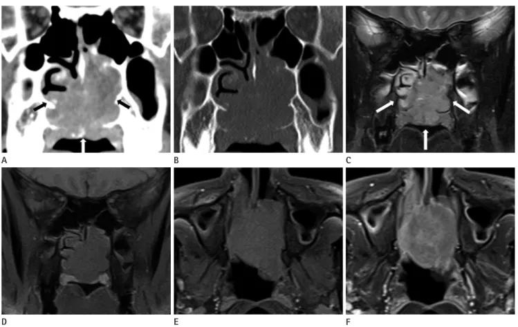
reached up to the anterior border of nasopharynx. The mass had soft tissue density with absence of calcification. The mass showed heterogeneous enhancement after intravenous bolus in- jection of contrast media (Fig. 2A, B).
The MRI examination was performed on 1.5 T using a Sie- mens Avanto equipment. On T2WI, an expansile lobulated mass in the left nasal cavity showed hyperintense signal intensi- ty compared with the muscle (Fig. 2C). On T1WI, the mass showed slight hyperintense signal intensity (Fig. 2D, E). Post- contrast T1-weighted image showed heterogeneous enhance- ment of the mass. As seen on CT scan, extension to adjacent structures was also being detected (Fig. 2F). Metastatic lymph- adenopathy was not seen.
The mass in the nasal cavity with extensive invasion into adja-
Fig. 2. Ewing’s sarcoma in the left nasal cavity in a 42-year-old man. Postcontrast coronal CT image (A) shows an expansile and well-defined soft tissue mass (arrows) in the left nasal cavity with inhomogeneous enhancement. The mass extends to the contralateral nasal cavity, left pos- terior ethmoid sinus and hard palate. Coronal CT image with bone algorithm (B) demonstrates better bony destructive change. On precontrast coronal T2-weighted image with fat suppression (C), an expansile lobulated mass (arrows) in the left nasal cavity shows hyperintense signal in- tensity compared with the muscle. Precontrast coronal and axial T1-weighted images with fat suppression (D, E) show slightly hyperintense sig- nal intensity. Postcontrast T1-weighted axial image with fat suppression (F) reveals heterogeneous enhancement of the mass. As seen on CT im- ages, extension to adjacent structures with bony destructive change is also noted.
E F
D
B
A C
and/or paranasal sinuses have been reported in the otolaryngol- ogy literature (1, 7, 9). In the radiology literature, only a single case report from 2003 by Harman et al. (3) has been described.
The prognosis for Ewing’s sarcoma depends on the presence of metastases, because Ewing’s sarcoma is highly malignant and metastasizes early to bones and lungs. Recently, it is accepted that prompt chemotherapy is necessary to treat occult metasta- sis, and a combination of surgical excision, radiotherapy and chemotherapy has significantly improved the 5-year survival rates, now reaching to 75% (1, 9).
Microscopically, Ewing’s sarcoma shows uniform, small, round cells with round to elongated nuclei, scanty cytoplasms and in- distinct cytoplasmic borders. Hemorrhagic areas and extensive necrotic lesions are common. The essential diagnostic examina- tion for Ewing’s sarcoma among many small round neoplasms is CD99 (O13) marker, the specific immunohistochemical exami- nation. In addition, molecular studies using PCR to detect char- acteristics of chromosomal translocations are definitive for the diagnosis of Ewing’s sarcoma. Specific genetic hallmarks of Ew- ing’s sarcoma is a gene sequence t(11;22)(q24;q12), which results in the fusion of the EWS gene with the FLI gene (1, 9, 10).
On the CT examination, Ewing’s sarcomas show similar find- ings regardless of the primary site. It is known to be a well-en- hancing, soft tissue density mass without calcification. If a tu- mor includes hemorrhage or necrosis, it may be detected as was consistent with Ewing’s sarcoma. Molecular studies using
polymerase chain reaction (PCR) confirmed EWS-FLI-1 [t(11;22) (q24;q12)].
Distant metastasis was excluded by fluorodeoxyglucose posi- tron emission tomography (not shown). After endoscopoic exci- sion, the patient was treated with chemotherapy, which consisted of 4 cycles of vincristine, actinomycin D and cyclophosphamide.
On a follow-up endoscopy performed 106 days after operation, tumor recurrence was not detected.
DISCUSSION
Ewing’s sarcoma is highly malignant, small, round cell tumor, originated from primitive neuroectodermal cells, as first de- scribed by James Ewing in 1921. Primary Ewing’s sarcoma is represented commonly in early childhood, adolescence but rarely in adulthood. There is slightly male predominance as 1.5 : 1 male to female ratio (4, 6, 7).
Ewing’s sarcoma is distinguished by two types: skeletal type and extraskeletal type. Most commonly, Ewing’s sarcoma arises from skeletal structures, especially long bones (35%), and pelvis (24%). The extraskeletal type of Ewing’s sarcoma usually occurs in the soft tissue of the lower extremities and the paravertebral region (8).
Thirteen cases of primary Ewing’s sarcoma in the nasal cavity Fig. 3. Pathologic features of Ewing sarcoma/Primitive neuroectodermal tumor.
A. The tumor is composed of densely distributed, uniform, small to medium sized, round cells with scanty cytoplasm. Mitotic figures are readily found (Hematoxylin & Eosin, × 400).
B. The tumor cells are immunoreactive with FLI-1 protein (Immunohistochemistry, × 400).
Note.-FLI-1 = Friend leukemia integration-1
A B
maxillary sinus. Rhinology 2008;46:75-78
2. Aferzon M, Wood WE, Powell JR. Ewing’s sarcoma of the ethmoid sinus. Otolaryngol Head Neck Surg 2003;128:
897-901
3. Harman M, Kirogˇlu F, Kösem M, Unal O. Primary Ewing’s sarcoma of the paranasal sinus with intracranial exten- sion: imaging features. Dentomaxillofac Radiol 2003;32:
343-346
4. Howarth KL, Khodaei I, Karkanevatos A, Clarke RW. A si- nonasal primary Ewing’s sarcoma. Int J Pediatr Otorhino- laryngol 2004;68:221-224
5. Javery O, Krajewski K, O’Regan K, Kis B, Giardino A, Jagan- nathan J, et al. A to Z of extraskeletal Ewing sarcoma family of tumors in adults: imaging features of primary disease, metastatic patterns, and treatment responses. AJR Am J Roentgenol 2011;197:W1015-W1022
6. Csokonai LV, Liktor B, Arató G, Helffrich F. Ewing’s sarcoma in the nasal cavity. Otolaryngol Head Neck Surg 2001;125:
665-667
7. Yeshvanth SK, Ninan K, Bhandary SK, Lakshinarayana KP, Shetty JK, Makannavar JH. Rare case of extraskeletal Ew- ings sarcoma of the sinonasal tract. J Cancer Res Ther 2012;8:142-144
8. Coskun BU, Cinar U, Savk H, Basak T, Dadas B. Isolated maxillary sinus Ewing’s sarcoma. Rhinology 2005;43:225- 228
9. Gupta S, Gupta OP, Mehrotra S, Mehrotra D. Ewing sarco- ma of the maxilla: a rare presentation. Quintessence Int 2009;40:135-140
10. Iezzoni JC, Mills SE. “Undifferentiated” small round cell tumors of the sinonasal tract: differential diagnosis up- date. Am J Clin Pathol 2005;124 Suppl:S110-S121
ill-defined bony destructive changes are common, and these re- flect the aggressive nature of the tumor (3, 5).
The MRI examination is more effective than the CT scan in determining the nature of tumors, delineating tumor margins, evaluating soft tissue involvement, and determining tumor stage and operative approach. On the MRI examination, Ewing’s sar- coma in the nasal cavity is known to be hypointense to isoin- tense on T1WI and varying signal intense on T2WI. It is hetero- geneously and markedly enhanced by gadolinium (3, 5).
In our study, the expansile lobulated mass in the left nasal cavity showed soft tissue density and inhomogeneous enhance- ment without calcification or cystic component on the CT scan.
On the MR imaging, the lesion showed slightly hyperintense signal relative to the muscles on T1WI, and hyperintense signal on T2WI with heterogenous enhancement. Extension to con- tralateral nasal cavity, ethmoidal sinus, nasopharynx and hard palate was detected with extensive bony destructive change.
These findings suggest malignant nature, and differential radio- logical diagnoses should be included malignant lymphoma, rhab- domyosarcoma, poorly-differentiated carcinomas and Ewing’s sarcoma. These tumors share radiologic findings and it is hard to conclude specific diagnosis without more examinations (1, 8).
In conclusion, primary Ewing’s sarcoma must be considered, when the expansile nasal mass is detected with extensive inva- sion into adjacent structures and bony destructive changes, al- though which is a very rare condition. Realizing the nature of Ewing’s sarcoma and understanding its diagnostic significance can lead to the approach of appropriate management.
REFERENCES
1. Kawabata M, Yoshifuku K, Sagara Y, Kurono Y. Ewing’s sar-
비강 내에서 발생한 골외 유잉 육종: 증례 보고1
정보영
1· 박석원
2· 김어진
3· 최종순
3· 이은자
1유잉 육종은 두경부의 매우 드문 종양이며 비강 내 혹은 부비동에서 특히 드물다. 우리는 코막힘과 코피를 증상으로 내원 하여 밝혀진 비강 내 유잉 육종 증례를 보고하고자 한다. CT와 MRI를 통해 왼쪽 비강에서 반대편 비강, 사골동, 비인두 까지 침범한 종양을 발견할 수 있었다. 비강 내 유잉 육종과 관련하여 본 증례의 영상 소견에 대해 살펴보도록 하겠다.
동국대학교 의과대학 일산병원 1영상의학과, 2이비인후과, 3병리과