http://dx.doi.org/10.3988/jcn.2012.8.2.109 J Clin Neurol 2012;8:109-115
Ischemic Stroke in Takayasu’s Arteritis: Lesion Patterns and Possible Mechanisms
Jaechun Hwang,a Suk Jae Kim,a Oh Young Bang,a Chin-Sang Chung,a Kwang Ho Lee,a Duk Kyung Kim,b Gyeong-Moon Kima
aDepartments of Neurology and bCardiology, Samsung Medical Center, Sungkyunkwan University School of Medicine, Seoul, Korea
Received July 12, 2011 Revised September 14, 2011 Accepted September 14, 2011 Correspondence Gyeong-Moon Kim, MD, PhD Department of Neurology, Samsung Medical Center, Sungkyunkwan University School of Medicine, 50 Irwon-dong, Gangnam-gu, Seoul 135-710, Korea Tel +82-2-3410-3598 Fax +82-2-3410-0052 E-mail [email protected]
Background and PurposezzThe purpose of the present study was to use brain magnetic reso- nance imaging (MRI) and magnetic resonance angiography (MRA) to identify the mechanism of stroke in patients with Takayasu’s arteritis (TA).
MethodszzAmong a retrospective cohort of 190 TA patients, 21 (3 males and 18 females) with a mean age of 39.9 years (range 15-68 years) who had acute cerebral infarctions were included in lesion pattern analyses. The patients’ characteristics were reviewed, and infarction patterns and the degree of cerebral artery stenosis were evaluated. Ischemic lesions were categorized into five subgroups: cortical border-zone, internal border-zone, large lobar, large deep, and small subcortical infarctions.
ResultszzIn total, 21 ischemic stroke events with relevant ischemic lesions on MRI were ob- served. The frequencies of the lesion types were as follows: large lobar (n=7, 33.3%), cortical border zone (n=6, 28.6%), internal border zone (n=1, 4.8%), small cortical (n=0, 0%), and large deep (n=7, 33.3%). MRA revealed that 11 patients had intracranial artery stenosis.
ConclusionszzHemodynamic compromise in large-artery stenosis and thromboembolic mech- anisms play significant roles in ischemic stroke associated with TA.
J Clin Neurol 2012;8:109-115 Key Wordszz vasculitis, thromboembolism, intracranial artery stenosis.
Open Access
cc This is an Open Access article distributed under the terms of the Cre- ative Commons Attribution Non-Commercial License (http://creative- commons.org/licenses/by-nc/3.0) which permits unrestricted non-com- mercial use, distribution, and reproduction in any medium, provided the ori- ginal work is properly cited.
Introduction
Takayasu’s arteritis (TA) is a chronic inflammatory disease of unknown etiology that primarily affects the aorta and its main branches.1 Vascular inflammation has been shown to cause ar- terial stenosis, occlusion, dilatation, and aneurysms. Clinical features such as claudication, renal hypertension, congestive heart failure, and coronary artery disease usually reflect limb or organ ischemia resulting from gradual stenosis of the in- volved arteries. Neurological manifestations that may accom- pany TA include headache, dizziness, visual disturbance or loss of vision, transient ischemic attacks (TIAs), and stroke.2
It has been reported that 10-20% of patients with TA will have an ischemic stroke or a TIA.3,4 Although cerebral isch- emic attacks are not common complications of TA, they can give rise to devastating neurological symptoms and be a major cause of morbid events and premature death.5 Antiplatelet agents and anticoagulants have been used for the prevention of stroke. In some cases surgical treatments including bypass surgery or stent insertion have also been advocated.6,7 How- ever, investigations into the mechanism of ischemic stroke in TA remain inadequate.1
Previous studies have indicated that the lesion patterns seen on magnetic resonance imaging (MRI) and magnetic resonance angiography (MRA) suggest particular stroke mechanisms.8,9 In the current study we determined some of the topographical characteristics of ischemic stroke in patients with TA in order to improve our understanding of the possible underlying mech- anisms.
Methods
Patients
We performed a retrospective analysis of 207 patients diag- nosed with TA at the Samsung Medical Center between Janu- ary 1993 and July 2009. TA patients were identified by re- viewing the registry in which data had been prospectively coll- ected. The local institutional review board approved this study.
The diagnosis of TA was assessed according to the American College of Rheumatology Criteria for the Classification of TA, which is based on the presence of symptoms and signs of isch- emic, inflammatory large-vessel disease, as well as supportive serological information and arteriographic findings.10 The char- acteristics and stroke risk factors of the patients were reviewed through previous medical records. Lesion patterns and the de- gree of stenosis in both the intracranial and extracranial arter- ies were evaluated through diffusion-weighted MRI (DWI), fluid-attenuated inversion recovery (FLAIR) MRI, and MRA.
We compared the age, gender, TA-related inflammatory markers, and other cerebrovascular risk factors such as hyper- tension, diabetes mellitus, dyslipidemia, ischemic heart disease, history of smoking, renal dysfunction, and prior transient mon- ocular blindness (TMB) between stroke TA patients and non- stroke TA patients. The history of previous hemispheric TIA was not considered because it is difficult to distinguish from extremity claudication caused by peripheral ischemia.
We included patients who had been confirmed with isch- emic stroke in stroke lesion pattern analysis. Stroke was de- fined as focal cerebral dysfunction relevant to an ischemic le- sion on MRI. We excluded patients who 1) did not have a stro- ke event, 2) had TIA or TMB without ischemic lesions on MRI, 3) had hemorrhagic stroke, 4) had been referred and whose MRI and MRA were not available to us, or 5) who had potential sources of cardioembolisms such as atrial fibrillation, mitral stenosis, a prosthetic valve, dilated cardiomyopathy, sick sinus syndrome, acute bacterial endocarditis, or patent foramen ovale.
Among the 207 TA patients (183 females) who were origi- nally surveyed, we ultimately included 190 patients (170 fe- males) in our study. We excluded four referral patients whose MRI and MRA were not available to us, three patients who had hemorrhagic stroke, and ten patients who had potential cardio- embolic sources.
MRI and MRA
Ischemic brain lesions were assessed and defined as hyperin- tense signal alterations on DWI or FLAIR MRI. We then clas- sified the distribution patterns of stroke lesions on MRI as well as the clinical manifestations. All ischemic lesions were cate- gorized into the following five subgroups according to lesion
size and topographical distribution based on published tem- plates:11,12
1. Cortical border-zone infarction: ischemic lesions mainly involving the border between the two main cerebral arteries, the border-zone area between the middle cerebral artery (MCA) and the anterior cerebral artery, or the posterior cerebral artery.
2. Internal border-zone infarction: hyperintense lesions at the border of the deep and superficial perforating arterial ter- ritories of the MCA.
3. Large lobar infarction: cortical infarction involving one or more divisions of a large vessel with or without subcortical lesions.
4. Large deep subcortical infarction: single large striatocap- sular lesions greater than 2 cm in diameter.
5. Small subcortical infarction: small single ischemic lesions less than 2 cm in diameter in the subcortical area suggesting lacunae.
A three-dimensional time-of-flight MRA scan of the intra- cranial arteries and gadolinium-enhanced MRA of the extra- cranial arteries was obtained from all patients. The standard Warfarin Versus Aspirin for Symptomatic Intracranial Disease Trial method was used to measure the degree of stenosis.13 The degree of arterial stenosis on MRA was graded as mild (<50%), moderate (50-69%), or severe (70-100%) according to the reduction in the diameter of the narrowest vessels. For intracranial artery stenosis (ICAS), the lumen reduction of the vertebrobasilar artery assessed on both the targeted maximal- intensity projection MRA and the source images to reduce the possibility of overestimation of stenosis inherent in the time- of-flight MRA technique. Two experienced observers who were blinded to the clinical data analyzed the images, and dis- crepancies were resolved through consensus.
Statistical analyses
Continuous data are presented as means and standard devia- tions, while categorical variables are presented as absolute and relative frequencies. We analyzed the differences between stroke and nonstroke patients using the chi-square test or Fisher’s exact test for categorical variables and Student’s t- test or the Mann-Whitney U-test for continuous variables. A probability value of p<0.05 was considered to be indicative of statistical significance. All statistical analyses were per- formed using commercially available software (SPSS for Win- dows, version 15.0; SPSS, Chicago, IL, USA).
Results
Of the 190 TA patients included in this study, 21 stroke patients (18 females) with a mean age of 39.9 years (range 15-68 years) were identified.
Conventional stroke risk factors and other TA risk factors
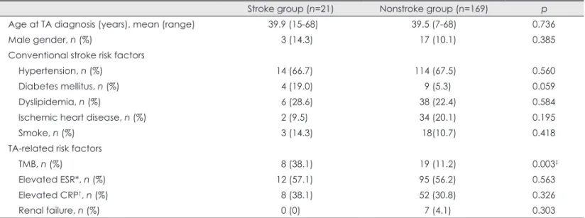
Conventional stroke risk factor profiles did not differ signifi- cantly between the stroke and nonstroke TA groups. Stroke and nonstroke patients had similar prevalence rates of hyper- tension, diabetes mellitus, dyslipidemia, and history of smok- ing (Table 1). There were also no differences when comparing TA-related factors such as erythrocyte sedimentation rate, C- reactive protein, and chronic renal failure. The frequency of previous TMB history was significantly higher in the stroke group.
MRI findings
In total, 21 first-ever ischemic stroke events with relevant isch- emic lesions on MRI were observed. Nine and 12 stroke events were identified by DWI and FLAIR imaging, respectively.
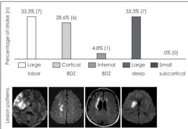
Most of the ischemic lesions were located within the MCA ter- ritory or internal/cortical border-zone area. Stroke patterns and angiographic features as well as clinical information for all pa- tients are provided in Table 2. The frequencies of the lesion types were as follows (Fig. 1): large lobar, n=7 (33.3%); corti- cal border zone, n=6 (28.6%); internal border zone, n=1 (4.8%); and large deep, n=7 (33.3%). There were no small sub- cortical infarctions in patients with TA.
MRA findings
As predicted, extracranial carotid artery stenosis or subclavian artery stenosis relevant to lesions were found in most ischemic stroke patients (71.4%; Table 2). Furthermore, MRA revealed that 11 patients had ICAS including the intracranial internal carotid artery, distal basilar artery, MCA, anterior cerebral ar- tery, and posterior cerebral artery. Fig. 2 shows all ICAS data related to the TA patients. ICAS relevant to stroke lesions
were found in 9 of 11 cases (81.8%); the degree of ICAS was mild in 4 (36.4%), moderate in 3 (27.3%), and severe in 4 (36.4%). The frequency of ICAS relevant to stroke was lower in the cortical border-zone infarctions (0%) than in large lobar (57.1%), internal border-zone (100%), and large deep (57.1%) infarctions (Table 3).
We examined the steno-occlusive state of the feeding artery in our nine patients with ICAS relevant to ischemic lesions separately according to stroke subdivision, and from this in- ferred the effect of ICAS in the stroke mechanism. ICAS was relatively common (57.1%) in large lobar infarction patterns, but every case exhibited an ipsilateral carotid artery steno-oc- clusive lesion. Conversely, ICAS lesions were not seen in six cortical border-zone infarction cases. Of the seven large deep patterns, four cases exhibited a relevant ICAS, but one was combined with carotid occlusion. The prevalence of ICAS rel- evant to ischemic lesions was higher in the large deep group (57.1%) when the cases of superimposed steno-occlusive ex- tracranial feeding arteries were excluded.
Discussion
We found that 11.1% of the TA patients in this study experi- enced stroke events, which is consistent with previous pro- spective studies.4 Stroke is an uncommon symptom for most TA patients and is rarely reported as the first manifestation;
however, Sikaroodi et al.14 reported one case of stroke as the first manifestation of TA. In our study, 52.3% of stroke pa- tients were first diagnosed with TA upon admission for an initial stroke event. It is recommended that if suspected, young stroke patients should be examined for TA based on nonspe- cific systemic symptoms such as fever, malaise, dizziness, and arm claudication.
Table 1. Stroke risk factor profiles of the stroke and nonstroke Takayasu’s arteritis (TA) groups
Stroke group (n=21) Nonstroke group (n=169) p
Age at TA diagnosis (years), mean (range) 39.9 (15-68) 39.5 (7-68) 0.736
Male gender, n (%) 3 (14.3) 17 (10.1) 0.385
Conventional stroke risk factors
Hypertension, n (%) 14 (66.7) 114 (67.5) 0.560
Diabetes mellitus, n (%) 4 (19.0) 9 (5.3) 0.059
Dyslipidemia, n (%) 6 (28.6) 38 (22.4) 0.584
Ischemic heart disease, n (%) 2 (9.5) 34 (20.1) 0.195
Smoke, n (%) 3 (14.3) 18(10.7) 0.418
TA-related risk factors
TMB, n (%) 8 (38.1) 19 (11.2) 0.003‡
Elevated ESR*, n (%) 12 (57.1) 95 (56.2) 0.563
Elevated CRP†, n (%) 8 (38.1) 52 (30.8) 0.326
Renal failure, n (%) 0 (0) 7 (4.1) 0.303
*ESR>27 mm/hour, †CRP>0.3 mg/dL, ‡p<0.05.
TMB: transient monocular blindness, ESR: erythrocyte sedimentation rate, CRP: C-reactive protein.
The mean age of diagnosis for TA was in the third decade, and did not significantly differ between the stroke and non- stroke TA groups. The mean time from TA diagnosis to stroke onset was 1.6 years (range 0-4 years). Strokes developed in TA patients at much a younger age than observed for strokes related to atherosclerotic mechanisms. The prevalence of con- ventional stroke risk factors, except for hypertension and dys- lipidemia, was lower among the TA patients than for tradition- al atherosclerotic stroke. The relatively high prevalence of hy- pertension is probably due to the involvement of renal artery stenosis.4,15 Therefore, stroke in TA patients seems to develop via a different mechanism than conventional atherosclerotic
changes. The TA-related serologic markers erythrocyte sedi- mentation rate and C-reactive protein, which represent disease activity, also did not differ significantly between the stroke and nonstroke groups. However, the time intervals from stroke on- set to laboratory studies were inconsistent and relatively long.
Therefore, a homogeneous large study of acute stroke in TA patients is needed to establish a correlation between TA dis- ease activity and ischemic stroke.
Known predispositions to stroke in TA patients were previ- ously limited to TMB history. Also known as amaurosis fugax, TMBs are TIAs involving the anterior cerebral circulation and are caused by thromboembolisms or hypoperfusion. Previous Table 2. Summary of clinical and imaging features of infarctions
Case Subgroup Sex/Age
(years) Main
symptom Lesion
site MRI ICAS Carotid artery stenosis Stroke risk factors
Site Degree Site Degree HTN DM Lipid
1 Large lobar F/25 Hemiparesis Rt MCA DWI Rt M1 Rt A1
Occlusion Severe
Rt CCA Lt CCA
Occlusion Moderate
N N N
2 Large lobar M/47 Hemiparesis Rt MCA FLAIR N N Rt CCA Moderate N N Y
3 Large lobar F/39 Hemiparesis Aphasia
Lt MCA FLAIR Lt M2 Mild Lt CCA Severe N N N
4 Large lobar F/24 Hemiparesis Rt MCA DWI Rt M1 Moderate Rt CCA Lt CCA
Occlusion Occlusion
Y N N
5 Large lobar F/34 Hemiparesis Hemianopsia
Rt MCA FLAIR N N Rt SCA Occlusion N N Y
6 Large lobar F/33 Hemiparesis Rt MCA FLAIR Rt M1 Mild Rt CCA Lt SCA
Moderate Occlusion
N N N
7 Large lobar F/15 Hemiparesis Aphasia
Lt MCA DWI N N Lt CCA Occlusion N N N
8 CBDZ F/15 Hemiparesis Lt BDZ DWI N N Rt CCA
Lt CCA
Occlusion Occlusion
Y N Y
9 CBDZ F/45 Hemiparesis Rt BDZ DWI N N Rt CCA
Lt CCA
Severe Severe
Y Y Y
10 CBDZ F/46 Hemiparesis Lt BDZ FLAIR N N Rt CCA
Lt CCA
Moderate Severe
Y N N
11 CBDZ F/17 Hemiparesis Rt BDZ FLAIR Lt dICA Moderate Rt CCA Lt CCA
Occlusion Occlusion
N N N
12 CBDZ M/34 Hemiparesis Rt BDZ FLAIR N N Rt SCA
Lt CCA
Mild Occlusion
Y N N
13 CBDZ F/49 Hypesthesia Bo BDZ DWI N N Lt SCA Severe Y Y Y
14 IBDZ F/49 Hemiparesis Rt BDZ DWI Rt M2 Mild N N Y N Y
15 Large deep F/51 General Weakness
Rt SC FLAIR Rt M1 Lt M1
Occlusion Severe
N N Y N N
16 Large deep F/33 Hemiparesis Lt SC DWI N N Rt CCA Mild Y N Y
17 Large deep F/38 Hemiparesis Rt SC FLAIR Rt P2 Mild Rt CCA Moderate Y Y N
18 Large deep F/43 Hemiparesis Rt SC DWI Rt M1 Severe N N Y N Y
19 Large deep M/43 Hemiparesis Rt SC FLAIR N N Rt CCA
Lt CCA
Occlusion Moderate
Y N Y
20 Large deep F/37 Hemiparesis Lt SC FLAIR Lt M1 Moderate Rt CCA Lt CCA
Severe Occlusion
Y N N
21 Large deep F/46 Hemiparesis Rt SC FLAIR Bo M1,2 Rt A1,2
Severe Mild
Lt CCA Lt ICA
Moderate Severe
Y Y Y
BDZ: border zone, Bo: both, CBDZ: cortical border zone, CCA: common carotid artery, dICA: distal internal carotid artery, DM: diabe- tes mellitus, DWI: diffusion-weighted imaging, F: female, FLAIR: fluid-attenuated inversion recovery, HTN: hypertension, IBDZ: internal border zone, ICA: internal carotid artery, ICAS: intracranial artery stenosis, Lt: left, M: male, MCA: middle cerebral artery, MRI: mag- netic resonance imaging, N: no, Rt: right, SC: striatocapsular, SCA: subclavian artery, Y: yes.
studies have suggested that TMB history is a warning sign for stroke.16,17 Our data indicated that previous TMB may also be related to future ischemic stroke in TA patients.
MRI analysis revealed that most of the ischemic lesions were located at MCA branches or in the internal/cortical bor- der-zone area. Large lobar type and cortical border-zone in- farctions were documented in 33.3% and 28.6% of cases, re-
spectively. Wedge-shaped large lobar and cortical border-zone infarctions are related to embolic mechanisms.18 In compari- son, internal border-zone infarctions - which are known to occur via a hemodynamic compromise mechanism19 - ac- counted for only 4.8% of all the stroke events. These results highlight not only the hemodynamic compromise in large- artery stenosis but also the thromboembolic mechanism in ischemic stroke of TA. There were no cases of a small sub- cortical infarction suggesting a lacunar infarction among our TA patients.
As noted above, most TA patients are young and rarely have conventional atherosclerotic risk factors. With respect to ath- erosclerotic carotid stenosis, embolic cerebral infarction is usually attributed to plaque rupture or thrombotically active carotid plaques associated with high inflammatory infil- trates.20,21 Some studies have revealed premature atherosclero- sis in TA patients based on ultrasonography or autopsy re- sults.22-24 In particular, Seyahi et al.15 reported that 27% of TA patients possessed atherosclerotic plaques.23 From this, we can surmise that a similar mechanism may be involved in cre- ating thromboembolisms, such as ruptures of atheromatous plaques or artery-to-artery embolisms.
Fig. 1. Comparison of stroke patterns in Takayasu’s arteritis. BDZ:
border zone infarction.
Percentage of stroke (n)Lesion patterns
0% (0) 33.3% (7)
Large lobar
Cortical BDZ
Internal BDZ
Large deep
Small subcortical 28.6% (6)
4.8% (1)
33.3% (7)
Fig. 2. Magnetic resonance angiography findings of intracranial artery stenosis in patients with Takayasu’s arteritis. Arrows indicate the site of intracranial artery stenosis.
Most previous reports have described TA as medium- or large-vessel arteritis involving the aorta and its main branch- es.1,4 To the best of our knowledge, only three case reports have described ICAS in TA, as discovered at autopsy or through ar- teriographic findings. Klos et al.26 described two patients with TA who had ischemic stroke due to intracranial involve- ment, in whom the cause of the infarction was proposed to be vasculitis.25 In our study revealed that more than half of the TA patients with stroke had ICAS. Based on our results, we be- lieve that intracranial involvement in TA may be under-recog- nized, and that cerebral angiography should be considered for patients diagnosed with TA. Given that TA patients are young and relatively free from atherosclerotic risk factors except hypertension and dyslipidemia, vascular inflammation may be an important risk factor in ICAS.22
From our analysis of the stroke subgroup distribution, ICAS seems to be more likely when patients have large deep subcor- tical infarctions and internal border-zone infarctions than oth- er types of infarction. A recent study regarding lesion patterns in cases of concurrent atherosclerosis of ICAS revealed a high prevalence of perforating artery infarctions and border-zone infarctions in the ischemic lesion distribution.27 Previous stud- ies have suggested that ICAS is related not only to artery-to- artery embolisms but also to hemodynamic compromise.18,28-31 Further studies with large case series are needed to confirm whether ICAS in TA patients is related to a diverse pattern of ischemic stroke.
This study was subject to several limitations. First, since many of the patients were referred to our hospital some time after their stroke events occurred, laboratory and image data were not homogeneous. Some patients were diagnosed by FLAIR imaging because no DWI images were available. Al- though FLAIR imaging is less sensitive than DWI, we were still able to diagnose and locate stroke lesions accurately using clinical correlations. Second, only a small number of stroke patients were included in order to analyze stroke risk factors and ICAS between lesion subtypes. The low prevalence of stroke in TA made it impossible to include a large number of
stroke patients in our analysis; however, this study currently represents the largest clinical series regarding stroke in TA pa- tients. Third, the prevalence of ICAS in TA patients with stroke was higher than in previous studies. Some partial reanalyzed steno-occlusive arterial states produced by a proximal arterial embolic source were thought to have contributed to the high prevalence of ICAS. Moreover, the measurable degree of ste- nosis may vary between MRA and conventional angiograms.
Although we analyzed the source images to reduce the possi- bility of overestimation of stenosis, MRA may overestimate the degree of ICAS. Directly comparing the ICAS prevalence between our study and previous studies is impossible due to different criteria being used for patient selection. We classified the lesion pattern as identified on DWI or FLAIR images;
however, the chronic large-artery steno-occlusive state may alter the collateral flow and cerebral artery territory, which may limit the ability to establish the stroke mechanism in TA pa- tients using lesion pattern analysis alone. Future investigations with perfusion images and conventional angiography would be helpful in determining the stroke mechanism. The final limi- tation of this study is the lack of pathologic proof of vasculitis.
However, considering that the American College of Rheuma- tology Criteria for the Classification of TA yielded a sensitivi- ty and specificity of more than 90%, there is little likelihood of overdiagnosis of TA in patients with conventional risk factors for stroke.10
The factor that was most associated with stroke in TA pa- tients was previous TMB history. The finding that large lobar, cortical border-zone, and large deep infarctions were common stroke types suggests that a thromboembolic mechanism un- derlies stroke in TA. Furthermore, ICAS may be more preva- lent in TA than was previously thought, which suggests that intracranial involvement is relatively common in TA. Future studies involving large numbers of subjects and focusing on the precise pathophysiological mechanisms leading to stenosis would help our understanding of its relationship with ischemic stroke in TA patients.
Conflicts of Interest
The authors have no financial conflicts of interest.
REFERENCES
1. Johnston SL, Lock RJ, Gompels MM. Takayasu arteritis: a review. J Clin Pathol 2002;55:481-486.
2. Kim HJ, Suh DC, Kim JK, Kim SJ, Lee JH, Choi CG, et al. Correla- tion of neurological manifestations of Takayasu’s arteritis with cerebral angiographic findings. Clin Imaging 2005;29:79-85.
3. Subramanyan R, Joy J, Balakrishnan KG. Natural history of aortoar- teritis (Takayasu’s disease). Circulation 1989;80:429-437.
4. Kerr GS, Hallahan CW, Giordano J, Leavitt RY, Fauci AS, Rottem M, et al. Takayasu arteritis. Ann Intern Med 1994;120:919-929.
5. Pfefferkorn T, Bitterling H, Hüfner K, Opherk C, Schewe S, Pfister Table 3. Relationships between ischemic patterns and relevant
arterial stenosis
Lesion pattern Total number Ipsilateral ICAS, n (%)
Ipsilateral ECAS, n (%) Large lobar 7 4 (57.1) 7 (100)
Cortical BDZ 6 0 (0) 5 (83.3)
Internal BDZ 1 1 (100) 0 (0)
Large deep subcortical
7 4 (57.1) 4 (57.1)
Small subcortical 0 0 0
BDZ: border zone, ICAS: intracranial artery stenosis, ECAS: ex- tracranial artery stenosis.
HW, et al. Malignant hemispheric infarction in Takayasu arteritis. J Neurol 2008;255:1425-1426.
6. Ogino H, Matsuda H, Minatoya K, Sasaki H, Tanaka H, Matsumura Y, et al. Overview of late outcome of medical and surgical treatment for Takayasu arteritis. Circulation 2008;118:2738-2747.
7. Liang P, Hoffman GS. Advances in the medical and surgical treatment of Takayasu arteritis. Curr Opin Rheumatol 2005;17:16-24.
8. Baird AE, Lövblad KO, Schlaug G, Edelman RR, Warach S. Multiple acute stroke syndrome: marker of embolic disease? Neurology 2000;
54:674-678.
9. Lee LJ, Kidwell CS, Alger J, Starkman S, Saver JL. Impact on stroke subtype diagnosis of early diffusion-weighted magnetic resonance im- aging and magnetic resonance angiography. Stroke 2000;31:1081- 1089.
10. Arend WP, Michel BA, Bloch DA, Hunder GG, Calabrese LH, Ed- worthy SM, et al. The American College of Rheumatology 1990 crite- ria for the classification of Takayasu arteritis. Arthritis Rheum 1990;33:
1129-1134.
11. Tatu L, Moulin T, Bogousslavsky J, Duvernoy H. Arterial territories of human brain: brainstem and cerebellum. Neurology 1996;47:1125- 1135.
12. Amarenco P, Bogousslavsky J, Caplan LR, Donnan GA, Hennerici MG. Classification of stroke subtypes. Cerebrovasc Dis 2009;27:493- 13. Chimowitz MI, Kokkinos J, Strong J, Brown MB, Levine SR, Silliman 501.
S, et al. The Warfarin-Aspirin Symptomatic Intracranial Disease Study.
Neurology 1995;45:1488-1493.
14. Sikaroodi H, Motamedi M, Kahnooji H, Gholamrezanezhad A, Youse- fi N. Stroke as the first manifestation of Takayasu arteritis. Acta Neurol Belg 2007;107:18-21.
15. Seyahi E, Ugurlu S, Cumali R, Balci H, Seyahi N, Yurdakul S, et al.
Atherosclerosis in Takayasu arteritis. Ann Rheum Dis 2006;65:1202- 1207.
16. Marshall J, Meadows S. The natural history of amaurosis fugax. Brain 1968;91:419-434.
17. Benavente O, Eliasziw M, Streifler JY, Fox AJ, Barnett HJ, Meldrum H, et al. Prognosis after transient monocular blindness associated with carotid-artery stenosis. N Engl J Med 2001;345:1084-1090.
18. Yong SW, Bang OY, Lee PH, Li WY. Internal and cortical border-zone
infarction: clinical and diffusion-weighted imaging features. Stroke 2006;37:841-846.
19. Howard R, Trend P, Russell RW. Clinical features of ischemia in cere- bral arterial border zones after periods of reduced cerebral blood flow.
Arch Neurol 1987;44:934-940.
20. Spagnoli LG, Mauriello A, Sangiorgi G, Fratoni S, Bonanno E, Schwartz RS, et al. Extracranial thrombotically active carotid plaque as a risk factor for ischemic stroke. JAMA 2004;292:1845-1852.
21. Masuda J, Yutani C, Ogata J, Kuriyama Y, Yamaguchi T. Atheroma- tous embolism in the brain: a clinicopathologic analysis of 15 autopsy cases. Neurology 1994;44:1231-1237.
22. Numano F, Kishi Y, Tanaka A, Ohkawara M, Kakuta T, Kobayashi Y.
Inflammation and atherosclerosis. Atherosclerotic lesions in Takayasu arteritis. Ann N Y Acad Sci 2000;902:65-76.
23. Filer A, Nicholls D, Corston R, Carey P, Bacon P. Takayasu arteritis and atherosclerosis: illustrating the consequences of endothelial dam- age. J Rheumatol 2001;28:2752-2753.
24. Numano F, Okawara M, Inomata H, Kobayashi Y. Takayasu’s arteritis.
Lancet 2000;356:1023-1025.
25. Molnár P, Hegedüs K. Direct involvement of intracerebral arteries in Takayasu’s arteritis. Acta Neuropathol 1984;63:83-86.
26. Klos K, Flemming KD, Petty GW, Luthra HS. Takayasu’s arteritis with arteriographic evidence of intracranial vessel involvement. Neu- rology 2003;60:1550-1551.
27. Man BL, Fu YP, Chan YY, Lam W, Hui AC, Leung WH, et al. Lesion patterns and stroke mechanisms in concurrent atherosclerosis of intra- cranial and extracranial vessels. Stroke 2009;40:3211-3215.
28. Gorelick PB, Wong KS, Bae HJ, Pandey DK. Large artery intracranial occlusive disease: a large worldwide burden but a relatively neglected frontier. Stroke 2008;39:2396-2399.
29. Caplan LR, Hennerici M. Impaired clearance of emboli (washout) is an important link between hypoperfusion, embolism, and ischemic stroke. Arch Neurol 1998;55:1475-1482.
30. Caplan LR. Intracranial large artery occlusive disease. Curr Neurol Neurosci Rep 2008;8:177-181.
31. Weber R, Kraywinkel K, Diener HC, Weimar C; German Stroke Study Collaboration. Symptomatic intracranial atherosclerotic stenoses:
prevalence and prognosis in patients with acute cerebral ischemia.
Cerebrovasc Dis 2010;30:188-193.