http://dx.doi.org/10.3988/jcn.2013.9.4.244 J Clin Neurol 2013;9:244-251
Introduction
Somatosensory evoked potentials (SSEPs) have been used dur- ing skull base,1,2 spine3-6 and brain tumor2,7 operations to pre-
dict and reduce post operative neurologic deficits.1,8 Intraop- erative monitoring of SSEPs allows for the dynamic assessment of the spinal cord dorsal columns, the brainstem medial lemn- sical pathways to the thalamus, and thalamic connections to the primary sensory cortex. Persistent changes in the SSEPs dur- ing these procedures can predict post operative neurologic defi- cits.1,9 Intraoperative restoration of SSEP changes may prevent post operative neurologic complications.1 Regional and hemi- spheric changes of cerebral blood flow (CBF) could potential-
Predictive Value of Somatosensory Evoked Potential Monitoring during Resection of Intraparenchymal and Intraventricular
Tumors Using an Endoscopic Port
Parthasarathy Thirumala,a Daniel Lai,b Jonathan Engh,a Miguel Habeych,a Donald Crammond,a Jeffrey Balzera
aCenter for Clinical Neurophysiology, Department of Neurologic Surgery, University of Pittsburgh Medical Center, Pittsburgh, PA, USA
bDepartment of Neurology, Stanford University, Palo Alto, CA, USA
Received August 27, 2012 Revised January 28, 2013 Accepted January 28, 2013 Correspondence
Parthasarathy Thirumala, MD, MS Center for Clinical Neurophysiology, Department of Neurologic Surgery, University of Pittsburgh Medical Center,
UPMC Presbyterian-Suite B-400, 200 Lothrop Street, Pittsburgh, PA 15213, USA
Tel +1-412-648-2228 Fax +1-412-383-9899 E-mail [email protected]
Background and PurposezzIntraoperative neurophysiological monitoring (IONM) using up- per and lower somatosensory evoked potentials (SSEPs) is an established technique used to pre- dict and prevent neurologic injury during intracranial tumor resections. Endoscopic port surgery (EPS) is a minimally-invasive approach to deep intraparenchymal and intraventricular brain tu- mors. The authors intended to evaluate the predictive value of SSEP monitoring during resection of intracranial brain tumors using a parallel endoscopic technique.
MethodszzA retrospective review was conducted of patients operated on from 2007-2010 uti- lizing IONM in whom endoscopic ports were used to remove either intraparenchymal or intra- ventricular tumors. Cases were eligible for review if an endoscopic port was used to resect an intracranial tumor and the electronic chart included all intraoperative monitoring data as well as pre- and post-operative neurologic exams.
Resultszz139 EPS cases met criteria for inclusion. Eighty five patients (61%) had intraparen- chymal and fifty four (39%) had intraventricular tumors or colloid cysts. SSEP changes were seen in eleven cases (7.9%), being irreversible in three (2.2%) and reversible in eight cases (5.8%). Seven patients (5.0%) with intraparenchymal tumors had SSEP changes which met our criterea for significant changes while there were four (2.9%) with intraventricular (p-val- ue=0.25). Five patients suffered post operative deficits, two reversible and two irreversible SSEP changes. Only one case exhibited post operative hemiparesis with no SSEP changes. The posi- tive predictive value of SSEP was 45.4% and the negative predictive value was 99.2%.
ConclusionszzBased on the high negative and low positive predictive values, the utility of SSEP monitoring for cylindrical port resections may be limited. However, the use of SSEP monitoring can be helpful in reducing the impact of endoscopic port manipulation when the tu- mor is closer to the somatosensory pathway. J Clin Neurol 2013;9:244-251 Key Wordszz endoscopic port, somatosensory, evoked potentials, somatosensory, monitoring.
Open Access
cc This is an Open Access article distributed under the terms of the Cre- ative Commons Attribution Non-Commercial License (http://creative- commons.org/licenses/by-nc/3.0) which permits unrestricted non-com- mercial use, distribution, and reproduction in any medium, provided the ori- ginal work is properly cited.
ly be identified with SSEP monitoring and thereby prevent neurovascular injury9-13. Additionally, SSEP monitoring can evaluate potential peripheral injury related to neck position,14-16 brachial plexus17 or peripheral nerve injury14-16 during surgery.
Endoscopic port surgery (EPS) presents an alternative ap- proach for resection of deep tumors. The port itself is a trans- parent cylindrical retractor measuring 4.00 mm in diameter;
the length of the port is tailored to the depth of the resection.
The port is introduced into the brain over a bullet shaped di- lator. In contrast to traditional microsurgical deep tumor re- section, this technique attempts to limit retraction and dissec- tion injury while improving the field of view at deep sites.18-20 The endoscopic port has been used to resect tumors within deep white matter and the ventricles. Our study will be the first to evaluate the utility of intraoperative SSEP monitoring to prevent postoperative neurologic deficits after endoscopic resection of brain tumors.
Methods
Between 2007 and 2010, we monitored 139 patients who un- derwent resection of either intraventricular or intraparenchymal tumors using EPS. We retrospectively reviewed all cases of en- doscopic tumor resections; inclusion criteria for the study in- cluded patients who underwent endoscopic tumor resections with intraoperative SSEP monitoring. Patients were excluded if the data was unavailable for analysis due to technical rea- sons or because of missing data. This study was approved by the local institutional review board for retrospective review of clinical outcomes.
Neurophysiologic monitoring
Intraoperative SSEP monitoring is standard at our institution for EPS cases.1 SSEP baseline values were obtained prior after anesthesia induction, but before the patients were positioned.
Upper and lower extremity SSEP responses were continu- ously obtained throughout the procedure. Physician oversight and interpretation was performed using a combined on-site and remote model utilized at the University of Pittsburgh Medi- cal Center.21 All patients underwent the procedure in a stereo- tactic frame for tumor localization. General inhalational an- esthesia was given to all patients with additional intravenous medications in some cases. In our institution we typically used inhalational anesthestics including sevoflurane, desflurane, isoflurane in concentrations with a Minimum Alveolar Con- centration between 0.5-0.75.
Upper extremity SSEPs
Median or ulnar nerve stimulation was performed bilaterally in an alternating fashion at the wrist with subdermal needle
electrode pairs. Recordings were obtained from the scalp and cervical region with subdermal electrodes. P4/Fz and P3/Fz scalp electrodes were used (per the international 10-20 system).
A cervical electrode was located at the C2 spinous process or mastoid and referenced to Fz. Stimulation frequency was 2.33- 2.45 Hz with duration of 0.2-0.3 milliseconds. Band pass filters were set at 10 to 300 Hz for cortical recordings and 30 to 1000 Hz for subcortical recordings. Averages were computed for ei- ther 128 or 256 trials, depending on the signal quality.
Lower extremity SSEPs
For the lower extremities, bilateral alternating tibial nerve stimulation, interleaved after the upper stimulation was used.
Peroneal nerve stimulation was used when reliable tibial nerve responses could not be elicited. Tibial nerve stimulation was performed at the medial malleolus of the ankle with subder- mal needle electrodes. The peroneal nerve was stimulated using pairs of subdermal needles located at the head of the fibula and medially in the popliteal fossa. Recordings were ob- tained from the scalp and cervical region with subdermal elec- trodes. Pz/Fz and P4/P3 scalp electrodes were used (per the in- ternational 10-20 system). A cervical electrode was located at the C2 spinous process or at the level of the mastoid and ref- erenced to Fz. Stimulation frequency was 2.33-2.45 Hz with duration of 0.2-0.3 milliseconds. Band pass filters were set at 10 to 300 Hz for cortical recordings and 30 to 1000 Hz for cer- vical recordings. Averages were computed for either 128 or 256 trials depending on the signal quality.21
Alarm criteria
A SSEP epoch average was collected approximately every 40 seconds and compared against the baseline. We defined as
‘significant’ a signal change persisting over at least two con- secutive epochs characterized by either a 50% reduction in amplitude or a prolongation of response latency by >10%
from baseline. This criterion is widely used in the literature, particularly in spine surgery cases.22-24
Medical record review
Medical records for all 139 patients were reviewed to deter- mine whether any new neurologic deficit was identified post- operatively. New motor/sensory deficits were defined as be- ing present if there was a new focal deficit noted in the chart during the immediate post operative period as compared to pre-operative neurologic exam. Similarly, the intraoperative records for all 139 patients were reviewed to identify any sig- nificant SSEP changes.
Data analysis
To determine the sensitivity and specificity of intraoperative
SSEP monitoring for detecting impending or resultant iatro- genic neurologic injury, we defined and then classified each of the 139 operative cases as one of the following.
True positive (TP): significant SSEP signal changes accom- panied by a new postoperative neurologic deficit; a case in which significant SSEP signal deterioration occurred as the re- sult of a recognized intraoperative cause, event, or complica- tion; or a case in which a significant SSEP signal deterioration improved to the baseline value after a specific intraoperative intervention.
True negative (TN): normal intraoperative SSEP signals in the absence of a new postoperative neurologic deficit.
False positive (FP): persistent significant SSEP signal de- terioration that did not improve with intraoperative interven- tions, or in which no intraoperative adjustments were made in cases of reversible changes and the patient woke up neurologi- cally intact.
False negative (FN): normal intraoperative SSEP signals with a new postoperative neurologic deficit.
Patients that were either TPs, FP of FNs were catagorized as ‘group 1’. TN patients were catagorized in ‘group 2’.
Quantitative analysis
The quantitative analysis has been described in detail previ- ously.21 The first part of analysis involved interpreting intra- operative SSEP data from patients who demonstrated signifi- cant changes. It was scrutinized to determine whether such a
change occurred in temporal proximity to specific intraoper- ative events such as changes in mean arterial pressure (MAP), manipulation of the endoscopic port, or a pharmacologic change in anesthesia administration (Table 1). In such cases, intraoperative records were reviewed to determine whether any subsequent intraoperative countermeasures such as in- creasing the MAP, repositioning the port, or decreasing the anesthesic dose were undertaken and whether the SSEP sig- nals improved after these actions. The second part of the anal- ysis details with measuring changes in latency and amplitude at the point of maximum change and at closure compared with baseline values at incision. Labels were used to refer to the SSEP responses, e.g., right cortical upper extremity amplitude (RCUA)-right (R), cortical (C), upper-limb response (U), amplitude (A)-and LBLL-left (L), brainstem (B), lower-limb response (L), latency (L)-refers to SSEP responses from the left upper-limb response in the brainstem and the contralater- al cortex. The latency and amplitude values were recorded at incision, maximum change, and closure. A percentage change in latency and amplitude from incision was calculated at 2 in- stances during surgery: at the maximum change and at clo- sure (Table 2). The percentage of change from incision (I) to maximum change (M) was calculated by the following for- mula: 100 [(I-M)/I]; similar change was calculated from in- cision to closure. The purpose of the calculation is to follow the trend of the responses and to correlate them with postop- erative outcomes.
Table 1. Neurophysiological changes, intraoperative interventions and neurologic status in patients who underwent endoscopic port surgery with SSEP monitoring
Patient Diagnosis Cannulation
route
Neurophysiologic
change Clinical sequelae Intraoperative
adjustment Statistics
1 Glioblastoma IP Reversible, LCUA Yes, weaker left
lower extremity
None FP
2 Glioblastoma IP Reversible, LCUA No None, resecting near
thalamus
TP
3 Glioblastoma IP Reversible, RCUA No None FP
4 Metastatic small cell carcinoma
IP Irreversible, RCUA, RCUL, RCLA, LCUA, LCUL
Yes, death Adjusted port trajectory TP
5 Anaplastic astrocytoma IP Irreversible RCUA, LCUA, LCLA
No Adjusted port trajectory TP
6 Glioblastoma IP Irreversible, RCUA, LCUA Yes, weaker right upper extremity
Gave lorazepam TP
7 Glioblastoma IP Reversible, RCUA, LCLA No None FP
8 Epidermoid tumor IV Reversible, RCUA, LCUA Yes, right hemiparesis Retractor traction TP
9 Cavernous malformation IV Reversible, RCUA, LCUA No Gave blood products FP
10 Colloid cyst IV Reversible, RCUA, LCUA No None-increased stimulation FP
11 Central neurocytoma IV Reversible, LCUA No None FP
12 Metastatic adenocarcinoma
IP None Yes, right hemiparesis None FN
FN: false negative, FP: false positive, IP: intraparenchymal, IV: intraventricular, LCLA: left cortical lower limb amplitude, LCUA: left cor- tical upper limb amplitude, LCUL: left cortical upper limb latency, RCLA: right cortical lower limb amplitude, RCUA: right cortical am- plitude upper limb amplitude, RCUL: left cortical upper limb latency, SSEP: somatosensory evoked potential, TP: true positive.
Statistical analysis
The Fischer’s exact test analysis was used to compare thein- cidence of SSEP changes between intraparenchymal and in- traventricular tumors. Analysis comparing demographic dif- ferences between groups one and two was complete with a two sample t test.
Results
We analyzed 139 port cases: 85 patients (61%) had intraparen- chymal tumors and 54 (39%) had intraventricular or colloid cysts. The average age of patients with SSEP changes was 52.7 years as compared to 49.6 without changes (p=0.60). De- mographics are reported in Table 3. Glioblastoma was the most common intracranial lesion in groups 1 and 2, though the inci- dence was somewhat higher in group 1. Each of the patients included in group 1 is listed in Table 2 which describes the changes observed during the surgery, the intraoperative man- agement and post operative sequelae. The resultant statistical
analysis is listed in Table 4.
Intraoperative SSEP changes
In total, SSEP changes were seen in eleven (7.9%) patients, eight cases (5.8%) had reversible waveform changes which were either at baseline or improving towards baseline at the conclusion of the surgery. Three (2.2%) had irreversible wave- form changes. Of the intraparenchymal tumors, seven patients (5.0%) had significant changes in the SSEPs, four were revers- ible and three were irreversible. Among the intraventricular tumors, four patients (2.9%) had significant SSEP changes, all of which were reversible.
Post operative neurologic deficits
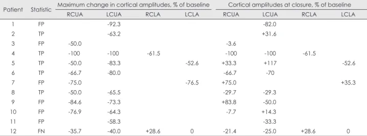
There were a total of five cases (3.6%) with post operative neu- rologic consequences. In two of the cases there were reversible SSEP changes, where permanent neurologic morbidity oc- curred. Another two cases had irreversible SSEP changes, with permanent neurologic morbidity. One patient had no Table 2. Quantitative analysis of cortical somatosensory evoked potential monitoring response amplitude and latency as a percentage of baseline and at the point of maximum change and closure
Patient Statistic Maximum change in cortical amplitudes, % of baseline Cortical amplitudes at closure, % of baseline
RCUA LCUA RCLA LCLA RCUA LCUA RCLA LCLA
1 FP -92.3 -82.0
2 TP -63.2 +31.6
3 FP -50.0 -3.6
4 TP -100 -100 -61.5 -100 -100 -61.5
5 TP -50.0 -83.3 -52.6 +33.3 +117 -52.6
6 TP -66.7 -80.0 -66.7 -70
7 FP -75.0 -76.5 +75.0 +35.3
8 TP -50.0 -65.5 -29.7 -29.3
9 FP -84.6 -73.3 +83.8 -50.0
10 FP -76.9 -64.3 -7.7 +14.3
11 FP -58.3 -33.3
12 FN -35.7 -40.0 +28.6 0 -21.4 -25.0 +28.6 0
FP: false positive, FN: false negative, LCLA: left cortical lower limb amplitude, LCUA: left cortical upper limb amplitude, RCLA: right cortical lower limb amplitude, RCUA: right cortical amplitude upper limb amplitude, TP: true positive.
Table 3. Demographics information of patients with and without changes in somatosensory evoked potentials
Group 1 (TP, FP, FN) Group 2 (TN)
Total 12 127
Age, mean 52.7 49.6
<50 years 6 61
≥50 years 6 67
Sex ratio (M : F) 1 : 1.4 1.1 : 1
Most common diagnosis (%) 5 glioblastoma (41.7) 28 glioblastoma (22.0)
Cannulation route (%) 8 intraparenchymal (5.8)
4 intraventricular (2.9)
77 intraparenchymal (55.4) 50 intraventricular (36.0) Neurologic deficits (%) 4 intraparenchymal (2.9)
1 intraventricular (0.7) FN: false negative, FP: false positive, TN: true negative, TP: true positive.
significant intraoperative SSEP change, but did show a new hemiparesis following the operation.
True positives
Patient 2 had reversible changes to the left cortical upper ex- tremity amplitude (LCUA) SSEP. At the time of the evoked potential change, the surgeons were operating close to the thalamus. The location of the ongoing surgical manipulation was thought to be the cause of the waveform change. After the procedure, the SSEP improved and the patient did not have any post operative neurologic deficit.
Patient 4 had irreversible changes to RCUA, the LCUA, and the right cortical lower extremity amplitude (RCLA). La- tencies in the right and left upper extremities became pro- longed until the waveforms were completely lost and could not be measured. The left cortical lower extremity amplitude (LCLA) was also diminished but was not significant. Cerebral cortex swelling was noted during the procedure. Post operative- ly, a CT scan demonstrated intraparenchymal and subarach- noid bleeding. The patient died postoperatively.
Patient 5 had irreversible changes to the LCLA, but had reversible changes to the LCUA and RCUA. The cortical la- tencies for the left arm, left leg and right arm did not change.
The SSEPs in the RCUA and LCUA improved with intraop- erative adjustment of the port. Moreover, by the end of the sur- gery, the RCUA and LCUA exceeded baseline values. How- ever, the LCLA did not improve with adjustment. Notably, the patient’s post operative exam was remarkable for improved weakness of the left lower extremity except for dorsiflexion of the tibialis anterior.
Patient 6 had irreversible SSEP waveform changes to the RCUA, but slight recovery of the LCUA at the conclusion of the operation. Intraoperative EEG was used during the pro- cedure in addition to SSEP monitoring in this case. There was concern that the EEG demonstrated seizures. He was given lo- razepam before the conclusion of the surgery. There was new weakness in the right arm post operatively.
Patient 8 had reversible changes to the RCUA and LCUA with subsequent improvement of both cortical amplitudes, but did not return to baseline. Latencies were not significantly affected. The trajectory of the endoscopic port was adjusted,
as it was believed that the changes were secondary to retraction.
The patient had a new right hemiparesis after the operation.
False positives
Patient 1 was noted to have a reversible change to the LCUA, without changes to the subcortical waveforms or left lower extremity waveforms. The waveforms most significantly de- creased after the withdrawal of the port from the brain. The pa- tient was found to have left lower extremity weakness after the surgery.
Patients 3 and 7 both had a reversible changes to the RCUA.
The LCLA in patient 7 also demonstrated a reversible change.
Patient 11 had a reversible change to the LCUA. For these cas- es, no cause was determined to have precipitated the waveform change in any patient. No intraoperative adjustments were made. At the conclusion of the surgery, the RCUA for patient 3 returned baseline as did both the RCUA and LCLA for pa- tient 7. The LCUA for patient 11 did improve, but was still 33% lower than baseline. Patients 3, 7 and 11 had no new post operative deficits.
Patients 9 and 10 both had significant changes to the RCUA and LCUA. In both cases there was no discernable cause for the change. Latencies were unaffected. Patient 9 did have significant blood loss requiring intraoperative transfusion, though blood pressure remained relatively constant. Howev- er, there was no temporal correlation between the neurophys- iological changes and blood transfusion. The changes were noted earlier during the surgery and transfusions were done late in the surgery. In addition, the blood transfusion was not an intraoperative measure suggested by the neurophysiology team. Otherwise, no intraoperative adjustments were made.
At the end of the procedure, the RCUA of patient 9 exceeded baseline while the LCUA improved, but was still 50% lower than baseline. The RCUA of patient 10 improved nearly to baseline and the LCUA slightly exceeded baseline. There were no post operative deficits noted for either patient.
False negative
Patient 12 did not have significant intraoperative waveform changes; however changes were noted in the LCUA and RCUA. The RCLA improved throughout the course of the surgery and had higher amplitude at the time of the proce- dure’s conclusion. The patient was noted to have new right hemiparesis in the immediate post operative period consis- tent with a left supplementary motor area syndrome. Over several weeks, the patient gradually regained strength in the affected side.
True negatives
The remaining 127 patients did not have intraoperative chang- Table 4. Intraoperative somatosensory evoked potential monitor-
ing results and postoperative neurologic status in a 2x2 table New sensory or motor deficit
SSEP changes Present Absent
Present 5 (TP) 6 (FP)
Absent 1 (FN) 127 (TN)
Sensitivity=83.3%, specificity=95.5%, positive predictive value:
45.4%, negative predictive value: 99.2%.
FN: false negative, FP: false positive, SSEP: somatosensory evoked potential, TN: true negative, TP: true positive.
es that met alarm criteria and did not have new post operative sensory or motor deficits.
Discussion
Our study aims to elucidate the utility of intraoperative SSEP monitoring during endoscopic brain tumor resection. In par- ticular, we are concerned with potential injury which may be prevented due to regional parenchymal compression second- ary to port manipulation and hemispheric changes in blood flow related to the procedure. The use of intraoperative SSEP monitoring has been shown to be effective in predicting post operative deficits under a number of circumstances including spine,3-6 carotid endarterectomies,10,13 skull based,1,2 and more recently minimally invasive skull based surgeries.21 This form of intraoperative monitoring can predict and prevent post op- erative morbidity by alerting the surgeon to intraoperative changes which may compromise the somatosensory pathway.
The introduction of an endoscopic port may cause injury due to mechanical strain during cannulation or withdrawal, local- ized ischemia due to regional compression, or injury due to intraoperative adjustments. Since the sensory and motor strip share a common vascular supply, motor deficits may also be detected.23 In our study there was no significant difference in the incidence of deficits between intraparenchymal and intra- ventricular tumor removal. The FN case in this study occurred with resection of an intraparenchymal tumor in the supplemen- tary motor area. Hence in our study, we performed a combined analysis of intraventricular and intraparenchymal endoscopic approaches for tumor removal.
In our study we had 5 cases with SSEP changes corre- sponding to potential or realized post operative deficits (TPs).
Amongst these TPs, irreversible changes were found in three cases (patients 4, 5, and 6). Irreversible SSEP changes have been associated with new post operative clinical deficits and were seen in patients 4 and 6 after the procedure.21,25 The waveform changes with patient 4 were severe, resulting com- plete loss of RCUA and LCUA. During the operation, the brain parenchyma was noted to become edematous. Post op- eratively, there was a significant intracranial hemorrhage which resulted in death. Patient 5 did have irreversible wave- form loss, but with adjustment of the port, a post operative deficit was potentially avoided. There were also 2 TP cases with reversible changes, one of which (patient 8) demonstrat- ed a new clinical deficit. Patient 2 was categorized as TP due to surgical manipulation near the thalamus at the time of the SSEP change. The majority of TP cases (patients 1, 4, 5, and 8) in our study exhibited SSEP changes related to manipulation of the port. It is possible that such manipulation can result in significant regional parenchymal compression, leading to
ischemia or even infarction.
Reversible and irreversible changes1,10,21,25 in SSEP wave- forms significantly increase the likelihood of post operative deficits and allow for possible intraoperative adjustments to prevent post operative complications. Animal models indicate that a drop in CBF below 16 to 20 mL/100g/minute causes a reversible decrease in the amplitude of cortical SSEP re- sponses.26,27 Additionally, animal studies have shown that an increase in the MAP, and consequently the CBF, after a 50%
reduction in SSEP amplitude can result in SSEP recovery to control values.28 In humans, persistent reduction of SSEP am- plitude by 50% is observed when CBF decreases below 14 mL/100 g/minute.29 Further animal studies have shown that there is a narrow hemodynamic window where a loss of cor- tical SSEP response does not imply loss of neuronal viability and that reversing the changes in CBF in a certain time frame reverses injurious or iatrogenic cellular ionic changes.30 Hence an early warning using alarm criteria might be helpful to re- adjust and reperfuse the brain parenchyma after port manipu- lations. This might prevent post operative neurologic compli- cations in the somatosensory or motor pathway which otherwise could not have been easily identified during port manipulation.
Significantly, there were six cases we recorded as being FPs.
Our observed positive predictive value of 45% has been ob- served in prior studies during scoliosis operations.31 The cause of FPs can be due to instrumentation error, anesthetic effects, transient alterations in MAP,28 or ineffective documentation of surgical or physiological changes. Most of the FPs with no documentation occurred during cases performed shortly after EPS was initially developed. The learning curve of the surgical, anesthesia and neurophysiology team might have contributed to this phenomenon as previously reported.21 A single case (patient 12) was recorded during which the SSEP waveforms did not reach alarm criteria, yet there was a new postoperative hemiparesis. Causes of FNs are of paramount importance as they represent lost opportunities to potentially intervene prior to development of the neurologic deficit. ‘De- layed onset’ neurologic deficits have been recognized in spi- nal surgeries32 as well as skull base procedures.2 These de- layed onset cases may be attributed to latent vascular or mechanical compression.32 More likely in our case, the affect- ed parenchyma was the supplementary motor area which was distant from the pathway which SSEPs monitor. Hence the ischemia in the region had some, but not significant, ef- fect on the somatosensory area resulting in changes that did not reach alarm criteria. A previous case series by Wiedemay- er et al.2 notes that one of the major reasons for FNs was the location of the lesion being outside the monitored pathway.
Taken together, SSEP monitoring during EPS may be able
to help prevent post operative deficits secondary to port ma- nipulation. During our procedure the patient is in a stereotactic frame for localization and has an endoport and a light source fitted for visualization. This instrumentation is never removed, which is necessary for us to perform a transcranial motor evoked potential study in our institution. Without the removal, any patient movement during the procedure might increase the chances of iatrogenic injury secondary to movement.33
In line with previous studies, we observed a robust nega- tive predictive value with expected specificity and sensitivi- ty.2,13,21,31,32 However, our positive predictive value was lower than expected. A low positive predictive value potentially re- sults in both needless interruptions to the surgical procedure and an inability to intervene on patients in whom there is an impending neurologic complication. This study is limited by its relatively small sample size, its retrospective nature and in- adequate documentation, making broader generalizations dif- ficult to make. The retrospective nature hindered assessment of patients’ post operative physical exams. As noted, delayed onset of neurologic deficits may remain latent and may only be realized until well after the surgery.2 The lack of long term follow up is a further limitation to this study.
Conclusions
Based on the high negative predictive and low positive pre- dictive value, the utility of SSEP monitoring during EPS for deep seated tumors appears genuine but limited. The use of SSEP monitoring can be helpful in reducing the impact of port manipulations when the tumor is close to somatosensory path- way. Alternatively, adjunctive use of transcranial motor evoked potentials may provide a useful supplement to the monitor- ing approach. A large patient series is needed to completely evaluate the utility of SSEPs during resection of deep brain tumors through a minimal access port.
Conflicts of Interest
The authors have no financial conflicts of interest.
REFERENCES
1. Bejjani GK, Nora PC, Vera PL, Broemling L, Sekhar LN. The pre- dictive value of intraoperative somatosensory evoked potential moni- toring: review of 244 procedures. Neurosurgery 1998;43:491-498;
discussion 498-500.
2. Wiedemayer H, Sandalcioglu IE, Armbruster W, Regel J, Schaefer H, Stolke D. False negative findings in intraoperative SEP monitoring:
analysis of 658 consecutive neurosurgical cases and review of pub- lished reports. J Neurol Neurosurg Psychiatry 2004;75:280-286.
3. Wilber RG, Thompson GH, Shaffer JW, Brown RH, Nash CL Jr.
Postoperative neurological deficits in segmental spinal instrumenta- tion. A study using spinal cord monitoring. J Bone Joint Surg Am 1984;66:1178-1187.
4. Jones SJ, Edgar MA, Ransford AO, Thomas NP. A system for the electrophysiological monitoring of the spinal cord during operations
for scoliosis. J Bone Joint Surg Br 1983;65:134-139.
5. Brown RH, Nash CL Jr, Berilla JA, Amaddio MD. Cortical evoked potential monitoring. A system for intraoperative monitoring of spi- nal cord function. Spine (Phila Pa 1976) 1984;9:256-261.
6. Bradshaw K, Webb JK, Fraser AM. Clinical evaluation of spinal cord monitoring in scoliosis surgery. Spine (Phila Pa 1976) 1984;9:636-643.
7. Kombos T, Picht T, Derdilopoulos A, Suess O. Impact of intraopera- tive neurophysiological monitoring on surgery of high-grade gliomas.
J Clin Neurophysiol 2009;26:422-425.
8. Gentili F, Lougheed WM, Yamashiro K, Corrado C. Monitoring of sensory evoked potentials during surgery of skull base tumours. Can J Neurol Sci 1985;12:336-340.
9. De Vleeschauwer P, Horsch S, Matamoros R. Monitoring of somato- sensory evoked potentials in carotid surgery: results, usefulness and limitations of the method. Ann Vasc Surg 1988;2:63-68.
10. Stejskal L, Kramár F, Ostrý S, Benes V, Mohapl M, Limberk B. Ex- perience of 500 cases of neurophysiological monitoring in carotid endarterectomy. Acta Neurochir (Wien) 2007;149:681-688; discus- sion 689.
11. Lopéz JR, Chang SD, Steinberg GK. The use of electrophysiological monitoring in the intraoperative management of intracranial aneu- rysms. J Neurol Neurosurg Psychiatry 1999;66:189-196.
12. Friedman WA, Kaplan BL, Day AL, Sypert GW, Curran MT. Evoked potential monitoring during aneurysm operation: observations after fifty cases. Neurosurgery 1987;20:678-687.
13. Lam AM, Manninen PH, Ferguson GG, Nantau W. Monitoring elec- trophysiologic function during carotid endarterectomy: a comparison of somatosensory evoked potentials and conventional electroenceph- alogram. Anesthesiology 1991;75:15-21.
14. Schwartz DM, Sestokas AK, Hilibrand AS, Vaccaro AR, Bose B, Li M, et al. Neurophysiological identification of position-induced neu- rologic injury during anterior cervical spine surgery. J Clin Monit Comput 2006;20:437-444.
15. Kamel IR, Drum ET, Koch SA, Whitten JA, Gaughan JP, Barnette RE, et al. The use of somatosensory evoked potentials to determine the relationship between patient positioning and impending upper extremity nerve injury during spine surgery: a retrospective analysis.
Anesth Analg 2006;102:1538-1542.
16. Chung I, Glow JA, Dimopoulos V, Walid MS, Smisson HF, Johnston KW, et al. Upper-limb somatosensory evoked potential monitoring in lumbosacral spine surgery: a prognostic marker for position-related ulnar nerve injury. Spine J 2009;9:287-295.
17. Schwartz DM, Drummond DS, Hahn M, Ecker ML, Dormans JP.
Prevention of positional brachial plexopathy during surgical correc- tion of scoliosis. J Spinal Disord 2000;13:178-182.
18. Kassam AB, Engh JA, Mintz AH, Prevedello DM. Completely endo- scopic resection of intraparenchymal brain tumors. J Neurosurg 2009;
110:116-123.
19. Harris AE, Hadjipanayis CG, Lunsford LD, Lunsford AK, Kassam AB. Microsurgical removal of intraventricular lesions using endoscop- ic visualization and stereotactic guidance. Neurosurgery 2005;56(1 Suppl):125-132; discussion 125-132.
20. Engh JA, Lunsford LD, Amin DV, Ochalski PG, Fernandez-Miranda J, Prevedello DM, et al. Stereotactically guided endoscopic port sur- gery for intraventricular tumor and colloid cyst resection. Neurosur- gery 2010;67(3 Suppl Operative):ons198-ons204; discussion ons204- ons205.
21. Thirumala PD, Kassasm AB, Habeych M, Wichman K, Chang YF, Gardner P, et al. Somatosensory evoked potential monitoring during endoscopic endonasal approach to skull base surgery: analysis of ob- served changes. Neurosurgery 2011;69(1 Suppl Operative):ons64- ons76; discussion ons76.
22. York DH, Chabot RJ, Gaines RW. Response variability of somato- sensory evoked potentials during scoliosis surgery. Spine (Phila Pa 1976) 1987;12:864-876.
23. Chen ZY, Wong HK, Chan YH. Variability of somatosensory evoked potential monitoring during scoliosis surgery. J Spinal Disord Tech 2004;17:470-476.
24. Balzer JR, Rose RD, Welch WC, Sclabassi RJ. Simultaneous somato- sensory evoked potential and electromyographic recordings during lumbosacral decompression and instrumentation. Neurosurgery 1998;
42:1318-1324; discussion 1324-1325.
25. Mizoi K, Yoshimoto T. Permissible temporary occlusion time in an- eurysm surgery as evaluated by evoked potential monitoring. Neuro- surgery 1993;33:434-440; discussion 440.
26. Branston NM, Symon L, Crockard HA, Pasztor E. Relationship be- tween the cortical evoked potential and local cortical blood flow fol- lowing acute middle cerebral artery occlusion in the baboon. Exp Neu- rol 1974;45:195-208.
27. Astrup J, Symon L, Branston NM, Lassen NA. Cortical evoked po- tential and extracellular K+ and H+ at critical levels of brain ischemia.
Stroke 1977;8:51-57.
28. Lopez JR. Intraoperative neurophysiological monitoring. Int Anes- thesiol Clin 1996;34:33-54.
29. Symon L. The relationship between CBF, evoked potentials and the clinical features in cerebral ischaemia. Acta Neurol Scand Suppl 1980;
78:175-190.
30. Branston NM, Symon L, Strong AJ. Reversibility of ischaemically induced changes in extracellular potassium in primate cortex. J Neu- rol Sci 1978;37:37-49.
31. Nuwer MR, Dawson EG, Carlson LG, Kanim LE, Sherman JE. So- matosensory evoked potential spinal cord monitoring reduces neuro- logic deficits after scoliosis surgery: results of a large multicenter survey. Electroencephalogr Clin Neurophysiol 1995;96:6-11.
32. Nuwer MR. Spinal cord monitoring. Muscle Nerve 1999;22:1620-1630.
33. Macdonald DB. Intraoperative motor evoked potential monitoring:
overview and update. J Clin Monit Comput 2006;20:347-377.