J Korean Soc Radiol 2016;74(3):189-194 http://dx.doi.org/10.3348/jksr.2016.74.3.189
INTRODUCTION
Mucosa-associated lymphoid tissue (MALT) lymphomas are an extranodal subset of marginal zone B-cell lymphomas and represent > 8% of all types of lymphomas (1). In 1983, Isaacson and Wright (2) initially described MALT lymphomas as a dis- tinctive type of B-cell lymphoma arising in the gastrointestinal tract. MALT lymphomas can be found at mucosal sites such as the stomach, lung, and salivary gland or from sites, which share an embryologic origin with mucosa such as the thyroid that normally lacks organized lymphoid tissue (3).
Primary hepatic MALT lymphoma is an extremely rare lesion, with only 45 cases reported in the English literature (4, 5). Be-
cause of its rarity, the radiologic findings in patients with this le- sion are seldom described, to our best knowledge. Herein, we re- ported a patient with pathologically proven primary MALT lymphoma who underwent ultrasonography, computed tomogra- phy (CT), magnetic resonance imaging (MRI), and fluorodeoxy- glucose positron emission tomography with CT (FDG PET/CT).
CASE REPORT
A 71-year-old woman with early-stage gastric cancer was ad- mitted to our hospital for metastasis work-up. She had a medi- cal history of diabetes mellitus, as well as cervical cancer for which she underwent radical hysterectomy with bilateral pelvic
Primary Hepatic Mucosa-Associated Lymphoid Tissue Lymphoma:
A Case Report
간의 원발성 점막관련 림프조직 림프종: 증례 보고
Kyung Su Kwag, MD
1, Suk Ki Jang, MD
1*, Jae Woo Yeon, MD
1, So Ya Paik, MD
2, Sang Jong Park, MD
3, Hyuk Jung Kim, MD
1Departments of 1Radiology, 2Pathology, 3Internal Medicine, Daejin Medical Center Bundang Jesaeng General Hospital, Seongnam, Korea
Primary hepatic mucosa-associated lymphoid tissue (MALT) lymphoma is an ex- tremely rare lesion. Primary hepatic lymphomas are known to present as a single mass in > 70% of cases, and in many instances with no specific features on imag- ing. Herein, we described a case of primary hepatic MALT lymphoma in a 71-year- old woman. A computed tomography (CT) scan revealed a mass, 4.5 × 3.0 cm, in liv- er segment 2 (S2) that was poorly defined, with subtle enhancement during the arterial phase. Gadoxetic acid-enhanced magnetic resonance imaging also showed an arterially enhancing mass in S2, with low signal intensity during the hepatobiliary phase and high signal intensity on diffusion-weighted imaging with a high b-value.
On fluorodeoxyglucose positron emission tomography/CT imaging, the mass showed a high standardized uptake value. Ultrasonography (US) revealed a hypoechoic mass, and US-guided core needle biopsy confirmed a hepatic MALT lymphoma.
Index terms
Mucosa-Associated Lymphoid Tissue Lymphoma Liver
Multidetector Computed Tomography Magnetic Resonance Imaging
Received August 25, 2015 Revised November 4, 2015 Accepted November 23, 2015
*Corresponding author: Suk Ki Jang, MD
Department of Radiology, Daejin Medical Center Bundang Jesaeng General Hospital, 20 Seohyeon-ro 180beon-gil, Bundang-gu, Seongnam 13590, Korea.
Tel. 82-31-779-3038 Fax. 82-31-779-0062 E-mail: [email protected]
This is an Open Access article distributed under the terms of the Creative Commons Attribution Non-Commercial License (http://creativecommons.org/licenses/by-nc/3.0) which permits unrestricted non-commercial use, distri- bution, and reproduction in any medium, provided the original work is properly cited.
lymphadenectomy 9 years earlier. On admission, a physical ex- amination showed no abnormal findings. Laboratory tests re- vealed leukopenia (white blood cell count = 2800/mm3) and thrombocytopenia (platelet count = 109000/mm3) related to cir- rhosis. Values on liver function tests were 49 U/L for aspartate aminotransferase and 49 U/L for alanine aminotransferase. The patient was positive for hepatitis B surface antigen, with a titer of 1921.8585 and a signal-to-cutoff ratio of > 50, and hepatitis B virus DNA [7.1 log (10) copies/mL). The serum alpha-fetopro- tein level was normal.
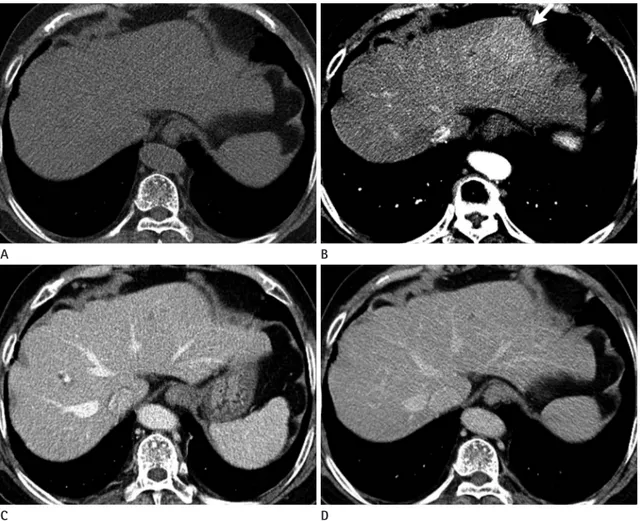
Liver CT (triple-phase CT) revealed a mass, 4.5 × 3.0 cm, in liver segment 2 (S2). The mass was poorly-defined with mild ar- terial enhancement (Fig. 1B) but not visualized on a the non-en- hanced scan (Fig. 1A) in the portal venous (Fig. 1C) or delayed phases (Fig. 1D). Significant regional lymphadenopathy or dis-
tant metastases were not detected.
Subsequently, we performed MRI using gadoxetic acid [Gd- EOB-DTPA (Primovist)] (Bayer-Schering, Berlin, Germany).
The tumor showed iso-signal intensity on T1-weighted images and homogeneous high signal intensity on T2-weighted images (Fig. 2A). Dynamic study of the tumor showed a homogeneous arterial enhancement and no distortion of vessels traversing the mass (Fig. 2B). The tumor demonstrated iso-signal intensity relative to normal liver parenchyma in the portal and delayed phases. In the hepatobiliary phase, the lesion had low signal in- tensity (Fig. 2C). On diffusion-weighted imaging, the intensity was high, with a high b-value (800 s/mm2) (Fig. 2D). The mass lesion was devoid of blood products or a fat component.
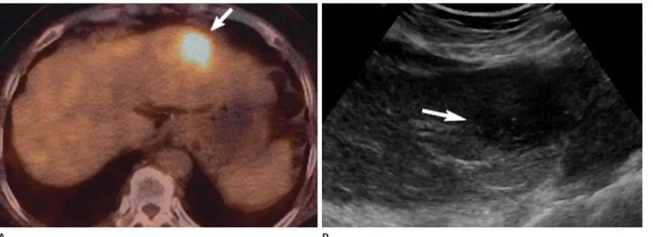
FDG PET/CT imaging revealed a high standardized uptake value (max SUV = 6.59) at the S2, suggesting a markedly hy-
Fig. 1. Hepatic MALT lymphoma in a 71-year-old woman on CT.
On unenhanced (A) and dynamic liver CT, the mass shows a mildly arterial enhancing mass (arrow) in liver segment 2 (B) in the arterial phase and iso-attenuation relative to the liver on unenhanced image (A), in the portal phase (C), and in the delayed phase (D).
CT = computed tomography, MALT = mucosa-associated lymphoid tissue A
C
B
D
permetabolic mass (Fig. 3A).
Considering the patient’s clinical history and radiologic find- ings, the differential diagnosis included a malignant hypervascu- lar mass, such as hepatocellular carcinoma (HCC), lymphoma, or metastatic hepatic tumors, and a benign hypervascular mass, such as an inflammatory pseudotumor (IPT) or a hepatic ade- noma. Our initial thought was that malignant hypervascular liver tumors are more likely than benign ones; furthermore, in a patient with cirrhosis, HCC could be suspected because it is the most common type of primary hypervascular liver cancer. He- patic lymphoma was excluded from the diagnosis based on dy- namic CT and MRI evidence of tumor penetration by existing blood vessels in hepatic malignant lymphoma (6).
Ultrasound-guided core needle biopsy was conducted to
confirm the histopathologic diagnosis. On ultrasound, the mass was hypoechoic, as compared with the liver parenchyma (Fig.
3B). Biopsy results demonstrated markedly intense, diffuse por- to-periportal atypical lymphocytic infiltration with lymphoid follicle formation. Immunohistochemical staining showed that the lesion was positive for CD20 and CD79a and negative for CD3, cyclin D1, BCL-6, and CD5, consistent with MALT lym- phoma (Fig. 4).
Treatment consisted of radiation therapy alone. At the 1-year follow-up, the primary hepatic MALT lymphoma had com- pletely disappeared on MRI, and there was no significant up- take of FDG in the liver on follow-up FDG PET/CT.
Fig. 2. Hepatic MALT lymphoma in a 71-year-old woman on gadoxetic acid-enhanced MRI.
A. T2-weighted axial MR image shows a homogeneous high signal instensity mass (arrowhead) in liver segment 2.
B, C. On gadoxetic acid-enhanced arterial phase (B), the mass shows homogeneous enhancement (arrowhead) and undistorted vessels travers- ing the mass (curved black arrow). The mass shows low signal intensity (arrowhead) in the hepatobiliary phase (C).
D. With diffusion-weighted imaging, the intensity is high (arrowhead), with a high b-value (800 s/mm2).
MALT = mucosa-associated lymphoid tissue, MRI = magnetic resonance imaging A
C
B
D
DISCUSSION
A primary hepatic MALT lymphoma is extremely rare. Jaffe (7) reported that hepatic malignant lymphomas make up < 1%
of all malignant lymphomas, and hepatic MALT lymphomas reportedly occur in only 3% cases of hepatic malignant lym- phoma.
The etiology of hepatic malignant lymphomas, especially MALT lymphoma, is not elucidated. MALT lymphomas origi- nate at sites normally devoid of organized lymphoid tissue, where- as lymphomas almost always arise in the setting of chronic in- flammatory disorders that are characterized by the accumulation of lymphoid tissue (8). A relationship between hepatitis C virus
(HCV) and hepatic malignant lymphoma has been previously reported, however, there are no reports on a relationship be- tween HCV and MALT lymphoma (4).
As is well known, primary hepatic lymphoma presents as a single mass in > 70% of cases, and in many cases MALT lym- phomas are solitary masses with diverse findings on radiologic imaging (4). Because the imaging findings are nonspecific, it is difficult to make a definite diagnosis based on imaging without histopathology.
To date, the characteristic diagnostic radiologic findings spe- cific to primary hepatic lymphoma, including MALT lympho- ma, are unknown. On CT scans, the hepatic MALT lymphoma is enhanced peripherally in the arterial phase and low attenuat- Fig. 3. Hepatic MALT lymphoma in a 71-year-old woman on FDG PET/CT and US.
A. FDG PET/CT scanning reveals a high standardized uptake value (max SUV = 6.59) (arrow) at liver segment 2, suggesting a hypermetabolic mass.
B. US demonstrates a hypoechoic solid mass (arrow) in liver segment 2.
FDG PET/CT = fluorodeoxyglucose positron emission tomography computed tomography, MALT = mucosa-associated lymphoid tissue, US = ul- trasonography
A B
Fig. 4. Histopathologic findings and immunohistochemical staining of hepatic MALT lymphoma in a 71-year-old woman.
A. The microscopic finding of the tumor includes a diffuse infiltration of small atypical lymphocytes expanding portal tracts, extending into lob- ules, and effacing normal hepatocytes (hematoxylin and eosin stain, × 100).
B. Immunohistochemical staining reveals CD20-positive lymphoid cells (× 400).
MALT = mucosa-associated lymphoid tissue
A B
ed in the delayed phase, according to an earlier report (4). An- other study indicated that hepatic MALT lymphoma shows pro- longed enhancement in the delayed phase on dynamic CT and is enhanced in the arterial phase with persisting enhancement in the portal phase on dynamic MRI (9). Similarly, in our case, en- hancement was evident in the arterial phase; however, unlike previous reports, our case showed iso-attenuation in the delayed phase and undistorted vessels traversing the liver mass (6).
When diagnosing a hypervascular liver tumor, various en- hancement patterns and ancillary imaging features such as cap- sule, hepatobiliary phase imaging, intralesional fat, hemorrhage, and restricted diffusion could be of help in characterizing the liver tumor. Our case showed a poorly defined mass with ho- mogeneous arterial enhancement, but according to a previous report, homogeneous arterial enhancement was not reported as an imaging feature of IPTs of the liver (6). One of the diagnostic features of HCC i.e., portal venous or delayed phase washout was not seen in this case. It is unlikely that our patient had a heterogeneous liver mass due to the presence of hemorrhage, lipid/fat, and rarely calcification, which are common imaging features of hepatic adenoma (5).
In conclusion, despite the difficulty involved, early radiologic diagnosis of primary hepatic MALT lymphoma should be con- sidered in the differential diagnosis of a hypervascular liver tu- mor, especially if tumor penetration by existing blood vessels can be visualized.
REFERENCES
1. Matasar MJ, Zelenetz AD. Overview of lymphoma diagno-
sis and management. Radiol Clin North Am 2008;46:175- 198, vii
2. Isaacson P, Wright DH. Malignant lymphoma of mucosa- associated lymphoid tissue. A distinctive type of B-cell lymphoma. Cancer 1983;52:1410-1416
3. Park JY, Choi MS, Lim YS, Park JW, Kim SU, Min YW, et al.
Clinical features, image findings, and prognosis of inflam- matory pseudotumor of the liver: a multicenter experi- ence of 45 cases. Gut Liver 2014;8:58-63
4. Doi H, Horiike N, Hiraoka A, Koizumi Y, Yamamoto Y, Hasebe A, et al. Primary hepatic marginal zone B cell lym- phoma of mucosa-associated lymphoid tissue type: case report and review of the literature. Int J Hematol 2008;88:
418-423
5. Grazioli L, Olivetti L, Mazza G, Bondioni MP. MR imaging of hepatocellular adenomas and differential diagnosis di- lemma. Int J Hepatol 2013;2013:374170
6. Apicella PL, Mirowitz SA, Weinreb JC. Extension of vessels through hepatic neoplasms: MR and CT findings. Radiolo- gy 1994;191:135-136
7. Jaffe ES. Malignant lymphomas: pathology of hepatic in- volvement. Semin Liver Dis 1987;7:257-268
8. Rodallec M, Guermazi A, Brice P, Attal P, Zagdanski AM, Frija J, et al. Imaging of MALT lymphomas. Eur Radiol 2002;
12:348-356
9. Shiozawa K, Watanabe M, Ikehara T, Matsukiyo Y, Kikuchi Y, Kaneko H, et al. A case of contiguous primary hepatic marginal zone B-cell lymphoma and hemangioma ulti- mately diagnosed using contrast-enhanced ultrasonogra- phy. Case Rep Oncol 2015;8:50-56
간의 원발성 점막관련 림프조직 림프종: 증례 보고
곽경수
1· 장석기
1* · 연재우
1· 백소야
2· 박상종
3· 김혁중
1간의 원발성 점막관련 림프조직 림프종은 매우 드문 질환이다. 간의 원발성 림프종의 70% 이상은 단일 종괴로 관찰되고 간의 점막관련 림프조직 림프종에서도 단일 종괴로 관찰되는데, 진단에 있어 영상학적 특징은 보고된 바가 없다. 저자들은 간의 원발성 점막관련 림프조직 림프종을 보인 71세 여자 환자의 증례를 보고하고자 한다. 역동적 조영증강 전산화단층촬 영의 동맥기에 약간의 조영증강을 보이는 4.5 × 3.0 cm 크기의 종괴가 간의 제2분절에서 관찰되었다. 역동적 자기공명 영상의 동맥기 영상에서 조영증강을 보이고, 간담도기 영상에서 저신호강도를 보이며, 확산강조영상에는 고신호강도를 보 이는 간종괴를 관찰하였다. Fluorodeoxyglucose (이하 FDG) 양전자방출단층촬영에서는 FDG의 섭취 증가를 보였다. 초 음파에서는 저에코성 병변으로 관찰되었고, 초음파 유도하 침조직검사를 시행하여 간의 원발성 점막관련 림프조직 림프 종으로 확진되었다.
분당제생병원 1영상의학과, 2병리과, 3내과