Copyrights © 2016 The Korean Society of Radiology
128
Case Report
pISSN 1738-2637 / eISSN 2288-2928 J Korean Soc Radiol 2016;74(2):128-131 http://dx.doi.org/10.3348/jksr.2016.74.2.128
INTRODUCTION
Neurofibromas are slow-growing, painless, benign nerve- sheath tumors. They occur most commonly in the dermis and subcutis, and are rarely found in the breast (1). Breast neurofibro- mas may develop as a result of inherited autosomal dominant neurofibromatosis type 1 (NF1) or randomly at a later time due to genetic mutations (2). They usually appear as well-circum- scribed oval masses on both mammography and ultrasonogra- phy (US), and are usually located on the nipple-areolar area (2-4).
We report a case of neurofibroma secondary to NF1 in the retro- mammary space resulting from endometrial cancer, which was detected on positron emission tomography-computed tomogra- phy (PET-CT) after hysterectomy.
CASE REPORT
A 50-year-old woman presented with a breast mass that showed mildly increased 18-fluorodeoxyglucose (FDG) uptake on outside PET-CT after hysterectomy for endometrioid adeno- carcinoma. She had no breast-related complaints or general symptoms.
On examination, she had a 3 × 2 cm firm mass in the right up- per outer (RUO) breast. She had multiple skin nodules since childhood, and her daughter and mother also had similar lesions.
Mammography (Fig. 1) revealed a well-circumscribed mass measuring 32 × 19 mm at its maximum diameter, adjacent to the right pectoralis major muscle. It was located in the upper breast tissue. Multiple well-defined oval lesions with smooth and par- tially indistinct margins were located in both breasts. They were
Neurofibroma of the Breast Detected on Positron Emission Tomography-CT in a Patient with Neurofibromatosis Type 1:
A Case Report
제1형 신경섬유종증 환자에서 양전자방출단층촬영으로 발견된 유방의 신경섬유종: 증례 보고
Hoseok Lee, MD
1, Hye Jung Kim, MD
2*, Bora Yang, MD
1, So Mi Lee, MD
2, Ji Yun Jeong, MD
31Department of Radiology, Kyungpook National University Hospital, Daegu, Korea
Departments of 2Radiology, 3Pathology, Kyungpook National University Medical Center, Daegu, Korea
Neurofibromas of the breast are rare. They are commonly found as solitary lesions or parts of neurofibromatosis type 1 and are usually located on the nipple-areolar area. We describe a case of neurofibroma of the breast in the retromammary space, which was detected on positron emission tomography-computed tomography after hysterectomy due to endometrial cancer.
Index terms Neurofibroma Neurofibromatosis Breast
Mammography Ultrasonography
Received June 23, 2015 Revised August 3, 2015 Accepted August 15, 2015
*Corresponding author: Hye Jung Kim, MD
Department of Radiology, Kyungpook National University Medical Center, 807 Hoguk-ro, Buk-gu, Daegu 41404, Korea.
Tel. 82-53-200-3376 Fax. 82-53-200-3349 E-mail: [email protected]
This is an Open Access article distributed under the terms of the Creative Commons Attribution Non-Commercial License (http://creativecommons.org/licenses/by-nc/3.0) which permits unrestricted non-commercial use, distri- bution, and reproduction in any medium, provided the original work is properly cited.
129
Hoseok Lee, et al
jksronline.org J Korean Soc Radiol 2016;74(2):128-131 surrounded by lucent halos of air, indicating that they were su- perficial. However, the mass in the RUO breast was inconsistent with a skin lesion.
US revealed a 30 × 12 mm well-circumscribed ovoid mass with posterior acoustic enhancement in the retromammary space of the RUO quadrant, corresponding to the mass on mam- mography and PET-CT (Fig. 2). It abutted the pectoralis major muscle with mild compression. Five more masses in the retro- mammary and interpectoral areas were detected by US examina-
tion. US-guided core biopsy of a mass in the RUO quadrant was performed. The core biopsy revealed a moderately cellular spin- dle-cell neoplasm resembling a neurogenic tumor phyllodes tu- mor with stromal overgrowth and liposarcoma. An excisional bi- opsy was done for accurate diagnosis.
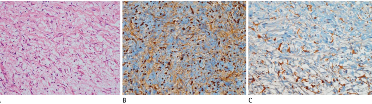
Histopathological examination of the mass showed a circum- scribed, moderately cellular spindle-cell neoplasm (Fig. 3). The mass consisted of evenly distributed spindle cells with wavy nu- clei. The spindle cells were intimately associated with ropy colla- gen bundles. Myxoid material separated the cells and collagen.
Some mononuclear cells, including mast cells and lymphocytes, were identified between the cells and the collagen. No mitosis or necrosis was observed. On immunochemical staining, most cells were positive for S100 and some stromal cells were positive for CD34. A histological diagnosis of neurofibroma was made.
DISCUSSION
Neurofibromas are common benign tumors that arise from ele- ments in the peripheral nervous system (2). They most frequently affect patients aged 20–30 years and have no sex predilection.
They are commonly found as solitary lesions or are secondary to NF1, which is considered a separate disease process. When asso- ciated with NF1, most neurofibromas occur as solitary lesions in the dermis or subcutis, and multiple lesions distributed evenly over the body surface or plexiform neurofibromas characterized by diffuse neural enlargement are often found (1). Neurofibro- mas of the breast are quite rare in patients with NF1, and they usually occur on the nipple-areolar area (2, 3).
Neurofibromas are considered to be benign tumors, but a Fig. 1. Mediolateral-oblique mammography shows a 32 × 19 mm cir-
cumscribed mass (arrows) with a partially obscured posterior margin in the posterior right upper breast. Multiple well-circumscribed ovoid lesions in both breasts are surrounded by lucent halos of air, reflecting their superficial location.
Fig. 2. US (A) and non-enhanced CT scan (B) show a well-circumscribed ovoid mass on the pectoralis major muscle (arrows). PET-CT scan (C) shows mildly increased FDG uptake. The mass (m) abuts the chest wall but is present within the breast.
FDG = fluorodeoxyglucose, PET-CT = positron emission tomography-computed tomography, US = ultrasonography
A B C
130
Neurofibroma of the Breast Detected on PET-CT in a Patient with Neurofibromatosis Type 1
jksronline.org
J Korean Soc Radiol 2016;74(2):128-131 small percentage, especially internal plexiform neurofibromas,
have malignant cellular features that make them more likely to evolve into malignant peripheral nerve sheath tumors (MPNSTs) (5). Individuals with NF1 have a 15% increased risk of malignan- cy, which is a fivefold higher for breast cancer before the age of 50 years than that seen in the general population (6). Presence of multiple neurofibromas in the breast may obscure a mass in the breast at palpation, leading to delayed clinical detection (7). Al- though current screening guidelines do not give specific con- siderations, careful screening and interpretation of mammo- graphic results for newly developing breast lesions is required.
The radiological appearance of neurofibromas varies and is known to depend on their histopathological characteristics. In the breast, neurofibromas usually appear as oval or round le- sions with circumscribed margins on both mammography and US (4). On US, they appear as hypoechoic lesions with posterior acoustic enhancement, resembling a cyst, which may cause mis- diagnosis. On magnetic resonance imaging, they demonstrate high signal intensity on T2-weighted images, especially if the tu- mor has a myxoid matrix. They may demonstrate either non- enhancement or gradual enhancement after contrast material injection (4).
In our case, a US image showed a well-circumscribed mass surrounded by extramammary fat in the retromammary area that widely abutted the pectoralis major muscle (Fig. 2A). On mammography and PET-CT, it appeared in the retromammary space and was separated from glandular tissue, indicating an ex- tramammary location (Figs. 1, 2B). The lesion was first detected on post-operative PET-CT with mild FDG uptake. The patient
had undergone a hysterectomy because of endometrioid carci- noma. Metastasis was a possible diagnosis, but most metastases of the breast are located in the parenchyma and superficially, in the fat-parenchymal interface (8). As described above, the risk of breast cancer in patients with NF1 is higher than that of the normal population (6). However, a breast cancer diagnosis was excluded because of the extramammary location of the lesion.
The patient’s history of NF1 and the extramammary location of the lesion were clues that led to a diagnosis of neurofibroma.
MPNST, a leading cause of death in NF1 patients, cannot be re- liably distinguished from neurofibroma by US (9). Therefore, if a lesion shows increased FDG uptake on PET-CT, MPNST should be considered and a histological diagnosis is necessary.
Neurofibromas are white-gray, soft, and well circumscribed but not encapsulated (10). They vary in shape and size, mostly measuring between 1 and 2 cm (4, 5). Neurofibromas are formed by a combined proliferation of all the elements of a peripheral nerve, with Schwann cells being the most predominant entities (5). Most are immunoreactive to S100 protein and, in keeping with their benign behavior, lack significant mitotic activity and necrosis (10). Schwannomas may be differentiated from neuro- fibromas by the presence of Verocay bodies, Antoni A and B areas, and a more diffuse and uniform S100 staining pattern (5, 10). These features were all absent in our case.
In conclusion, neurofibroma of the breast is rare but should be included in the differential diagnosis of breast lesion in NF1 patients. Though it can have nonspecific imaging features, its ex- tramammary location on mammography and US is useful in distinguishing it from parenchymal lesions.
Fig. 3. Microscopic examination of the specimen.
A. A microphotograph of the specimen shows evenly distributed spindle cells with wavy nuclei (hematoxylin-eosin stain, original magnification × 200).
B, C. Immunochemical stain for S100 (B) and CD34 (C) show S100 positivity in most, but not all, cells (dark cells) and CD34 positivity in some stromal cells (original magnification × 200).
A B C
131
Hoseok Lee, et al
jksronline.org J Korean Soc Radiol 2016;74(2):128-131
REFERENCES
1. Fink D, Schneider C, Wight E, Perucchini D, Haller U. [Neu- rofibromatosis of the breast in a patient with Morbus von Recklinghausen]. Gynakol Geburtshilfliche Rundsch 2000;
40:47-49
2. Sherman JE, Smith JW. Neurofibromas of the breast and nipple-areolar area. Ann Plast Surg 1981;7:302-307 3. Bongiorno MR, Doukaki S, Aricò M. Neurofibromatosis of
the nipple-areolar area: a case series. J Med Case Rep 2010;
4:22
4. Gokalp G, Hakyemez B, Kizilkaya E, Haholu A. Myxoid neurofibromas of the breast: mammographical, sono- graphical and MRI appearances. Br J Radiol 2007;80:e234- e237
5. Tucker T, Wolkenstein P, Revuz J, Zeller J, Friedman JM. As- sociation between benign and malignant peripheral nerve sheath tumors in NF1. Neurology 2005;65:205-211
6. Sharif S, Moran A, Huson SM, Iddenden R, Shenton A, Howard E, et al. Women with neurofibromatosis 1 are at a moderately increased risk of developing breast cancer and should be considered for early screening. J Med Genet 2007;44:481-484
7. Millman SL, Mercado CL. An unusual presentation of neu- rofibromatosis of the breast. Breast J 2004;10:45-47 8. Lee SH, Park JM, Kook SH, Han BK, Moon WK. Metastatic
tumors to the breast: mammographic and ultrasono- graphic findings. J Ultrasound Med 2000;19:257-262 9. Reynolds DL Jr, Jacobson JA, Inampudi P, Jamadar DA,
Ebrahim FS, Hayes CW. Sonographic characteristics of pe- ripheral nerve sheath tumors. AJR Am J Roentgenol 2004;
182:741-744
10. Jeyaretna DS, Oriolowo A, Smith ME, Watkins RM. Solitary neurofibroma in the male breast. World J Surg Oncol 2007;5:23
제1형 신경섬유종증 환자에서 양전자방출단층촬영으로 발견된 유방의 신경섬유종: 증례 보고
이호석
1· 김혜정
2* · 양보라
1· 이소미
2· 정지윤
3유방에서 발생한 신경섬유종은 드물다. 이 병변들은 주로 단발성으로 발견되거나 신경섬유종증 1형의 일부로 나타나고 보 고된 증례들에서 주로 유두-유륜 부위에 위치한다. 저자들은 자궁내막암 수술 후에 양전자방출단층촬영으로 발견된 유 방 후부에서 발생한 신경섬유종의 증례를 보고하고자 한다.
1경북대학교병원 영상의학과, 칠곡경북대학교병원 2영상의학과, 3병리과