Introduction
The association between androgens and cardiovascular diseases (CVDs) remains controversial. For many years, an- drogenic hormones have been associated with an increased risk of CVDs [1]. This has been largely based on the gender disparity in the incidences of CVDs, with a higher male pre- disposition [1]. Further, abuse of anabolic steroid by athletes has been linked to various adverse effects in the cardiovascu-
lar system including thrombosis, hypertension as well as heart failure [2]. However, recent evidence from various popula- tion-based and animal studies demonstrates beneficial effects of physiological levels of androgens on the cardiovascular system.
These beneficial effects are consistent with the higher in- cidence of atherosclerosis [3, 4], aneurysms [5], heart failure and coronary artery disease [6] among hypogonadal males.
The hypogonadism may be due to age-related decline in endogenous androgen levels [7, 8], androgen deprivation therapies [9], and disorders that either damage the testis or re- duce gonadotropin stimulation [10, 11]. The basis for higher prevalence of CVD among hypogonadal men has been ex- plored in biochemical and physiological models and has been attributed to adverse effects of androgen insufficiency on lipid profiles [3]. Low androgen levels are associated with increased
Corresponding author:
Isaac Cheruiyot
Department of Human Anatomy, University of Nairobi, P.O. Box 30197- 00100, Nairobi, Kenya
Tel: +254-721353833, Fax: +254-02318262, E-mail: [email protected]
Histomorphological changes in the common carotid artery of the male rat in induced
hypogonadism
Isaac Cheruiyot, Beda Olabu, Martin Kamau, Kevin Ongeti, Pamela Mandela
Department of Human Anatomy, University of Nairobi, Nairobi, Kenya
Abstract: The role of androgens in the development of cardiovascular diseases remains controversial. The current study therefore sought to determine the changes in the histomorphology of the common carotid artery of the male rat in orchidectomy-induced hypogonadism. Twenty-two Rattus norvegicus male rats aged 2 months were used. The rats were randomly assigned into baseline (n=4), experimental (n=9), and control (n=9) groups. Hypogonadism was surgically induced in the experimental group by bilateral orchiectomy under local anesthesia. At experiment weeks 3, 6, and 9, three rats from each group (experimental and control) were euthanized, their common carotid artery harvested, and routine processing was done for paraffin embedding, sectioning, and staining. The photomicrographs were taken using a digital photomicroscope for morphometric analysis. Orchidectomy resulted in the development of vascular fibrosis, with a significant increase in collagen fiber density and decrease in smooth muscle and elastic fiber density. Moreover, there was development of intimal hyperplasia, with fragmentation of medial elastic lamellae in the common carotid artery of the castrated rats. Orchidectomy induces adverse changes in structure of the common carotid artery of the male rat. These changes may impair vascular function, therefore constituting a possible structural basis for the higher incidences of cardiovascular diseases observed in hypogonadism.
Key words: Androgens, Cardiovascular disease, Hypogonadism, Common carotid artery, Orchidectomy
Received April 30, 2018; 1st Revised June 25, 2018; 2nd Revised June 30, 2018; 3rd Revised July 2, 2018; Accepted July 3, 2018
in hypogonadism is associated with endothelial dysfunction [9, 15], structural changes in the vascular wall that may con- stitute an anatomical basis for the higher prevalence of CVD among hypogonadal men are largely unexplored. This study therefore aims to describe structural changes in the common carotid artery (CCA) of male rat following orchiectomy- induced hypogonadism.
Materials and Methods
This was a non-randomized trial (quasi-experimental) us- ing the rat model. A total of 22 male rats aged 2 months were used in this study. Rats were used as the study model because of their ease of handling, low maintenance cost and close physiological resemblance to man. Further, the structure of the rat CCA is similar to that in man. Two-month-old rats were used because this is the age at which they attain sexual maturity. Rats with visible neck or scrotal pathology were ex- cluded. The animals were housed in cages floored with wood shavings that were changed regularly. They were kept in their cages for 2 weeks prior to commencement of the study for acclimatization. The animals were placed under a normal 12 hours’ light/dark diurnal cycle and provided with standard rat pellets and water ad libitum. Ethical approval to conduct the study was granted by the Biosafety, Animal Care and Use committee of Nairobi University, Nairobi, Kenya (Ethical ap- proval number: FVMBAUEC/2016/96).
Four rats were chosen using simple random sampling tech- nique to demonstrate the baseline (day 0) histomorphology of the CCA. The remaining rats were divided randomly into two groups (11 experimental, 11 controls). Hypogonadism was induced in the experimental via bilateral orchiectomy under local anesthesia. The animals were placed in the dorsal recumbent position and under physical restrain. The scrotal skin disinfected using iodine solution. Two milliliters of 1%
lignocaine was injected in the around the scrotal sac to pro- vide local anesthesia. A 1.5-cm incision was made at the base of the hemi-scrotal sacs. Subcutaneous tissue was bluntly dis- sected to reveal the vaginal processes. This was then excised to access the testis and the spermatic cord which were then
wool soaked in iodine and secured with a bandage.
On experimental week 3, 6, and 9, three animals from both groups were picked randomly, euthanized and perfused with normal and formal saline solutions. Their CCA were harvested and processed for paraffin embedding and section- ing. The rats’ CCA were fixed in 10% formalin for twelve hours. This was followed by dehydration in increasing grades of alcohol (70% up to absolute alcohol) at one hour intervals, and clearing in toluene. Thereafter, the vessels were placed in the memmert oven for wax infiltration. The CCA were em- bedded in paraffin wax and oriented for transverse section- ing. After cooling, the embedded tissues were blocked using wooden blocks and then serially cut into 7-μm sections using a microtome. Fifteen 7-μm sections were randomly obtained from the ten ribbons, floated on a 60°C water bath and picked on a glass slide, then dried in an oven for 12 hours. Masson’s Trichrome was used to display smooth muscle cells and col- lagen fibers while Wiegerts stain was used to display elastic fiber profile. Hematoxylin and eosin was used to display the smooth muscle cell nuclei in the CCA.
Photomicrographs of the sections were taken using a digi- tal camera (Canon Powershot A640, 12 mp, Beijing, China) mounted on a photomicroscope (Carl Zeiss, Axiostar Plus Microimaging, Jena, Germany) for stereological analysis using the Fiji-ImageJ. This is an open source software developed by the United States National Institute of Health for processing and analyzing images. The variables obtained include volu- metric densities of collagen fibers, elastic fibers, and smooth muscle cells (in %). Histomorphological changes of the vessel wall were also described. The collected were entered into the SPSS software version 21 (IBM Corp., Armonk, NY, USA) for coding, tabulation and statistical analysis. Volumetric densi- ties are expressed in frequencies (%). The data were groupd:
control group (non-castrated) and experimental (castrated) group. After confirming that the data was not normally dis- tributed (using box plots and histograms), non-parametric tests were used for univariate analysis. Kruskal‒Wallis H-test was used to compare the medians of the volumetric densities of collagen fibers, elastic fibers, and smooth muscle cell nuclei along the various harvesting periods within each group while
Man‒Whitney U test was used to compare the medians of the above variables between control and experimental groups. A P-value <0.05 was considered significant at 95% confidence interval. Data are presented in tables and photomicrographs.
Results
The CCA displayed features of an elastic artery with three conventional tunics; tunica intima (TI), media (TM), and ad- ventitia (TA). TI consisted of a single layer of endothelial cells lying on a thin sub-endothelial connective tissue. TM was the most prominent layer, being made up of 3‒5 thick continuous elastic lamellae with smooth muscle cells and collagen fibers interspersed between them. TA comprised of thick collagen fiber bundles running circumferentially around the vessel wall. Elastic fibers were also identified in this layer.
Intima hyperplasia
Androgen deprivation resulted in the development of focal
intimal thickenings in the CCA of castrated animals. These thickenings were composed of collagen fibers and smooth muscle cells (Fig. 1D, F).
Decreased smooth muscle cell density
There was a progressive decrease in the smooth muscle cells count in the CCA of the castrated animals from 48.84%
at baseline to 34.38%, 28.35%, and 24.25% in week 3, 6, and 9, respectively (P<0.001) (Fig. 2), with that in the control animals remaining fairly constant, being 48.79%, 48.1%, and 46.3% in week 3, 6, and 9, respectively (P=0.414). Moreover, castration resulted in reduced thickness of the smooth muscle bundles, coupled with a decrease in medial smooth muscle cell nuclei as demonstrated with hematoxylin and eosin stain (Fig. 1A‒D).
Increased deposition of collagen fibers
Castration resulted in vascular fibrosis with a progressive increase in the CCA collagen fiber density (Fig. 1C, D), from
A B C D
E F G H
Fig. 1. Photomicrographs of common carotid artery (CCA). (A) A photomicrograph showing the structure of the CCA of a normogonadic rat seen at week 6. Note the numerous spindle shaped smooth muscle cell nuclei (black arrows) in the tunica media (TM). TA, tunica adventitia. Hematoxylin and eosin (H&E) staining. (B) A photomicrograph showing structure of the CCA of a hypogonadic rat seen 6 weeks after castration. Note the decrease in smooth muscle cell nuclei (black arrows). H&E staining. (C) A photomicrograph of CCA of a normogonadic rat seen a normogonadic rat at week 9 of the study. Note the abundant smooth muscle cells in the TM. IMT, intimamedial thickness. Masson’s trichrome staining. (D) A photomicrograph of the CCA of a hypogonadic rat seen a normogonadic rat at week 9 of the study. Note that collagen:smooth muscle ratio is higher compared to controls rats. Also not the disruption of collagen fibers in the TA due to wider spaces between the fibers. TI, tunica intima. Masson’s trichrome staining. (E) A photomicrograph showing the structural organization of the CCA of a normogonadic rat at week 6 of the study. Note the continuous wavy elastic lamellae in the TM and abundant dark staining elastic fibers in the TA. Wiegerts’ elastic staining. (F) A photomicrograph showing the structural organization of the CCA of a hypogonadic rat seen 6 weeks after castration. Note the decreased quantity of elastic fibers in the TA (arrowheads).
Wiegerts’ elastic staining. (G) A photomicrograph showing the organization of TM of CCA of a normogonadic rat at week 9 of the study. Note the abundant elastic fibers (black arrows) in the TA. Although the elastic lamellae did not take in enough stain, note that they are thick continuous wavy. Wiegerts’ elastic staining. (H) A photomicrograph showing the structural organization of TM of CCA of a hypogonadic rat seen 9 weeks after castration. Note the loss elastic fibers, and the loss of continuity and concentric arrangement of elastic lamellae (black arrows). Wiegerts’ elastic staining.
Scale bars=200 μm (A–H).
33.22% at baseline to 61%, 70.52%, and 74.23% at week 3, 6, and 9 after castration respectively (P<0.001) (Fig. 3). There were however no statistically significant changes in the col- lagen fiber density in control rats across the study period (P=0.216). Further, there was disruption of the lamellae orga- nization of collagen fiber bundles in the TA of castrated rats as evidenced by wider spaces between the collagen fibers.
Disruption of elastic lamellae organization
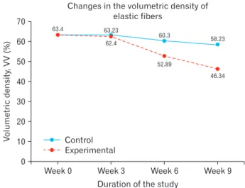
Androgen deprivation resulted in the disruption of the lamella organization of medial elastic lamellae in the CCA of castrated rats (Fig. 1E, F). While the CCA of control rats had thick continuous medial elastic lamellae running circumfer- entially around the vessel wall, there was a reduction in the
thickness coupled with fragmentation of these elastic lamel- lae in the TM of castrated. Further, there was a significant decrease in the elastic fiber density in the CCA of castrated rats (Fig. 1E, F), from 63.4% at baseline to 62.4%, 52.89%, and 46.34% at week 3, 6, and 9, respectively (P=0.014) (Fig.
4). There were however no statistically significant changes in the elastic fiber densities in control animals across the study period (P=0.074).
Discussion
Higher incidences of CVD have been reported among hypogonadal males compared to their normogonadic coun- terparts. The structural basis for this predisposition however remains relatively underexplored.
In the current study, orchidectomy resulted in the devel- opment of intimal hyperplasia (IH). Non-induced IH in hy- pogonadal states remains hitherto undescribed to the best of our knowledge. Previous studies focused mainly on effects of exogenous testosterone on the size of mechanically-induced carotid atherosclerotic plaque, reporting a reduction in the plaque area following treatment of the animals with exog- enous testosterone [12, 16]. Similarly, population-based stud- ies have reported an inverse correlation between the levels of endogenous androgens and the presence of carotid and aortic atherosclerotic plaques [17, 18]. Since IH normally precedes the formation of an atherosclerotic plaque, all are therefore in agreement that that low levels of androgens are associated with IH.
The IH observed in the current study could be attributed
Volumetricdensity 30 20
10
Week 9 Duration of the study
0
Control Experimental
Week 6 Week 3
Week 0
24.25
Fig. 2. Line graph showing trends in smooth muscle cell density in control and experimental animals.
Volumetricdensity,VV(%)
80
50 40 30 20 10
Week 9 Duration of the study
0
Control Experimental
Changes in the volumetric density of collagen fibers
70 60
Week 6 Week 3
Week 0 33.22
61.01
37.21
70.52
38.98
74.23
40.33
Fig. 3. Line graph showing trends in collagen fiber density in control and experimental animals.
Volumetricdensity
30 20 10
Week 9 Duration of the study
0
Control Experimental
Week 6 Week 3
Week 0
Fig. 4. Line graph showing trends in elastic fiber density in control and experimental animals.
to the increased production of pro-inflammatory cytokines [19] and urotensin II [20] that normally occurs in hypogo- nadal states. The former is known to cause endothelial dam- age [19], while the latter is involved in the stimulation of vas- cular smooth muscle cell proliferation and migration [21] as well as the recruitment of macrophages into the vascular wall [22]. Both processes are crucial in the pathogenesis of IH.
Since the rate of development of the IH in the current study was proportional to the duration of exposure of the animals to hypogonadism; and that IH normally precedes atheroma formation, it is plausible to postulate the IH would have de- veloped into an atherosclerotic plaque had the animals been followed up for a longer duration of time. The progressive occlusion of the vessel lumen in atherosclerosis is known to compromise vascular supply to vital organs such as the brain and heart. The observations of the current study may there- fore partly explain the higher incidences of myocardial isch- emia [6] as well as stroke [23] among hypogonadal men.
The present study reports a decrease in vascular smooth muscle composition following orchidectomy-induced hypo- gonadism. This is in agreement with previous studies that reported decreased penile trabeculae smooth muscle cells [24, 25] as well as cardiomyocytes [26, 27] in hypogonadal states.
Studies have also reported disorganization of smooth muscle cells and decreased myofilament quantity in castrated animals [24, 28]. This decrease may be attributed to increased pro- grammed cell death, atrophy and dedifferentiation into other phenotypes [28]. Androgen deprivation is known to result in the upregulation of angiotensin II (AT2) receptors within the vascular wall [27, 29]. These receptors are known to acti- vate caspases [26], the primary mediators of apoptosis, while causing the downregulation of anti-apoptotic molecules, B- cell lymphoma 2 [27]. Another possible explanation for the decrease in smooth muscle cells may be androgen deficiency- associated atrophy. Although we could not find any published literature detailing atrophy of vascular smooth muscles in hypogonadal states, androgen deprivation in animals have shown to induce atrophy of penile cavernosal smooth muscle cells [24, 30, 31]. This is supported by skeletal muscle atrophy and fatigue normally observed in hypogonadal men [4, 32].
The decrease in smooth muscle cells can also attributed to their dedifferentiation into other phenotypes. Unlike skel- etal or cardiac muscle cell that are terminally differentiated, smooth muscle cells retain remarkable plasticity and can undergo phenotypic changes in conditions such as vascular wall injury [33]. Concordant with this, surgical castration as
well as blockade of 5α reductase has been demonstrated to cause dedifferentiation of prostate smooth muscle cells into fibroblasts [34, 35]. Furthermore, it is known that androgens induce mesenchymal stem cells into the smooth muscle cell lineage, and androgen deficiency accordingly causes these stem cells to follow adipocyte lineage [31]. However, we did not identify any deposition of adipose tissue within the vessel wall. Vascular smooth muscle cells are important in regula- tion of vascular tone, blood pressure and wall integrity [36].
Decreased smooth muscle cell quantity is associated with various arterial pathologies such as aneurysms [37], hyper- tension [36], vascular calcification as well as rapture of ath- erosclerotic plaque [38]. Therefore, decreased smooth muscle density observed in this study may provide a structural basis for the higher incidences of aneurysms as well as hyperten- sion among hypogonadal men [5].
The present study also demonstrates that castration re- sults in increased volumetric density of vascular collagen fibers, proportional to the duration of hypogonadism. Similar changes have been reported to occur in the penis [24, 25, 39]
and myocardium [40, 41] in studies involving androgen hor- mone deficiency. These results supported by the observation that androgen receptor knockout in mice exacerbates cardiac fibrosis [42]. Increased collagen fiber could be attributed to accumulation of fibroblasts within the vessel wall or their differentiation into a more proliferative and synthetic phe- notype, myofibroblasts [43]. Various pathways involved in increased collagen fiber synthesis in hypogonadal states have been described. It is known that androgens have an inhibi- tory effect on the production of transforming growth factor β (TGF-β) by various inflammatory cells such as macrophages and monocytes [44]. Thus, androgen ablation results in the upregulation of TGF-β production [19, 45], resulting in the activation of fibroblasts, with increased production of extra- cellular matrix [46]. TGF-β also induces the differentiation of these fibroblasts into more synthetic types, the myofibroblasts [47]. Another possible explanation for the decrease in smooth muscle cells is the upregulation of AT2 production and ex- pression in cardiomyocytes and vascular smooth muscle cells [27]. AT2 promotes fibrosis by activating fibroblasts as well as enhancing their differentiation into myofibroblasts, with increased production of extracellular matrix [48]. Collagen fibers are important for structural support of the vessel wall by preventing overstretching during systole [49]. Their excess production however leads to stiffening of arteries, reducing vessel compliance [50]. This also occurs with aging and has
tial elastic fibers, coupled with fragmentation of medial elastic lamellae. Changes in vascular elastic fibers and medial elastic lamellae with hypogonadism remain hitherto undescribed, although similar findings have been reported in the penile elastic fiber system [25, 39, 51] and in vascular aging [52, 53].
The molecular mechanisms underlying elastic fiber reduc- tion and fragmentation of medial elastic lamellae in hypogo- nadism remain relatively unexplored. However, matrix metal- loproteinases (MMPs), particularly MMP2 and MMP9 have been shown to have high affinity for elastin and are involved in their degradation during extracellular matrix remodeling [54-56]. These MMPs can be activated by various cytokines such as TGF-β and tumor necrosis factor α [57, 58]. Since expression of these cytokines is normally upregulated in hy- pogonadal states [19], it is possible that the decrease in elastic fibers as well as elastic lamellae fragmentation observed in this study is attributed to cytokine-mediated MMP degrada- tion. Furthermore, some workers have argued that androgens regulated protein synthesis of connective tissue, and decrease in their production could therefore give rise to the switch from elastic fibers to collagen fibers, forming another possible basis for the decreased elastic fiber density [59]. The decrease in volumetric density of elastic fibers suggests a higher rate of degradation by MMPs compared to the rate of deposition by fibroblasts.
Together with collagen fibers, elastic fibers and lamellae are important in maintaining the compliant nature of dy- namic structures such as arteries [60]. They are integral to the windkessel mechanism that enables conduit arteries such as the aorta to deform during systole and use the stored energy to recoil to their original states during diastole, thereby driv- ing blood through the circulatory system [53, 56]. However, with aging, there is loss of elastic fibers as well as fragmenta- tion of elastic lamellae [61, 62]. As such, the load is trans- ferred to collagen fibers, which are stiffer, resulting in loss of vessel compliance [56]. Such changes are known to cause an increase in systolic blood pressure, a key risk factor for the development of heart failure [53, 61]. Also, fragmentation of elastic lamellae results in loss of integrity of the vessel wall, which contributes to aneurysmal dilatations [63, 64].
[5] in hypogonadal men.
Limitations
Castration is a surgical procedure that causes tissue injury.
Therefore, some of the changes observed in this study may have been due to reactive processes to tissue injury. However, based on the fact that the changes observed in this study were proportional to the duration of androgen deficiency, they are most likely to be due to gonadal hormone deficiency rather than surgical trauma. We were unable to determine whether the decrease in smooth muscle composition was as a result of atrophy, apoptosis or both.
Nonetheless, this is the first study to the best of our knowl- edge, to detail changes in the vascular structure in induced hypogonadism. The results of the current study may consti- tute an anatomical basis for the higher incidences of CVDs among hypogonadal males.
Conclusion
Orchidectomy induces adverse changes in structure of the CCA of the male rat. These changes may impair vascular function, therefore constituting a possible structural basis for the higher incidences of CVDs observed in hypogonadism.
References
1. McGrath KC, McRobb LS, Heather AK. Androgen therapy and atherosclerotic cardiovascular disease. Vasc Health Risk Manag 2008;4:11-21.
2. Maravelias C, Dona A, Stefanidou M, Spiliopoulou C. Adverse effects of anabolic steroids in athletes: a constant threat. Toxicol Lett 2005;158:167-75.
3. Haring R, Baumeister SE, Völzke H, Dörr M, Felix SB, Kroemer HK, Nauck M, Wallaschofski H. Prospective association of low total testosterone concentrations with an adverse lipid profile and increased incident dyslipidemia. Eur J Cardiovasc Prev Re- habil 2011;18:86-96.
4. Fahed AC, Gholmieh JM, Azar ST. Connecting the lines between hypogonadism and atherosclerosis. Int J Endocrinol 2012;2012:
793953.
5. Yeap BB, Hyde Z, Almeida OP, Norman PE, Chubb SA, Jamrozik K, Flicker L, Hankey GJ. Lower testosterone levels predict inci-
dent stroke and transient ischemic attack in older men. J Clin Endocrinol Metab 2009;94:2353-9.
6. Martín-Merino E, Johansson S, Morris T, García Rodríguez LA. Androgen deprivation therapy and the risk of coronary heart disease and heart failure in patients with prostate cancer:
a nested case-control study in UK primary care. Drug Saf 2011;
34:1061-77.
7. Khaw KT, Dowsett M, Folkerd E, Bingham S, Wareham N, Luben R, Welch A, Day N. Endogenous testosterone and mor- tality due to all causes, cardiovascular disease, and cancer in men: European prospective investigation into cancer in Norfolk (EPIC-Norfolk) Prospective Population Study. Circulation 2007;
116:2694-701.
8. Menke A, Guallar E, Rohrmann S, Nelson WG, Rifai N, Kanarek N, Feinleib M, Michos ED, Dobs A, Platz EA. Sex steroid hor- mone concentrations and risk of death in US men. Am J Epide- miol 2010;171:583-92.
9. Karakitsos D, Patrianakos AP, De Groot E, Boletis J, Karabinis A, Kyriazis J, Samonis G, Parthenakis FI, Vardas PE, Daphnis E.
Androgen deficiency and endothelial dysfunction in men with end-stage kidney disease receiving maintenance hemodialysis.
Am J Nephrol 2006;26:536-43.
10. Kumar N, Swamy R, Patil J, Guru A, Aithal A, Shetty P. Presence of arteriovenous communication between left testicular ves- sels and its clinical significance. Case Rep Vasc Med 2014;2014:
160824.
11. Christe N, Meier CA. Hypotestosteronaemia in the aging male:
should we treat it? Swiss Med Wkly 2015;145:w14216.
12. Alexandersen P, Haarbo J, Byrjalsen I, Lawaetz H, Christiansen C. Natural androgens inhibit male atherosclerosis: a study in cas- trated, cholesterol-fed rabbits. Circ Res 1999;84:813-9.
13. Muraleedharan V, Jones TH. Testosterone and the metabolic syndrome. Ther Adv Endocrinol Metab 2010;1:207-23.
14. Bobjer J, Naumovska M, Giwercman YL, Giwercman A. High prevalence of androgen deficiency and abnormal lipid profile in infertile men with non-obstructive azoospermia. Int J Androl 2012;35:688-94.
15. Castela A, Vendeira P, Costa C. Testosterone, endothelial health, and erectile function. ISRN Endocrinol 2011;2011:839149.
16. Hanke H, Lenz C, Hess B, Spindler KD, Weidemann W. Effect of testosterone on plaque development and androgen receptor expression in the arterial vessel wall. Circulation 2001;103:1382- 5.
17. Chan YX, Knuiman MW, Hung J, Divitini ML, Handelsman DJ, Beilby JP, McQuillan B, Yeap BB. Testosterone, dihydrotestoster- one and estradiol are differentially associated with carotid inti- ma-media thickness and the presence of carotid plaque in men with and without coronary artery disease. Endocr J 2015;62:777- 86.
18. Farias JM, Tinetti M, Khoury M, Umpierrez GE. Low testoster- one concentration and atherosclerotic disease markers in male patients with type 2 diabetes. J Clin Endocrinol Metab 2014;99:
4698-703.
19. Gilliver SC, Ashworth JJ, Mills SJ, Hardman MJ, Ashcroft GS.
Androgens modulate the inflammatory response during acute wound healing. J Cell Sci 2006;119(Pt 4):722-32.
20. Pelletier G, Lihrmann I, Dubessy C, Luu-The V, Vaudry H, Lab- rie F. Androgenic down-regulation of urotensin II precursor, urotensin II-related peptide precursor and androgen receptor mRNA in the mouse spinal cord. Neuroscience 2005;132:689-96.
21. Rodríguez-Moyano M, Díaz I, Dionisio N, Zhang X, Avila- Medina J, Calderón-Sánchez E, Trebak M, Rosado JA, Ordóñez A, Smani T. Urotensin-II promotes vascular smooth muscle cell proliferation through store-operated calcium entry and EGFR transactivation. Cardiovasc Res 2013;100:297-306.
22. Zhao J, Zhang SF, Shi Y, Ren LQ. Effects of urotensin II and its specific receptor antagonist urantide on rat vascular smooth muscle cells. Bosn J Basic Med Sci 2013;13:78-83.
23. Morgentaler A. Testosterone deficiency and cardiovascular mor- tality. Asian J Androl 2015;17:26-31.
24. Traish A, Kim N. The physiological role of androgens in penile erection: regulation of corpus cavernosum structure and func- tion. J Sex Med 2005;2:759-70.
25. Olabu BO. Structural changes in the rabbit penile architecture in induced hypogonadism [thesis]. Nairobi: University of Nairobi;
2014.
26. Sánchez-Más J, Turpín MC, Lax A, Ruipérez JA, Valdés Chávarri M, Pascual-Figal DA. Differential actions of eplerenone and spironolactone on the protective effect of testosterone against cardiomyocyte apoptosis in vitro. Rev Esp Cardiol 2010;63:779- 87.
27. Kang NN, Fu L, Xu J, Han Y, Cao JX, Sun JF, Zheng M. Testos- terone improves cardiac function and alters angiotensin II recep- tors in isoproterenol-induced heart failure. Arch Cardiovasc Dis 2012;105:68-76.
28. Traish AM, Goldstein I, Kim NN. Testosterone and erectile func- tion: from basic research to a new clinical paradigm for manag- ing men with androgen insufficiency and erectile dysfunction.
Eur Urol 2007;52:54-70.
29. Ikeda Y, Aihara K, Yoshida S, Sato T, Yagi S, Iwase T, Sumitomo Y, Ise T, Ishikawa K, Azuma H, Akaike M, Kato S, Matsumoto T.
Androgen-androgen receptor system protects against angioten- sin II-induced vascular remodeling. Endocrinology 2009;150:
2857-64.
30. Wespes E. Smooth muscle pathology and erectile dysfunction.
Int J Impot Res 2002;14 Suppl 1:S17-21.
31. Traish AM, Kim N. Weapons of penile smooth muscle destruc- tion: androgen deficiency promotes accumulation of adipocytes in the corpus cavernosum. Aging Male 2005;8:141-6.
32. Dandona P, Rosenberg MT. A practical guide to male hypogo- nadism in the primary care setting. Int J Clin Pract 2010;64:682- 96.
33. Rzucidlo EM, Martin KA, Powell RJ. Regulation of vascular smooth muscle cell differentiation. J Vasc Surg 2007;45 Suppl A:
A25-32.
34. Arnold JT, Isaacs JT. Mechanisms involved in the progression of androgen-independent prostate cancers: it is not only the cancer cell's fault. Endocr Relat Cancer 2002;9:61-73.
37. Ailawadi G, Eliason JL, Upchurch GR Jr. Current concepts in the pathogenesis of abdominal aortic aneurysm. J Vasc Surg 2003;38:584-8.
38. Clarke MC, Figg N, Maguire JJ, Davenport AP, Goddard M, Lit- tlewood TD, Bennett MR. Apoptosis of vascular smooth muscle cells induces features of plaque vulnerability in atherosclerosis.
Nat Med 2006;12:1075-80.
39. El-Sakka AI. Reversion of penile fibrosis: Current information and a new horizon. Arab J Urol 2011;9:49-55.
40. Chung CC, Kao YH, Chen YJ, Chen YJ. Androgen modulates cardiac fibrosis contributing to gender differences on heart fail- ure. Aging Male 2013;16:22-7.
41. Čulić V. Androgens in cardiac fibrosis and other cardiovascular mechanisms. Int J Cardiol 2015;179:190-2.
42. Ikeda Y, Aihara K, Sato T, Akaike M, Yoshizumi M, Suzaki Y, Izawa Y, Fujimura M, Hashizume S, Kato M, Yagi S, Tamaki T, Kawano H, Matsumoto T, Azuma H, Kato S, Matsumoto T. An- drogen receptor gene knockout male mice exhibit impaired car- diac growth and exacerbation of angiotensin II-induced cardiac fibrosis. J Biol Chem 2005;280:29661-6.
43. Darby IA, Hewitson TD. Fibroblast differentiation in wound healing and fibrosis. Int Rev Cytol 2007;257:143-79.
44. Chipuk JE, Cornelius SC, Pultz NJ, Jorgensen JS, Bonham MJ, Kim SJ, Danielpour D. The androgen receptor represses trans- forming growth factor-beta signaling through interaction with Smad3. J Biol Chem 2002;277:1240-8.
45. Placencio VR, Sharif-Afshar AR, Li X, Huang H, Uwamariya C, Neilson EG, Shen MM, Matusik RJ, Hayward SW, Bhowmick NA. Stromal transforming growth factor-beta signaling mediates prostatic response to androgen ablation by paracrine Wnt activ- ity. Cancer Res 2008;68:4709-18.
46. Petrov VV, Fagard RH, Lijnen PJ. Stimulation of collagen pro- duction by transforming growth factor-beta1 during differentia- tion of cardiac fibroblasts to myofibroblasts. Hypertension 2002;
39:258-63.
47. Pohlers D, Brenmoehl J, Löffler I, Müller CK, Leipner C, Schul- tze-Mosgau S, Stallmach A, Kinne RW, Wolf G. TGF-beta and fi- brosis in different organs: molecular pathway imprints. Biochim Biophys Acta 2009;1792:746-56.
48. Sorescu D. Smad3 mediates angiotensin II- and TGF-beta1-in- duced vascular fibrosis: Smad3 thickens the plot. Circ Res 2006;
98:988-9.
49. Chow MJ, Turcotte R, Lin CP, Zhang Y. Arterial extracellular matrix: a mechanobiological study of the contributions and in- teractions of elastin and collagen. Biophys J 2014;106:2684-92.
bly. Birth Defects Res C Embryo Today 2007;81:229-40.
53. Sherratt MJ. Tissue elasticity and the ageing elastic fibre. Age (Dordr) 2009;31:305-25.
54. Chung AW, Au Yeung K, Sandor GG, Judge DP, Dietz HC, van Breemen C. Loss of elastic fiber integrity and reduction of vas- cular smooth muscle contraction resulting from the upregulated activities of matrix metalloproteinase-2 and -9 in the thoracic aortic aneurysm in Marfan syndrome. Circ Res 2007;101:512-22.
55. Lau AC, Rosenberg H, Duong TT, McCrindle BW, Yeung RS.
Elastolytic matrix metalloproteinases and coronary outcome in children with Kawasaki disease. Pediatr Res 2007;61:710-5.
56. Wagenseil JE, Mecham RP. Elastin in large artery stiffness and hypertension. J Cardiovasc Transl Res 2012;5:264-73.
57. Han YP, Tuan TL, Wu H, Hughes M, Garner WL. TNF-alpha stimulates activation of pro-MMP2 in human skin through NF- (kappa)B mediated induction of MT1-MMP. J Cell Sci 2001;
114(Pt 1): 131-9.
58. Gordon GM, Ledee DR, Feuer WJ, Fini ME. Cytokines and sig- naling pathways regulating matrix metalloproteinase-9 (MMP-9) expression in corneal epithelial cells. J Cell Physiol 2009;221:402- 11.
59. Traish AM, Guay AT. Are androgens critical for penile erections in humans? Examining the clinical and preclinical evidence. J Sex Med 2006;3:382-404.
60. Cecelja M, Chowienczyk P. Role of arterial stiffness in cardiovas- cular disease. JRSM Cardiovasc Dis 2012;1:cvd.2012.012016.
61. O’Rourke MF, Hashimoto J. Mechanical factors in arterial aging:
a clinical perspective. J Am Coll Cardiol 2007;50:1-13.
62. Fleenor BS. Large elastic artery stiffness with aging: novel trans- lational mechanisms and interventions. Aging Dis 2013;4:76-83.
63. Jacob T, Hingorani A, Ascher E. Role of apoptosis and proteoly- sis in the pathogenesis of iliac artery aneurysms. Vascular 2005;
13:34-42.
64. Isenburg JC, Simionescu DT, Starcher BC, Vyavahare NR. Elas- tin stabilization for treatment of abdominal aortic aneurysms.
Circulation 2007;115:1729-37.
65. Oka R, Utsumi T, Endo T, Yano M, Kamijima S, Kamiya N, Shirai K, Suzuki H. Effect of androgen deprivation therapy on arterial stiffness and serum lipid profile changes in patients with prostate cancer: a prospective study of initial 6-month follow-up.
Int J Clin Oncol 2016;21:389-96.
66. Dockery F, Bulpitt CJ, Agarwal S, Vernon C, Rajkumar C. Ef- fect of androgen suppression compared with androgen receptor blockade on arterial stiffness in men with prostate cancer. J An- drol 2009;30:410-5.