A Multicenter Retrospective Analysis of the Clinical Features of Pernicious Anemia in a Korean Population
To determine the approximate incidence and clinical features of pernicious anemia in a Korean population, we retrospectively analyzed clinical data for patients with pernicious anemia who were diagnosed between 1995 and 2010 at five hospitals in Chungnam province. Ninety-seven patients were enrolled, who accounted for 24% of patients with vitamin B12 deficiency anemia. The approximate annual incidence of pernicious anemia was 0.3 per 100,000. The median age was 66 (range, 32-98) yr, and the male/female ratio was 1.25. Anemia-associated discomfort was the most common symptom (79.4%), followed by gastrointestinal and neurological symptoms (78.4% and 38.1%, respectively).
Pancytopenia was found in 36 patients (37.1%), and autoimmune disorders were found in 15 patients (15.5%). Antibody to intrinsic factor was detected in 62 (77.5%) of 80 patients examined, and antibody to parietal cells was detected in 35 (43.2%) of 81 patients examined. Of the 34 patients who underwent tests for Helicobacter pylori, 7 (12.5%) were positive. The anemia-associated and gastrointestinal symptoms resolved completely in all patients after intramuscular injection of cobalamin, whereas neurological symptoms remained in some. In conclusion, pernicious anemia is less frequent in Koreans than in Western populations; however, the clinical features of this disorder in Koreans do not differ from those of Western cases.
Key Words: Anemia, Pernicious; Vitamin B12; Anti-Intrinsic Factor Antibody; Anti-Parietal Cell Antibody; Helicobacter Pylori
Ik-Chan Song,1 Hyo Jin Lee,1 Han-Jo Kim,2 Sang-Byung Bae,2 Kyu-Taek Lee,2 Young-Jun Yang,3 Suk-Young Park,3 Do-Yeun Cho,4 Nae Yu Kim,5 In-Sung Cho,5 and Deog-Yeon Jo1
1Department of Internal Medicine, Chungnam National University Hospital, Daejeon; 2Department of Internal Medicine, Soonchunhyang University Hospital, Cheonan; 3Department of Internal Medicine, Daejeon St Mary’s Hospital, Daejeon; 4Department of Internal Medicine, Konyang University Hospital, Daejeon; 5Department of Internal Medicine, Eulji University Hospital, Daejeon, Korea
Received: 14 May 2012 Accepted: 16 November 2012 Address for Correspondence:
Deog-Yeon Jo, MD
Department of Internal Medicine, Chungnam National University Hospital, 33 Munwha-ro, Jung-gu, Daejeon 301-721, Korea
Tel: +82.42-280-7162, Fax: +82.42-257-5753 E-mail: [email protected]
http://dx.doi.org/10.3346/jkms.2013.28.2.200 • J Korean Med Sci 2013; 28: 200-204
INTRODUCTION
Pernicious anemia is a megaloblastic anemia caused by intrin- sic factor deficiency secondary to autoimmune destruction of the acid- and pepsin-secreting portion of the gastric mucosa.
Pernicious anemia is the most common cause of vitamin B12
deficiency and one of the most common autoimmune diseases in Western countries (1, 2). Pernicious anemia was previously thought to be a rare disease in East Asia, but is now regarded as a disorder affecting virtually all racial and ethnic groups (3). Al- though reports describing pernicious anemia in the East Asia are quite rare, there is some information on the clinical features of this disorder in Japan and China. For example, Sugihara and Yawata (4) reported the clinical statistical data of pernicious ane- mia in a Japanese population in 1992. Recently, Chan et al. (5) reported the clinical features of 181 patients with pernicious anemia and demonstrated that pernicious anemia is a major cause of megaloblastic anemia in Chinese individuals. In Korea, the first case of pernicious anemia was reported in 1966 (6). Since then, only case reports or small series have been reported (7-9).
For example, Song et al. (8) described eight patients with perni-
cious anemia among 45 patients with vitamin B12 deficiency megaloblastic anemia, and Chun et al. (9) reported the clinical features of 22 patients with pernicious anemia. The incidence, prevalence, and clinical features of this disorder remain largely unknown in Korea. Thus, we performed a multicenter retro- spective study to estimate the approximate incidence and de- fine the clinical features of pernicious anemia in a Korean pop- ulation.
MATERIALS AND METHODS Study subjects
We retrospectively analyzed the medical records of patients who were newly diagnosed with pernicious anemia between 1995 and 2010 at five university hospitals in Chungnam province. The diagnosis of pernicious anemia was made when serum vitamin B12 levels were < 200 pg/mL in patients with anemia (hemoglo- bin level of < 13.0 g/dL in men and < 12.0 g/dL in women) who displayed one of the following abnormalities: positive anti-in- trinsic factor antibody, positive anti-parietal cell antibody with moderate to severe atrophic gastritis of the stomach body with-
out Helicobacter pylori infection, histologically confirmed severe atrophic gastritis of the stomach body, or the presence of other autoimmune disorders.
Data acquisition and analysis
We collected and analyzed the results of clinical presentations, complete blood counts (CBC), peripheral blood (PB) smear mor- phology, chemistry, bone marrow (BM) studies, endoscopy and biopsy, anti-intrinsic factor antibody, anti-parietal cell antibody, the presence of autoimmune disorders, and response to cobal- amin replacement therapy. In most cases, serum vitamin B12
levels were measured by eletrochemiluminescence immuno- assay. Anti-intrinsic factor antibody and anti-parietal cell anti- body were measured by immunoradioassay and indirect immu- nofluorescence, respectively. Responses to cobalamin replace- ment therapy were assessed by changes in the CBC and resolu- tion of symptoms after 3 months of treatment.
Ethics statement
The study protocol was approved by the institutional review board of Chungnam National University Hospital (IRB No. 2012- 04-007). Informed consents were waived because the study was retrospective chart analysis.
RESULTS
Patient enrollment
Between January 1, 1995, and December 31, 2010, a total of 416 patients was diagnosed with megaloblastic anemia. Among these patients, 405 and 11 were associated with vitamin B12 de- ficiency and folate deficiency, respectively. Among the 405 pa- tients with vitamin B12 deficiency, 97 (23%) were diagnosed
with pernicious anemia, and the cause in 243 (60.0%) was de- termined to be gastrectomy. The remaining 65 patients were not fully evaluated for etiology (Table 1). Most cases of pernicious anemia were diagnosed after 2000, and only four (4.1%) cases were diagnosed before 2000. The median age of the 97 patients (54 men and 43 women) at diagnosis was 66 yr (range, 32-98 yr) (Table 2). The years in which patient recruitment began differed among participating hospitals, but all hospitals enrolled patients who were diagnosed between 2005 and 2010 (61 patients in to- tal). If all newly developed cases of pernicious anemia in Chun- gnam province were seen by the five hospitals between 2005 and 2010, the approximate annual incidence of pernicious ane- mia would be 0.3 per 100,000 during this period (Fig. 1).
Symptoms and signs
The presenting complaints included generalized weakness (66.0
%), sore tongue (53.6%), anorexia (42.3%), numbness (37.1%), exertional dyspnea (36.1%), vertigo (33.0%), involuntary weight
Table 1. Causes of vitamin B12 deficiency (N = 405)
Causes No. of patients (%)
Pernicious anemia 97 (24)
Gastrectomy 243 (60)
Others 65 (16)
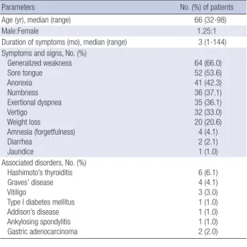
Table 2. Characteristics of patients with pernicious anemia (N = 97)
Parameters No. (%) of patients
Age (yr), median (range) 66 (32-98)
Male:Female 1.25:1
Duration of symptoms (mo), median (range) 3 (1-144) Symptoms and signs, No. (%)
Generalized weakness Sore tongue Anorexia Numbness Exertional dyspnea Vertigo Weight loss
Amnesia (forgetfulness) Diarrhea
Jaundice
64 (66.0) 52 (53.6) 41 (42.3) 36 (37.1) 35 (36.1) 32 (33.0) 20 (20.6) 4 (4.1) 2 (2.1) 1 (1.0) Associated disorders, No. (%)
Hashimoto’s thyroiditis Graves’ disease Vitiligo
Type I diabetes mellitus Addison’s disease Ankylosing spondylitis Gastric adenocarcinoma
6 (6.1) 4 (4.1) 3 (3.0) 1 (1.0) 1 (1.0) 1 (1.0) 2 (2.0)
No. of patients
Year
’95 ’96 ’97 ’98 ’99 ’00 ’01 ’02 ’03 ’04 ’05 ’06 ’07 ’08 ’09 ’10
70 60 50 40 30 20 10 0
5 0
6 2 5
1 3 1 4
0 14
4 17
5 20
5 18
5 48
13 40
9 38
8 50
12 62
15 28
6 47
11 Cobalamin deficiency anemia
Pernicious anemia
Fig. 1. Annual distribution of the numbers of newly diagnosed patients with cobalamin deficiency anemia and pernicious anemia in Daejeon City and Chungnam Province.
loss of > 4.5 kg or > 5% of one’s body weight over the period of 6 months (20.6%), forgetfulness (4.1%), diarrhea (2.0%), and jaundice (1.0%). Anemia-associated discomfort was the most common symptom (79.4%); followed by gastrointestinal symp- toms that included anorexia, sore tongue, and weight loss (78.4
%); and neurological symptoms (38.1%). The median duration of symptoms at diagnosis was 3 months (range, 1-144 months) (Table 2).
Associated disorders
At presentation, Hashimoto’s thyroiditis was found in six (6.1%) patients, vitiligo in three (3.0%), and Graves’ disease in four (4.1
%). Type 1 diabetes mellitus, Addison’s disease, and ankylosing spondylitis were found in one patient each. Two (2.0%) patients already had stomach cancer at the time of diagnosis of perni- cious anemia (Table 2).
Hematologic features
CBC at presentation revealed anemia with a hemoglobin of
< 7.0 g/dL in 46 (47.2%) patients and between 7.0 and 10.0 g/dL in 35 (36.1%) patients. The median hemoglobin level was 7.3 g/
dL (range, 3.1-12.9 g/dL). White blood cell (WBC) counts were
< 4 × 10⁹/L in 44 (45.4%) patients. The median white blood cell count was 4.14 × 10⁹/L (range, 1.4-12.58 × 10⁹/L). Platelets counts were < 50 × 109/L in 12 (12.4%) patients, 50 to 150 × 109/L in 51 (52.6%) patients, and > 150 × 109/L in 34 (35.1%) patients. The median platelet count was 113 × 109/L (range, 23-373 × 109/L).
Pancytopenia was observed in 36 (37.1%) patients. Macrocyto- sis with a mean corpuscular volume of > 99 fL was noticed in
86 (88.7%) patients (median, 118; range, 84-144 fL). Hyperseg- mented neutrophils, defined as the presence of ≥ 5% of five- lobed neutrophils or the presence of six-lobed neutrophils and macro-ovalocytes, were detected in 54 (55.7%) and 86 (88.7%) patients, respectively. Serum levels of vitamin B12 were < 50 pg/
mL in 39 (40.2%) patients, 50 to 100 pg/mL in 33 (34.0%) pa- tients, and 101 to 200 pg/mL in 25 (25.8%) patients. The median serum vitamin B12 level was 64.5 pg/mL (range, 10-198 pg/mL) (Table 3).
Serum antibodies to intrinsic factor and parietal cells and endoscopic findings
Serum antibody to intrinsic factor was detected in 62 (77.5%) of 80 patients examined. Serum antibody to parietal cells was de- tected in 35 (43.2%) of 81 patients examined. Both serum anti- body to intrinsic factor and serum antibody to parietal cells were detected in 27 (34.6%) of 78 patients examined. Either one of the two serum antibodies was detected in 71 (85.5%) of 83 pa- tients examined (Table 4). Of the 97 patients, 84 underwent en- doscopic examination. Fifty-six of 57 (98.2%) patients who un- derwent gastric biopsy had atrophic gastritis of the fundus or body of the stomach. One patient had gastric adenoma. H. py- lori was demonstrated in 7 (20.6%) of 34 patients examined by Giemsa staining of endoscopic biopsy specimens (Table 4).
Responses to cobalamin therapy
Eighty-three (94.3%) of 88 patients who were treated by intra- muscular injection of cobalamin normalized their hemoglobin levels within 3 months of treatment. The WBC counts returned
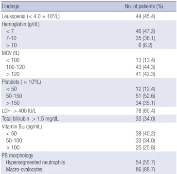
Table 3. Laboratory findings at the time of diagnosis (N = 97)
Findings No. of patients (%)
Leukopenia (< 4.0 × 10⁹/L) 44 (45.4)
Hemoglobin (g/dL) < 7
7-10 > 10
46 (47.2) 35 (36.1) 6 (6.2) MCV (fL)
< 100 100-120 > 120
13 (13.4) 43 (44.3) 41 (42.3) Platelets ( × 109/L)
< 50 50-150 > 150
12 (12.4) 51 (52.6) 34 (35.1)
LDH > 400 IU/L 78 (80.4)
Total bilirubin > 1.5 mg/dL 33 (34.0)
Vitamin B12 (pg/mL) < 50
50-100 > 100
39 (40.2) 33 (34.0) 25 (25.8) PB morphology
Hypersegmented neutrophils Macro-ovalocytes
54 (55.7) 86 (88.7) MCV, mean corpuscular volume; LDH, lactate dehydrogenase; PB, peripheral blood.
Table 4. Findings of serum antibodies and H. pyroli testing at the time of diagnosis (N = 97)
Findings No. of patients (%)
Antibodies
Anti-intrinsic factor antibody (+) Anti-parietal cell antibody (+) Both antibodies (+) Any antibodies (+)
62/80 (77.5) 35/81 (43.2) 27/78 (34.6) 71/83 (85.5) H. pylori testing
Positive Negative
7/34 (20.6) 27/34 (79.4)
Table 5. Responses to cobalamin therapy (N = 88)
Parameters No. of patients (%)
Correction of anemia 83/88 (94.3)
Correction of leukopenia 40/44 (90.9)
Correction of thrombocytopenia 56/63 (88.9)
Resolution of symptoms Generalized weakness Exertional dyspnea Vertigo Sore tongue Anorexia Numbness
60/60 (100) 32/32 (100) 30/30 (100) 49/49 (100) 39/39 (100) 17/36 (47.2)
to normal values in 40 (90.9%) of 44 patients, and platelet counts returned to normal values in 56 (88.9%) of 63 patients. After co- balamin therapy, the vast majority of symptoms, including gen- eralized weakness, exertional dyspnea, vertigo, sore tongue, and anorexia, were improved in all patients; however, numbness was improved in only 17 (47.2%) of 36 patients (Table 5).
DISCUSSION
The present study describes the largest number of Korean indi- viduals with pernicious anemia. Between 1995 and 2010, a total of 97 patients with pernicious anemia were enrolled from five university hospitals in one Korean province, which was nearly one-fourth of patients with megaloblastic anemia induced by vitamin B12 deficiency. In Western countries, prevalence and annual incidence of pernicious anemia have been reported to be about 150 per 100,000 and approximately 10 per 100,000, re- spectively (10, 11). During the period between 2005 and 2010 in the present study, the approximate annual incidence of perni- cious anemia was 0.3 per 100,000, indicating that this disorder is much less frequent in Korea than in Western countries. Based on the current clinical practice situation in Chungnam prov- ince, we believe that the vast majority of patients with perni- cious anemia were seen at the five hospitals that participated in the present study. However, one university hospital did not par- ticipate in the study, and some patients may have visited hospi- tals outside this area. Thus, the incidence of pernicious anemia is likely to be even higher than that estimated here.
In the present study, total gastrectomy was found to be a lead- ing cause of vitamin B12 deficiency anemia, which differs from Western reports. One possible explanation for this discrepancy is that gastrectomy has been practiced more frequently in Korea than in Western countries because of a higher incidence of gas- tric cancer and H. pylori-associated gastric ulcers. Our study revealed that a proportion of patients who undergo total gas- trectomy have not been placed on appropriate cobalamin re- placement, despite the fact that monitoring and management for vitamin B12 deficiency is a well-known practice in individu- als who undergo total gastrectomy.
The present study showed that most patients had symptoms of anemia and gastrointestinal manifestations, and about one- third of patients had neurologic symptoms at presentation. After cobalamin treatment the gastrointestinal symptoms resolved completely in all patients, whereas neurological symptoms re- mained in some. These features in Korean patients do not differ from those in Western cases (1). Pernicious anemia may be as- sociated with autoimmune endocrinopathies and antireceptor autoimmune disease such as chronic autoimmune thyroiditis (Hashimoto’s thyroiditis), insulin-dependent diabetes mellitus, Addison’s disease, primary ovarian failure, primary hypopara- thyroidism, Graves’ disease, vitiligo, myasthenia gravis, and the
Lambert-Eaton syndrome (1). In the present study, pernicious anemia was accompanied by several autoimmune disorders.
However, the prevalence of autoimmune disorders in our study population was lower than that in Western populations. For ex- ample, autoimmune thyroid diseases were present in 10.2% of our study population, which is much lower than the 24% to 27%
in Western populations (12, 13). At the present time, we have no explanation for this difference; thus, further studies are warranted.
Pernicious anemia is known to be associated with gastric ad- enocarcinoma as well as gastric carcinoid tumors. In a popula- tion-based cohort study in Sweden, the risk of gastric carcinoma was increased 3-fold and that of gastric carcinoid tumors was increased 13-fold in patients with pernicious anemia (14, 15).
We found gastric adenocarcinoma in two patients at the time of diagnosis of pernicious anemia and in two more patients dur- ing follow-up. Because the present study was a retrospective analysis, some cases of gastric cancer were possibly not detect- ed. Given the prevalence of gastric cancer in general population in Korea (16), it is suggested that pernicious anemia might be a risk factor for the development of gastric cancer in Korea as well.
No institutes that participated in this study used the Schilling test for the diagnosis of pernicious anemia. As an antibody as- say is noninvasive, simple, and capable of differentiating cobal- amin malabsorption due to intrinsic factor deficiency from other causes such as cobalamin malabsorption, measurement of se- rum antibody to intrinsic factor now supersedes the Schilling test in terms of confirmation of the diagnosis of pernicious ane- mia (1, 17). The frequencies of serum antibodies to intrinsic fac- tor and parietal cells in this study were 77.5% and 43.2%, respec- tively. These profiles are similar to those among Western cases with pernicious anemia, in which the frequencies of serum an- tibodies to intrinsic factor and parietal cells are about 70% and 35% to 40%, respectively (1). In the past, anti-parietal cell anti- body was detected more frequently than anti-intrinsic factor antibody (18, 19). The disparities could be explained, at least in part, by recent improvement in the detection of anti-intrinsic factor antibody. Furthermore, it has been hypothesized that the frequency of anti-parietal cell antibody is decreased by loss of antigen, which is caused by loss of parietal cells according to the progression of autoimmune gastritis (20).
There has been a notion that H. pylori infection provokes type A gastritis and thus induces pernicious anemia (21, 22). On the other hand, contradictory observations have also been reported (23, 24). In the present study, only 7 (20.6%) of 34 patients were positive in H. pylori testing, which is a rather low positivity com- pared with the 60% to 70% positivity in the general Korean pop- ulation (25, 26). Thus, H. pylori infection is unlikely to be associ- ated with pernicious anemia.
In conclusion, pernicious anemia is much less frequent in Koreans than in Western populations; however, the clinical fea- tures of this disorder in Koreans do not differ from those of West-
ern cases.
ACKNOWLEDGMENTS
The authors have no conflicts of interest to disclose.
REFERENCES
1. Toh BH, van Driel IR, Gleeson PA. Pernicious anemia. N Engl J Med 1997; 337: 1441-8.
2. Jacobson DL, Gange SJ, Rose NR, Graham NM. Epidemiology and esti- mated population burden of selected autoimmune diseases in the Unit- ed States. Clin Immunol Immunopathol 1997; 84: 223-43.
3. Carmel R. Ethnic and racial factors in cobalamin metabolism and its disorders. Semin Hematol 1999; 36: 88-100.
4. Sugihara T, Yawata Y. Japanese clinical statistical data of patients with pernicious anemia. Nihon Rinsho 1992; 50: 771-86.
5. Wun Chan JC, Yu Liu HS, Sang Kho BC, Yin Sim JP, Hang Lau TK, Luk YW, Chu RW, Fung Cheung FM, Tat Choi FP, Kwan Ma ES. Pernicious anemia in Chinese: a study of 181 patients in a Hong Kong hospital. Med- icine (Baltimore) 2006; 85: 129-38.
6. Yim PS, Woo MS, Chung SJ, Yoon YS, Jeen TH, Lee YB. A case report of pernicious anemia. Korean J Hematol 1969; 4: 23-31.
7. Lee DG, Do IH, Kim DW, Park JI, Chung SY, Moon SK. A case of perni- cious anemia associated with chronic atrophic gastirtis. Korean J Hema- tol 1987; 22: 115-21.
8. Song HH, Kwon JH, Kim JH, Jeong JY, Kim HJ, Lee KS, Jang DY, Ahn JS, Shin DH, Kang SH, et al. Causes and clinical features of vitamine B12 de- ficiency megaloblastic anemia. Korean J Hematol 2004; 39: 243-8.
9. Chun JM, Park NS, Park NH, Yun GW, Yang YJ, Park SE, Yun HJ, Jo DY, Kwon GC, Kim S. Pernicious anemia: a retrospective analysis of 22 cases.
Korean J Hematol 2005; 40: 219-25.
10. Jacobson DL, Gange SJ, Rose NR, Graham NM. Epidemiology and esti- mated population burden of selected autoimmune disease in the United States. Clin Immunol Immunopathol 1997; 84: 223-43.
11. Pedersen AB, Mosbech J. Morbidity of pernicious anemia: incidence, prevalence, and treatment in a Danish country. Acta Med Scand 1969;
185: 449-52.
12. Carmel R, Spencer CA. Clinical and subclinical thyroid disorders associ- ated with pernicious anemia. Observations on abnormal thyroid-stimu-
lating hormone levels and on a possible association of blood group O with hyperthyroidism. Arch Intern Med 1982; 142: 1465-9.
13. Ottesen M, Feldt-Rasmussen U, Andersen J, Hippe E, Schouboe A. Thy- roid function and autoimmunity in pernicious anemia before and dur- ing cyanocobalamin treatment. J Endocrinol Invest 1995; 18: 91-7.
14. Hsing AW, Hansson LE, McLaughlin JK, Nyren O, Blot WJ, Ekbom A, Fraumeni JF Jr. Pernicious anemia and subsequent cancer. A population based cohort study. Cancer 1993; 71: 745-50.
15. Karlson BM, Ekbom A, Wacholder S, McLaughlin JK, Hsing AW. Cancer of the upper gastrointestinal tract among patients with pernicious ane- mia: a case-cohort study. Scand J Gastroenterol 2000; 35: 847-51.
16. Jung KW, Park S, Kong HJ, Won YJ, Lee JY, Park EC, Lee JS. Cancer statis- tics in Korea: incidence, mortality, survival and prevalence in 2008. Can- cer Res Treat 2011; 43: 1-11.
17. Carmel R. Pepsinogens and other serum markers in pernicioua anemia.
Am J Clin Pathol 1988; 90: 442-5.
18. Irvine WJ. Immunologic aspects of pernicious anemia. N Engl J Med 1965; 273: 432-8.
19. Samloff IM, Kleinman MS, Turner MD, Sobel MV, Jeffries GH. Blocking and binding antibodies to intrinsic factor and parietal call antibody in pernicious anemia. Gastroenterology 1968; 55: 575-83.
20. Toh BH, Alderuccio F. Pernicious anaemia. Autoimmunity 2004; 37:
357-61.
21. Kaptan K, Beyan C, Ural AU, Cetin T, Avcu F, Gülşen M, Finci R, Yalçín A. Helicobacter pyroli is it a novel causative agent in vitamin B12 defi- ciency? Arch Intern Med 2000; 160: 1349-53.
22. Stopeck A. Links between Helicobacter pylori infection, cobalamin defi- ciency, and pernicious anemia. Arch Intern Med 2000; 160: 1229-30.
23. Haruma K, Komoto K, Kawaguchi H, Okamoto S, Yoshihara M, Sumii K, Kajiyama G. Pernicious anemia and Helicobacter pyroli infection in Japan. evaluation in a country with a high prevalence of infection. Am J Gastroenterol 1995; 90: 1107-10.
24. Fong TL, Dooley CP, Dehesa M, Cohen H, Carmel R, Fitzgibbons PL, Perez-Perez GI, Blaser MJ. Helicobacter pyroli infection in pernicious ane- mia. a prospective controlled study. Gastroenterology 1991; 100: 328-32.
25. Kim HS, Lee YC, Lee HW, Yoo HM, Lee CG, Kim JM, Lee KJ, Kim PS, Moon BS, Park HJ, et al. Seroepidemiologic study of Helicobacter pyroli infection in Korea. Korean J Gastroenterol 1999; 33: 170-82.
26. Yim JY, Kim N, Choi SH, Kim YS, Cho KR, Kim SS, Seo GS, Kim HU, Baik GH, Sin CS, et al. Seroprevalence of Helicobacter pyroli in South Korea.
Helicobacter 2007; 12: 333-40.