ORIGINAL ARTICLE
국소 진행성 위암에서 2제, 3제 선행항암화학 요법의 임상 결과
김주석, 강선형, 문희석, 성재규, 정현용, 설지영1
충남대학교 의과대학 내과학교실 소화기내과, 외과학교실1
Clinical Outcome of Doublet and Triplet Neoadjuvant Chemotherapy for Locally Advanced Gastric Cancer
Ju Seok Kim, Sun Hyung Kang, Hee Seok Moon, Jae Kyu Sung, Hyun Yong Jeong, and Ji Young Sul1
Division of Gastroenterology, Department of Internal Medicine, Department of Surgery1, Chungnam National University School of Medicine, Daejeon, Korea
Background/Aims: In gastric cancer, the rate of recurrence and metastasis following radical resection is high, necessitating improvement in survival and cure rates. Neoadjuvant chemotherapy (NAC) has potential benefits for locally advanced gastric cancer; however, the surgical benefits and effects on survival are unclear. This study evaluates the effectiveness of NAC in locally advanced gastric cancer and compares clinical outcomes of doublet and triplet regimens.
Methods: We reviewed patient medical records of 383 patients who underwent NAC (n=41) or surgery only (n=342) for treatment of locally advanced gastric cancer. The baseline characteristics and clinical outcomes were compared between the groups.
Chemotherapy patients were classified according to regimen, doublet (n=28) and triplet (n=13), and NAC-related clinical response, safety, and toxicity were analyzed.
Results: The baseline characteristics did not differ significantly between groups. After NAC, the tumor downstage rate was 51.2% (21/41); however, overall survival (p=0.205) and disease-free survival (p=0.415) were not significantly different between the groups. On subgroup analysis, no significant differences in drug toxicity (p=0.604) or clinical response (p=0.374) were found between outcomes of doublet and triplet chemotherapy regimens.
Conclusions: In patients with locally advanced gastric cancer, NAC showed tolerable drug toxicity and increased tumor downstage, but NAC failed to increase the survival rate, which may be caused by a high D2-lymphadenectomy rate. Therefore, NAC was found to be a therapeutic option for select gastric cancer patients. (Korean J Gastroenterol 2016;68:245-252)
Key Words: Neoadjuvant therapy; Drug therapy; Stomach; Adenocarcinoma
Received August 22, 2016. Revised October 12, 2016. Accepted October 14, 2016.
CC This is an open access article distributed under the terms of the Creative Commons Attribution Non-Commercial License (http://creativecommons.org/licenses/
by-nc/4.0) which permits unrestricted non-commercial use, distribution, and reproduction in any medium, provided the original work is properly cited.
Copyright © 2016. Korean Society of Gastroenterology.
교신저자: 문희석, 35015, 대전시 중구 문화로 282, 충남대학교 의과대학 내과학교실 소화기내과
Correspondence to: Hee Seok Moon, Division of Gastroenterology, Department of Internal Medicine, Chungnam National University School of Medicine, 282 Munhwa-ro, Jung-gu, Daejeon 35015, Korea. Tel: +82-42-280-7163, Fax: +82-42-257-5753, E-mail: [email protected]
Financial support: None. Conflict of interest: None.
INTRODUCTION
Gastric cancer is the most frequently occurring cancer in Korea.1 In a phase III study involving patients with stage 2 or higher resectable stomach cancer, patients treated with ad- juvant chemotherapy demonstrated a 15% increase in the disease-free survival (DFS) rate and a 10% increase in the
overall survival (OS) rate, compared with those treated with only surgery.2 Adjuvant chemotherapy after D2 lymphade- nectomy is considered standard treatment for gastric cancers.2,3 Despite these efforts, compared with early stage gastric cancer with 5-year survival rates over 90%, locally ad- vanced gastric cancer entails a high risk of lymph node meta- stasis, and even with complete resection, the prognosis is
poor due to relapse.4 Thus, several methods to increase treatment effectiveness have been attempted, including ra- diation therapy after D2 lymphadenectomy or consolidation adjuvant chemotherapy. However, none of these methods re- duce relapse or increase survival, and they are no longer rec- ommended for treatment.5,6
Neoadjuvant chemotherapy (NAC) is a potential treatment regimen for locally advanced gastric cancer. NAC can reduce tumor size, decrease clinical stage, enhance drug sensitivity, and reduce micrometastasis.7,8 However, it is not effective for all patients, which may delay treatment, so ideal surgery tim- ing may be missed due to disease progression. Many studies evaluating the effects of NAC report that it reduces the clin- ical stage and increases the curative resection (R0) rate when compared to surgery alone. However, there is no clear evidence that NAC increases the OS rate, the ultimate goal of treatment; therefore, it is a controversial treatment method.9,10 In addition, several of these studies do not use a consistent NAC regimen, so it is unclear which chemo- therapy agent should be used in NAC treatment.11 Thus, this study evaluates the effectiveness of NAC in locally advanced gastric cancer, and compares a doublet regimen with a triplet regimen to propose criteria for the selection of a chemo- therapy agent.
SUBJECTS AND METHODS
1. Study design and patient selection
Charts were reviewed for patients who were histologically diagnosed with gastric adenocarcinoma at the Chungnam National University School of Medicine between January 2008 and June 2014. The subjects were patients who re- ceived ongoing care for at least for one year after treatment for gastric cancer in this hospital.
The majority of cases were gastric cancer, although cases of gastroesophageal junction cancer were included. Patients were 18 years or older, with a World Health Organization (WHO) performance status score of 0 or 1. In order to target patients with locally advanced gastric cancer, those with clin- ical stage higher than T3 or lymph node metastasis were enrolled. Patients who received cancer treatment previously or those with distant metastasis, secondary malignancy, and inadequate cardiac or renal function (serum creatinine clear- ance rate ≤60 mL/min) were excluded. Pretreatment clin-
ical evaluation included a complete blood cell count with dif- ferential and serum multichannel chemical analysis. For clin- ical staging, upper gastrointestinal endoscopy with biopsy, abdominal CT, and chest radiography were conducted.
2. Neoadjuvant chemotherapy
Preoperative chemotherapy was administered over three cycles, with changes to dosage or time dictated by tumor re- sponse or safety. Chemotherapy regimens were classified in- to doublet and triplet regimens according to the number of the cytotoxic agents used. The doublet regimen was FOLFOX (oxaliplatin [100 mg/m2], leucovorin [200 mg/m2 of body surface area], intravenously on day 1; and 5-fluorouracil [5-FU, 2,400 mg/m2] continuous infusion over 48 hours, re- peated every two weeks), while the triplet regimen was DCF (docetaxel [75 mg/m2], cisplatin [60 mg/m2] of body surface area, intravenously on day 1; and 5-FU [750 mg/m2] con- tinuous infusion on each day 1-5, repeated every three weeks). Before each cycle of chemotherapy, a complete blood count and liver function test were performed, and elec- trolyte and serum creatinine levels were determined. DCF was reduced in patients with myelosuppression or thrombo- cytopenia, and 5-FU dosage was adjusted if mucositis or diar- rhea occurred. In addition, if serum creatinine increased, cis- platin dosage was reduced or suspended according to the de- gree of renal function. The severity of toxicity or adverse ef- fects was defined according to the National Cancer Institute Common Toxicity Criteria version 4.0.
Assessment of response to neoadjuvant therapy was based on reduction of primary tumor size as measured by en- doscopy and abdominal CT. Complete disappearance of le- sions on endoscopy and CT was considered a clinically com- plete response (CR). A tumor size reduction of greater than 50% compared with the initial findings was defined as a parti- al response (PR). Patients with a minor response or no change in the condition were defined as having stable disease (SD).
The presence of new lesions or an increase of 25% or more in primary tumor size was considered progressive disease (PD). CR and PR were designated as responders, while SD and PD were designated as non-responders. Changed stage was defined if either T or N stage was up or down.
3. Surgery
Surgery was scheduled two to four weeks after completion
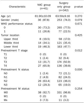
Table 1. Patiens Baseline Characteristics
Characteristic NAC group (n=41)
Surgery only group
(n=342)
p-value
Age (yr) 61.90±10.09 63.59±9.16 0.270
Gender (male) 36 (87.8) 253 (74.0) 0.079
WHO performance status 0.671
0 30 (73.2) 265 (77.5)
1 11 (26.8) 77 (22.5)
Tumor location 0.425
Upper third 8 (19.5) 58 (17.0)
Middle third 14 (34.1) 121 (35.4)
Lower third 19 (46.3) 163 (47.7)
Pretreatment T stage 0.012
T1 0 (0) 0 (0)
T2 1 (2.4) 32 (9.4)
T3 13 (31.7) 174 (50.9)
T4 27 (65.9) 136 (39.8)
Pretreatment N status 0.000
N0 1 (2.4) 72 (21.1)
N1 2 (4.9) 82 (24.0)
N2 26 (63.4) 123 (36.0)
N3 12 (29.3) 65 (19.0)
Pretreatment M status 0.254
M0 38 (92.7) 331 (96.8)
M1 0 (0) 0 (0)
Mx 3 (7.3) 11 (3.2)
Values are presented as mean±SD or n (%).
NAC, neoadjuvant chemotherapy; WHO, World Health Organization.
of the last cycle of chemotherapy in the NAC group, while op- erations for the surgery only group were performed immedi- ately after diagnosis. For patients with curative resection, to- tal or distal subtotal gastrectomy was performed depending on the location and macroscopic type of the gastric cancer.
An extended D2 lymphadenectomy was performed, accord- ing to the rules of the Japanese Research Society for Gastric Cancer. En bloc resection of adjacent organs was performed when their involvement was questionable. All resected speci- mens were examined at local pathology laboratories accord- ing to the standard protocol. The pathological tumor, lymph node, metastasis (pTNM) stage were assessed according to the guidelines of the Union for International Cancer Control (UICC)/American Joint Committee on Cancer (AJCC). R0 re- section was defined as the removal of all gross tumor materi- al and a histopathological examination of proximal, distal, and circumferential margins that revealed the absence of malignant cells more than 2 mm from the edge.
4. Ongoing care
Each patient was assessed via a complete physical exami- nation, routine lab work, chest radiography, abdominal CT, and tumor marker analysis, every six months for five years, then annually or until death. Twenty-seven patients were lost, and 199 patients (52.0%) died while under continuing care.
5. Statistical analysis
DFS was calculated from diagnosis to the first event (local recurrence or progression, or distant recurrence), and OS was calculated from diagnosis to death. Kaplan-Meier curves for DFS and OS were compared with the log-rank test on an intention-to-treat basis. Categorical variables were analyzed using chi-square or Fisher’s exact tests. All analyses were conducted using IBM SPSS Statistics for Windows, version 19.0 (Released 2010., IBM Corp., Armonk, NY, USA). Two-sid- ed null hypotheses of no difference were rejected if p-values were less than 0.05.
RESULTS
1. Patient characteristics
Three hundred and eighty-three patients were enrolled in this study. Forty-one of the 383 patients were administered preoperative chemotherapy followed by surgery, while 342
received only surgery. There were 289 men (75.5%), and the mean patient age was 63.40 years (standard deviation [SD]=
9.61 years). The baseline characteristics of the two groups are shown in Table 1, which illustrates that the patient dis- tribution according to age, gender, WHO performance status, tumor location, and pretreatment M status was well balanced between treatment groups. However, pretreatment clinical T stage (p=0.012) and N status (p=0.000) were significantly higher in the NAC group than in the surgery only group.
2. Clinical outcome of neoadjuvant chemotherapy Forty-one patients received NAC and were further classi- fied according to the chemotherapy regimen. The doublet group consisted of 28 patients (68.3%) and the triplet group consisted of 13 patients (31.7%). The mean age (SD) of the doublet group was 64.96 years (9.67 years), while that of the triplet group was 55.31 years (7.75 years). The triplet group patients were younger than those in the doublet group, but not significantly different (p=0.602). The sex distribution of the two groups was similar (p=0.659). The NAC group was an-
Table 2. Grade 3 or 4 Toxicity and Clinical Response Assessment during Neoadjuvant Chemotherapy
Variable Total
(n=41)
Doublet (n=28)
Triplet
(n=13) p-value Toxicity (total) 15 (36.6) 9 (32.1) 6 (46.2) 0.604
Neutropenia 5 (12.2) 4 (14.3) 1 (7.7) Thrombocytopenia 3 (7.3) 1 (3.6) 2 (15.4)
Anemia 2 (4.9) 1 (3.6) 1 (7.7)
Nausea/vomiting 2 (4.9) 2 (7.1) 0 (0)
Mucositis 1 (2.4) 0 (0) 1 (7.7)
Fever 1 (2.4) 0 (0) 1 (7.7)
Nephrotoxicity 1 (2.4) 1 (3.6) 0 (0)
Clinical response 0.374
Complete response 5 (12.2) 3 (10.7) 2 (15.4) Partial response 14 (34.1) 9 (32.1) 5 (38.5) Stable disease 19 (46.3) 15 (53.6) 4 (30.8) Progressive disease 3 (7.3) 1 (3.6) 2 (15.4) Values are presented as n (%).
Table 3. Surgical and Pathologic Results
Characteristic NAC group (n=41)
Surgery only group
(n=342)
p-value
Type of surgery 0.726
Total gastrectomy 6 (14.6) 39 (11.4) Distal gastrectomy 35 (85.4) 303 (88.6)
Resection margin 0.829
R0 38 (92.7) 321 (93.9)
R1 2 (4.9) 20 (5.8)
R2 0 (0) 1 (0.3)
Open & closure 1 (2.4) 0 (0)
Postoperative complication 6 (14.6) 61 (17.8) 0.770 Pathologic results
Tumor stage 0.001
T1 6 (14.6) 0 (0)
T2 4 (9.8) 8 (2.3)
T3 3 (7.3) 128 (37.4)
T4 28 (68.3) 206 (60.2)
Nodal status 0.000
N0 12 (29.3) 4 (1.2)
N1 8 (19.5) 72 (21.1)
N2 5 (12.2) 96 (28.1)
N3 16 (39.0) 170 (49.7)
Metastasis status 0.127
M0 39 (95.1) 338 (98.8)
M1 2 (4.9) 4 (1.2)
Values are presented as n (%).
NAC, neoadjuvant chemotherapy.
alyzed for drug toxicities, and clinical responses were eval- uated with endoscopy and CT after cancer treatment. These results are shown in Table 2. Fifteen patients (36.6%) experi- enced grade 3 or 4 toxicity (nine patients [32.1%] in the dou- blet group and six patients [46.2%] in the triplet group), but there was no significant difference in toxicity according to the NAC regimen (p=0.604). The most common chemotherapy toxicity was neutropenia (12.2%), followed by thrombocyto- penia, anemia, nausea/vomiting, and mucositis. Twelve pa- tients (29.3%) stopped cancer treatment or reduced the che- motherapy agent dosage due to the drug’s side effects (seven patients [25.0%] in the doublet group and five pa- tients [38.5%] in the triplet group), although these differ- ences were not statistically significant (p=0.608).
Of the 41 NAC group patients, five patients achieved CR, and the doublet and triplet regimens did not differ sig- nificantly in clinical response rate (p=0.374). According to the response categories mentioned earlier, 19 patients (46.3%) were responders, 22 patients (53.7%) were non-responders, and the two groups showed similar results (p=0.749).
3. Surgical findings and surgical pathology
The surgical and pathological results were compared be- tween the NAC and surgery-only groups, and the results are in Table 3. All 383 patients in the study received surgery, with 41 patients (10.7%) receiving preoperative chemotherapy and 342 patients (89.3%) receiving surgery only. There were no significant differences in surgery type (p=0.726), post-
operative complications (p=0.770), and R0 resection rate (p=0.829) between groups. Three hundred and fifty-nine pa- tients (93.7%) had R0 resection (92.7% in the NAC group, and 93.9% in the surgery only group), a high R0 resection rate in both groups. Massive peritoneal seeding was found in one patient in the NAC group, which resulted in the termination of the operation without gastric resection.
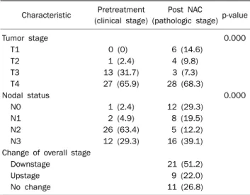
Based on postoperative pathological results, pathologic stage was re-evaluated in all patients and compared with the preoperative clinical stage. The results showed no difference in M status between groups (p=0.127), but T stage (p=0.001) and N status (p=0.000) were significantly lower in the NAC group than in the surgery only group, in contrast to the pre- treatment clinical stage under the influence of downstaging after chemotherapy. In the NAC group only, preoperative clin- ical stage was compared with postoperative pathologic stage, and the difference was analyzed (Table 4). Both tumor stage and nodal status significantly decreased in pathologic stage after NAC, as compared to the pretreatment clinical stage (p=0.000). In overall staging, 21 patients (51.2%) were downstaged, nine patients (22.0%) upstaged, and 11 pa-
Table 4. Stage Change of the Pretreatment vs. Post Neoadjuvant Chemotherapy
Characteristic Pretreatment (clinical stage)
Post NAC
(pathologic stage) p-value
Tumor stage 0.000
T1 0 (0) 6 (14.6)
T2 1 (2.4) 4 (9.8)
T3 13 (31.7) 3 (7.3)
T4 27 (65.9) 28 (68.3)
Nodal status 0.000
N0 1 (2.4) 12 (29.3)
N1 2 (4.9) 8 (19.5)
N2 26 (63.4) 5 (12.2)
N3 12 (29.3) 16 (39.1)
Change of overall stage
Downstage 21 (51.2)
Upstage 9 (22.0)
No change 11 (26.8)
Values are presented as n (%).
NAC, neoadjuvant chemotherapy.
Table 5. Pattern of Recurrence, Survival Status, and Cause of Death
Characteristic NAC group (n=41)
Surgery only group (n=342) p-value
Recurrence 20 (48.8) 218 (63.7) 0.119
Locoregional only 1 (2.4) 30 (8.8) Distant only 5 (12.2) 36 (10.5)
Both 14 (34.1) 152 (44.4)
Death 17 (41.5) 182 (53.2) 0.209
Cancer related 13 (31.7) 161 (47.1) Surgery related 2 (4.9) 12 (3.5)
Others 2 (4.9) 9 (2.6)
Values are presented as n (%).
NAC, neoadjuvant chemotherapy.
Fig. 1. Kaplan-Meier estimates of overall survival rate.
Fig. 2. Kaplan-Meier estimates of disease-free survival rate.
tients (26.8%) were unchanged.
4. DFS and OFS
The median follow-up (SD) period was 32.03 months (14.91 months) for all patients, 33.39 months (19.63 months) for the NAC group, and 31.86 (17.14) months for the surgery only group. The survival and recurrence during the observation period were analyzed, as shown in Table 5. One hundred and ninety-nine patients (52.0%) died (17 patients [41.5%] in the NAC group and 182 patients [53.2%] in the sur- gery only group); there was no difference in the OS between the two groups (p=0.209). The deaths of 174 (87.4%) of the 199 patients were cancer-related. A recurrence was ob- served in 238 patients (62.1%), with the most recurrence oc- curring in locoregional and distant regions simultaneously,
as seen in 166 patients (69.7%). There was no significant dif- ference in the recurrence rate between the NAC group and the surgery only group (p=0.119).
The estimated median OS was 48.84 months (95% CI, 37.22 to 60.45) in the NAC group versus 42.76 months (95%
CI, 38.37 to 47.15) in the surgery only group (Fig. 1). The OS rate at two years was 61.2% in the NAC group and 52.4% in the surgery only group. On a log-rank test, there was no differ- ence in the OS between groups (p=0.205). The estimated median DFS was 46.08 months (95% CI, 34.32 to 57.84) in the NAC group versus 38.32 months (95% CI, 33.79 to 42.85) in the surgery only group (Fig. 2). There was no differ- ence in DFS between groups (p=0.415).
DISCUSSION
In this study, NAC significantly decreased clinical T stage
and N stage, and 51.2% of patients treated with NAC experi- enced downstaging. In addition, NAC treatment did not in- crease postoperative complications, and the side effects of the chemotherapy agent were tolerable. However, in stom- ach cancer cases, the operation range cannot be reduced even with a decrease in tumor size or clinical stage after NAC, as the operation range is determined by the location of the primary lesion.12 In addition, the determination of clinical stage through imaging examination is inaccurate. It is re- ported that CT has an accuracy of approximately 77-89% for T stage and approximately 59-78% for N stage.13,14 Thus, the effect of NAC in locally advanced gastric cancer is meaningful only when the OS improves, which is the ultimate goal of treatment. However, in this study, the observed decrease in stage did not lead to an improvement in OS (p=0.205) or DFS (p=0.415). In the Medical Research Council (MRC) Adjuvant Gastric Infusional Chemotherapy (MAGIC) trial that com- pared a group with preoperative ECF administration to a sur- gery only group with 503 patients, preoperative chemo- therapy decreased T stage and N stage, which led to an im- provement in survival (hazard ratio [HR], 0.75; 95% CI, 0.60 to 0.93; p=0.009).15 However, there are several differences between our study and the MAGIC trial. Our study conducted D2 lymphadenectomy with all patients except one who did not have resection due to peritoneal seeding, while the MAGIC trial conducted D2 lymphadenectomy in just 43% of the patients. Therefore, we hypothesize that the effect of NAC was less because of the more extensive lymphadenectomy.
In addition, there was a difference in the clinical stage of the enrolled patients, and the patient group of the MAGIC trial was in an earlier clinical stage.
In a randomized trial that showed a high D2 lymphadenec- tomy rate of 92%, similar to that reported in our study, there was no increase in postoperative complications after NAC.
The R0 resection rate improved (81.9% vs. 66.7%; p=0.035), but the OS did not improve compared to the surgery only group (HR, 0.84; 95% CI, 0.52 to 1.35; p=0.466).16 Similar to our study, in this trial, tumor downstaging was caused by NAC, but there was a question about whether this led to the increase in survival. In a meta-analysis with 1,249 patients, there was tumor downstaging after NAC (OR, 1.77; 95% CI, 1.27 to 2.49; p=0.0009), which led to an increase in the R0 resection rate (OR, 1.38; 95% CI, 1.03 to 1.85; p=0.03) and eventually significant increases in the OS rate (OR, 1.40; 95%
CI, 1.11 to 1.76; p=0.005) and progression-free survival (OR, 1.62; 95% CI, 1.21 to 2.15; p=0.001).17 However, in a sub- group analysis, Western countries favored NAC more than Asian countries (OR, 1.40; 95% CI, 1.07 to 1.83), indicating that treatment results differ by region, most likely caused by the differences in D2 lymphadenectomy rate and location of the gastric cancer.18
In NAC, the selection of chemotherapy regimen is an im- portant consideration, but a standard regimen is not esta- blished. The response rate of the doublet regimen in meta- static gastric cancer is approximately 40%, and the possi- bility of tumor progression within two months is approx- imately 20%, considered high. However, the response rate of the triplet regimen is 50-70% and the possibility of pro- gression within two months was approximately 5%.19,20 In a phase II trial with DOS triplet therapy as the NAC regimen, 54% patients responded with no drug side effects, and the two-year DFS rate was high (89.7%).21
The effect of NAC on surgical complications was also con- sidered, as it is important that the risk not be higher than with surgery alone. This study compared the doublet and triplet regimens, and found no difference in drug toxicity (p=0.604) or postoperative complications (p=0.770) between groups.
Seven patients (25.0%) in the doublet regimen and five pa- tients (38.5%) in the triplet regimen stopped cancer treat- ment or reduced anti-cancer drug dosage due to intolerable drug side effects, yet there was no statistical difference be- tween groups (p=0.608). In the analysis of clinical re- sponses, the response rate was higher in the triplet regimen, with a rate of 53.9% in the triplet and 42.8% in the doublet regimen, but there was no statistically significant difference between the two (p=0.749). This may be a result of small sam- ple size, especially in the triplet group, which had only 13 patients. The triplet regimen showed similar toxicity to the doublet regimen, but as it had a higher response rate, the trip- let regimen was determined to be the preferred NAC treatment.
In NAC, cisplatin- or 5-FU-based regimens have been wide- ly used with favorable results. According to multicenter randomized studies, the response rate of cisplatin, leucovor- in, and 5-FU was 35.2% (95% CI, 23.7% to 45.7%).22 The re- sults of treatment with more modern cytotoxic agents, such as capecitabine, oxaliplatin, or docetaxel, were similar in sev- eral studies.23,24 In addition, targeted therapies, such as epi-
dermal growth factor receptor inhibitors or anti-angiogenetic agents, have been attempted as alternative therapy.
However, further studies are needed for a proper character- ization of these therapies.25,26
Our study has a few limitations. First, it was a retrospective, single center study, so it is difficult to generalize the results of this study. Second, the sample size was small, especially for the triplet regimen subgroup in the larger NAC group, which may create a selection bias. In addition, endoscopic ul- trasound was not conducted in all patients in pretreatment clinical staging. As we only evaluated the clinical staging us- ing CT, the T or N stage may be inaccurate, which is likely to have influenced the change in stage after NAC. It is a limi- tation of a retrospective study, but in many other studies, only CT evaluation was performed for the clinical stage.
NAC has clear effects on some cancers, but its effects on gastric cancer are still controversial, and results differ be- tween Eastern countries and Western countries. In the treat- ment of locally advanced gastric cancer, preoperative che- motherapy is preferred in Europe, while adjuvant chemo- therapy after D2 lymphadenectomy is recommended in East Asia, including South Korea and Japan.27 It was reported that NAC and D2 lymphadenectomy increase survival, but this trend was not supported by our study, so combined NAC and D2 surgery therapy is not yet recommended.28 In this study, NAC decreased clinical stage in patients, but this did not lead to an increase in the OS, which may be a result of the high D2 lymphadenectomy rate.
In conclusion, our study demonstrated that NAC treatment successfully downstaged tumors, did not increase post-oper- ative complications, and showed a tolerable toxicity. More- over, it can be considered a therapeutic option in locally ad- vanced gastric cancer. Additionally, the higher response rate of the triplet NAC regimen, despite similar side effects to the doublet regimen, may be important in NAC treatment determination. However, NAC treatments should be chosen with consideration of patient characteristics or operation condition. Although these results have provided new insights into NAC treatment regimens, additional studies are still needed to provide further insights into NAC treatment.
REFERENCES
1. Chung MW, Jeong O, Park YK, et al. Comparison on the long term outcome between endoscopic submucosal dissection and sur-
gical treatment for undifferentiated early gastric cancer. Korean J Gastroenterol 2014;63:90-98.
2. Sakuramoto S, Sasako M, Yamaguchi T, et al. Adjuvant chemo- therapy for gastric cancer with S-1, an oral fluoropyrimidine. N Engl J Med 2007;357:1810-1820.
3. Kim YH. Chemotherapy for advanced gastric cancer: slow but further progress. Cancer Res Treat 2005;37:79-86.
4. Nashimoto A, Nakajima T, Furukawa H, et al. Randomized trial of adjuvant chemotherapy with mitomycin, Fluorouracil, and Cytosine arabinoside followed by oral Fluorouracil in serosa-neg- ative gastric cancer: Japan Clinical Oncology Group 9206-1. J Clin Oncol 2003;21:2282-2287.
5. Ronellenfitsch U, Schwarzbach M, Hofheinz R, et al. Preoper- ative chemo(radio)therapy versus primary surgery for gastro- esophageal adenocarcinoma: systematic review with meta- analysis combining individual patient and aggregate data. Eur J Cancer 2013;49:3149-3158.
6. Ajani JA, Komaki R, Putnam JB, et al. A three-step strategy of in- duction chemotherapy then chemoradiation followed by surgery in patients with potentially resectable carcinoma of the esoph- agus or gastroesophageal junction. Cancer 2001;92:279-286.
7. Ross P, Nicolson M, Cunningham D, et al. Prospective random- ized trial comparing mitomycin, cisplatin, and protracted ve- nous-infusion fluorouracil (PVI 5-FU) With epirubicin, cisplatin, and PVI 5-FU in advanced esophagogastric cancer. J Clin Oncol 2002;20:1996-2004.
8. Li W, Qin J, Sun YH, Liu TS. Neoadjuvant chemotherapy for ad- vanced gastric cancer: a meta-analysis. World J Gastroenterol 2010;16:5621-5628.
9. Bang YJ, Kim YW, Yang HK, et al. Adjuvant capecitabine and ox- aliplatin for gastric cancer after D2 gastrectomy (CLASSIC): a phase 3 open-label, randomised controlled trial. Lancet 2012;379:315-321.
10. Wang LB, Shen JG, Xu CY, Chen WJ, Song XY, Yuan XM. Neoad- juvant chemotherapy versus surgery alone for locally advanced gastric cancer: a retrospective comparative study. Hepatoga- stroenterology 2008;55:1895-1898.
11. Sumpter K, Harper-Wynne C, Cunningham D, et al. Report of two protocol planned interim analyses in a randomised multicentre phase III study comparing capecitabine with fluorouracil and ox- aliplatin with cisplatin in patients with advanced oesophagogas- tric cancer receiving ECF. Br J Cancer 2005;92:1976-1983.
12. Roukos DH, Kappas AM. Perspectives in the treatment of gastric cancer. Nat Clin Pract Oncol 2005;2:98-107.
13. Shimizu K, Ito K, Matsunaga N, Shimizu A, Kawakami Y.
Diagnosis of gastric cancer with MDCT using the water-filling method and multiplanar reconstruction: CT-histologic correla- tion. AJR Am J Roentgenol 2005;185:1152-1158.
14. Chen CY, Hsu JS, Wu DC, et al. Gastric cancer: preoperative local staging with 3D multi-detector row CT--correlation with surgical and histopathologic results. Radiology 2007;242:472-482.
15. Cunningham D, Allum WH, Stenning SP, et al. Perioperative che- motherapy versus surgery alone for resectable gastro- esophageal cancer. N Engl J Med 2006;355:11-20.
16. Schuhmacher C, Gretschel S, Lordick F, et al. Neoadjuvant che- motherapy compared with surgery alone for locally advanced
cancer of the stomach and cardia: European Organisation for Research and Treatment of Cancer randomized trial 40954. J Clin Oncol 2010;28:5210-5218.
17. Ge L, Wang HJ, Yin D, et al. Effectiveness of 5-flurouracil-based neoadjuvant chemotherapy in locally-advanced gastric/gastro- esophageal cancer: a meta-analysis. World J Gastroenterol 2012;18:7384-7393.
18. Hartgrink HH, van de Velde CJ, Putter H, et al. Neo-adjuvant che- motherapy for operable gastric cancer: long term results of the Dutch randomised FAMTX trial. Eur J Surg Oncol 2004;30:
643-649.
19. Kang YK, Ryu MH, Yoo C, et al. Phase I/II study of a combination of docetaxel, capecitabine, and cisplatin (DXP) as first-line che- motherapy in patients with advanced gastric cancer. Cancer Chemother Pharmacol 2011;67:1435-1443.
20. Menges M, Schmidt C, Lindemann W, et al. Low toxic neo- adjuvant cisplatin, 5-fluorouracil and folinic acid in locally ad- vanced gastric cancer yields high R-0 resection rate. J Cancer Res Clin Oncol 2003;129:423-429.
21. Park I, Ryu MH, Choi YH, et al. A phase II study of neoadjuvant docetaxel, oxaliplatin, and S-1 (DOS) chemotherapy followed by surgery and adjuvant S-1 chemotherapy in potentially resect- able gastric or gastroesophageal junction adenocarcinoma.
Cancer Chemother Pharmacol 2013;72:815-823.
22. Fink U, Schuhmacher C, Stein HJ, et al. Preoperative chemo- therapy for stage III-IV gastric carcinoma: feasibility, response
and outcome after complete resection. Br J Surg 1995;82:
1248-1252.
23. Van Cutsem E, Moiseyenko VM, Tjulandin S, et al. Phase III study of docetaxel and cisplatin plus fluorouracil compared with cis- platin and fluorouracil as first-line therapy for advanced gastric cancer: a report of the V325 Study Group. J Clin Oncol 2006;24:
4991-4997.
24. Cunningham D, Starling N, Rao S, et al. Capecitabine and ox- aliplatin for advanced esophagogastric cancer. N Engl J Med 2008;358:36-46.
25. Valverde CM, Macarulla T, Casado E, Ramos FJ, Martinelli E, Tabernero J. Novel targets in gastric and esophageal cancer. Crit Rev Oncol Hematol 2006;59:128-138.
26. Shah MA, Ramanathan RK, Ilson DH, et al. Multicenter phase II study of irinotecan, cisplatin, and bevacizumab in patients with metastatic gastric or gastroesophageal junction adenocar- cinoma. J Clin Oncol 2006;24:5201-5206.
27. Sano T, Sasako M, Yamamoto S, et al. Gastric cancer surgery:
morbidity and mortality results from a prospective randomized controlled trial comparing D2 and extended para-aortic lympha- denectomy--Japan Clinical Oncology Group study 9501. J Clin Oncol 2004;22:2767-2773.
28. Hartgrink HH, van de Velde CJ, Putter H, et al. Extended lymph node dissection for gastric cancer: who may benefit? Final re- sults of the randomized Dutch gastric cancer group trial. J Clin Oncol 2004;22:2069-2077.