36 Copyright © 2014 The Korean Brain Tumor Society and The Korean Society for Neuro-Oncology
INTRODUCTION
Neurofibromatosis type 1 (NF1) is an autosomal dominant- ly inherited familial tumor syndrome. The incidence of NF1 is about 1 in 3500 individuals [1]. Benign brain tumors such as pilocytic astrocytomas, optic-hypothalamic gliomas are the majority of intracranial neoplasms found in patient with NF1. However, there is an increased risk of developing ma- lignant tumors in the central and peripheral nervous system [2,3]. Glioblastoma is a common malignant brain tumor in adults but glioblastoma in patients with NF1 is rare. Huttner et al. [4] reported that children with NF1 may be at risk of glioblastoma and that the prognosis of glioblastoma in chil- dren with NF1 might be better than those without NF1. We present rare occurrence of glioblastoma with NF1 in a 32-year- old man and its treatment.
CASE REPORT
A 32-year-old man presented with progressive headache.
His neurologic examination was normal. He had multiple
Glioblastoma in a Patient with Neurofibromatosis Type 1:
A Case Report and Review of the Literature
Tae-Seok Jeong, Gi-Taek Yee
Department of Neurosurgery, Gil Medical Center, Gachon University, Incheon, Korea
Received August 8, 2013 Revised November 8, 2013 Accepted March 7, 2014 Correspondence Gi-Taek Yee
Department of Neurosurgery, Gil Medical Center, Gachon University, 21 Namdong-daero 774beon-gil, Namdong-gu, Incheon 405-760, Korea Tel: +82-32-460-3304
Fax: +82-32-460-3899 E-mail: [email protected]
Neurofibromatosis type 1 (NF1) is an autosomal dominantly inherited familial tumor syndrome. Benign tumors such as pilocytic astrocytoma, optic glioma make up the majority of intracranial neoplasms in patients with NF1. There have only been a handful of cases in which adult glioblastoma presented with NF1. A 32-year-old male presented with headache and radiological studies showing a high grade intra- axial tumor. The patient underwent gross total surgical excision and the pathology revealed glioblasto- ma. After the surgery, he received concomitant chemo-radiotherapy with temozolomide and adjuvant temozolomide chemotherapy. We report a NF1 patient who developed glioblastoma and reviewed re- lated articles.
Key Words Glioblastoma; Neurofibromatosis type 1; CCRT; Temozolomide.
soft masses on his body including the scalp, and also had multiple café-au-lait spots over his chest, abdomen, back, and arms. The patient had no family history of neurofibro- matosis. The magnetic resonance imaging (MRI) showed a huge well enhancing mass with peri-tumoral edema in the right frontal lobe and the apparent diffusion coefficient maps demonstrated iso or slightly low signal intensity compared to the brain parenchyme (Fig. 1). The patient underwent a right frontal craniotomy and gross total tumor removal was per- formed. We removed some of the scalp masses as we made the scalp incision for the craniotomy. The pathological examina- tion of the intracranial and scalp mass revealed glioblastoma and plexiform neurofibroma respectively (Fig. 2). On immu- nohistochemistry, the intracranial specimen was strongly posi- tive for glial fibrillary acidic protein (GFAP), and negative for epidermal growth factor receptor (EGFR). The proliferation marker Ki-67 was 10% and the methylguanine methyltrans- ferase (MGMT) gene was unmethylated on methylation-spe- cific polymerase chain reaction. The patient underwent stan- dard concomitant chemo-radiotherapy followed by adjuvant chemotherapy with temozolomide. After finishing 6-cycles of adjuvant chemotherapy, the patient showed no remarkable symptoms and follow-up MRI obtained 9 months after sur- gery showed no evidence of recurrence (Fig. 3).
CASE REPORT Brain Tumor Res Treat 2014;2(1):36-38 / pISSN 2288-2405 / eISSN 2288-2413 http://dx.doi.org/10.14791/btrt.2014.2.1.36
This is an Open Access article distributed under the terms of the Creative Commons Attribution Non-Commercial License (http://creativecommons.org/licenses/by-nc/3.0) which permits unrestricted non-commercial use, distribution, and reproduction in any medium, provided the original work is properly cited.
TS Jeong et al.
37
DISCUSSION
NF1, also known as von Recklinghausen disease, is char-
acterized by multiple skin lesions (café-au-lait spots, axillary and/or inguinal freckling), Lisch nodules, and various tu- mors including optic glioma, brain tumors, soft-tissue sarco-
Fig. 1. Preoperative magnetic resonance imaging (MRI) reveals large mass in the frontal lobe cross the midline and compress both frontal horns of ventricle. A: T1-weighted image shows large tumor with peripheral cyst component. B: Gd-contrast enhanced MRI shows well en- hancement. C: Apparent diffusion coefficient maps demonstrates iso or slight low signal intensity.
A B C
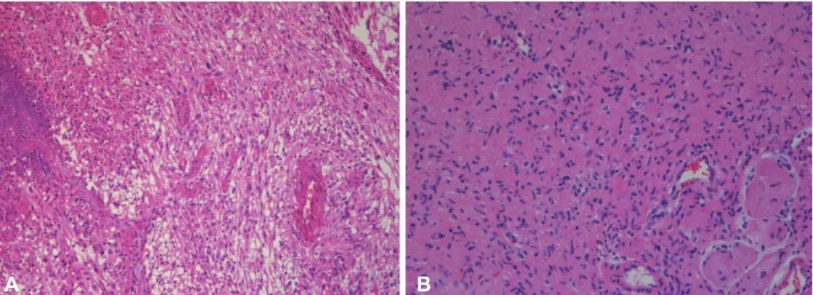
A B
Fig. 2. Histologic examination. A: H&E stain (×100) shows characteristic features of glioblastoma. B: Scalp mass is revealed plexiform neu- rofibroma (H&E stain, ×200).
A B
Fig. 3. Post-adjuvant chemotherapy with temozolomide, 9 months after surgery. Gd-enhanced axial (A) and coronal (B) T1-weighted MR im- ages reveal no evidence of tumor recurrence.
38 Brain Tumor Res Treat 2014;2(1):36-38 GBM in a Patient with NF1
mas, carcinomas (breast, lungs, gastrointestinal tract, thyroid, pharynx, ovaries), malignant melanoma, pheochromocyto- mas [5-7]. The majority of intracranial tumors with NF1 are pilocytic astrocytomas and optic gliomas which are thought to be pathologically low grade. Up to the year 2010, 12 pa- tients with NF1 associated with glioblastoma had been re- ported [4]. Among them four occurred in adults. One patient was clinically represented with progressive multiple sclerosis and neurofibromatosis but glioblastoma remained silent dur- ing the patient’s life. The pathology of glioblastoma was re- vealed by autopsy [8]. Table 1 shows the characteristics of the glioblastomas with NF1 in adults [8-11]. Huttner et al. [4] re- viewed five glioblastoma patients in children with NF1 and reported their clinicopathologic features. This study suggest- ed that pathological features such as increased p53 expres- sion, increased EGFR amplification, and increased prolifera- tive indices might have predicted a worse overall outcome.
They also provided that the survival of glioblastoma patient with NF1 was better than those without NF1. The median overall survival of patients with and without NF1 was 9.25 and 1.08 years respectively. However, because of the limited num- ber of patients, further investigation is needed. In adults, glioblastoma is the most common and aggressive primary brain tumor. The methylation status of the MGMT gene pro- moter is currently a promising molecular prognostic marker.
There is a significant association between MGMT promoter methylation and the outcome of glioblastoma patients treated with temozolomide. Molecular analysis of glioblastomas aris- ing in NF1 patients showed the presence of genetic alterations such as p16INK4A/ARF deletion and p53 mutations [12]. Some studies have reported the cooperation of mutations in NF1 and p53 in the development astrocytomas in transgenic mice [13,14]. In our case, the promoter methylation of MGMT gene was not observed. Immunohistochemical findings were positive for GFAP, and negative for EGFR. Preliminary stud- ies reported that the prognosis of glioblastoma with NF1 in children may differ from patient without NF1 [4]. In adults, there is limited information about glioblastoma with NF1
cases. In this present case, the patient was treated using sur- gical excision and chemoradiotherapy with temozolomide.
Further investigation for the glioblastoma patients with NF1 in adults would be necessary.
Conflicts of Interest
The authors have no financial conflicts of interest.
REFERENCES
1. Friedman JM. Epidemiology of neurofibromatosis type 1. Am J Med Genet 1999;89:1-6.
2. Blatt J, Jaffe R, Deutsch M, Adkins JC. Neurofibromatosis and child- hood tumors. Cancer 1986;57:1225-9.
3. Korf BR. Malignancy in neurofibromatosis type 1. Oncologist 2000;
5:477-85.
4. Huttner AJ, Kieran MW, Yao X, et al. Clinicopathologic study of glio- blastoma in children with neurofibromatosis type 1. Pediatr Blood Cancer 2010;54:890-6.
5. Farmer JP, Khan S, Khan A, et al. Neurofibromatosis type 1 and the pediatric neurosurgeon: a 20-year institutional review. Pediatr Neuro- surg 2002;37:122-36.
6. Guillamo JS, Créange A, Kalifa C, et al. Prognostic factors of CNS tu- mours in Neurofibromatosis 1 (NF1): a retrospective study of 104 pa- tients. Brain 2003;126(Pt 1):152-60.
7. Walther MM, Herring J, Enquist E, Keiser HR, Linehan WM. von Recklinghausen’s disease and pheochromocytomas. J Urol 1999;162:
1582-6.
8. Pál E, Gömöri E E, Gáti I. Neurofibromatosis and glioblastoma in a case of multiple sclerosis. Eur J Neurol 2001;8:717-8.
9. Mehta RS, Abraham M, Plesa C, Ennis P. Glioblastoma multiforme in an adult with von Recklinghusen disease. Commun Oncol 2008;5:544-8.
10. Hakan T, Aker FV. Case report on a patient with neurofibromatosis type 1 and a frontal cystic glioblastoma. Neurol Neurochir Pol 2008;
42:362-5.
11. Broekman ML, Risselada R, Engelen-Lee J, Spliet WG, Verweij BH.
Glioblastoma multiforme in the posterior cranial fossa in a patient with neurofibromatosis type I. Case Rep Med 2009;2009:757898.
12. Gutmann DH, James CD, Poyhonen M, et al. Molecular analysis of as- trocytomas presenting after age 10 in individuals with NF1. Neurology 2003;61:1397-400.
13. Reilly KM, Loisel DA, Bronson RT, McLaughlin ME, Jacks T. Nf1;
Trp53 mutant mice develop glioblastoma with evidence of strain-spe- cific effects. Nat Genet 2000;26:109-13.
14. Zhu Y, Guignard F, Zhao D, et al. Early inactivation of p53 tumor sup- pressor gene cooperating with NF1 loss induces malignant astrocyto- ma. Cancer Cell 2005;8:119-30.
Table 1. The characteristics of previous cases of Glioblastoma with NF1 in adults
Authors Reference year Age (years)/sex Location Treatment Post-treatment course
Pál et al. [8] 2001 37/female Occipital No treatment
Mehta et al. [9] 2008 63/male Parietal Bx only
Refuse any intervention
Dead 2 months after his initial presentation
Hakan and Aker [10] 2008 28/female Frontal SR+RT+CTx
(temozolomide) Dead 41 months after surgery
Broekman et al. [11] 2009 28/female Cerebellum SR+RT+CTx Dead 6 months after surgery
Present case 32/male Frontal SR+RT+CTx Alive
Bx: biopsy, SR: surgical resection, RT: radiation therapy, CTx: chemotherapy, NF1: neurofibromatosis type 1