연부조직의 거대 세포종은 국소형과 미만형으로 분류되는 무통 성의 양성종양으로 활액막 세포에서 기원한다. 발생 위치에 따라 관절강내와 관절강외로 나뉘고 성장 양상에 따라 국소형과 미만 형으로 나뉘게 된다. 국소형의 거대세포종은 주로 관절주위와 점 액낭에서 발생하며 수부에 가장 흔한 것으로 보고 되었다.1) 미만 형의 거대세포종은 상대적으로 드물며 관절내 발생이 흔한 것으 로 알려져 있으며 관절외의 피하 혹은 근육내 발생하는 것은 매우 드물다.1) 저자들은 9세 남아로 삼각근내의 미만형 거대세포종으 로 진단된 증례에 대해 경험한 바를 보고하고자 한다.
증례 보고
9세 남자 환아가 6개월 전 우측 어깨에서 우연히 발견한 종괴를 주소로 내원하였다. 신체 검진상 종괴는 우측 어깨의 삼각근 외측 으로 관찰되었으며 통증 및 압통은 없었다. 딱딱하게 고정된 양상 으로 둥근 모양이었고 3×4 cm 정도의 크기로 6개월간의 크기 변 화는 없었으며 색소침착 등은 관찰되지 않았다. 어깨 및 종괴 원 위부의 감각 이상이나 운동제한 또한 관찰되지 않았다. 우측 어깨 의 단순 방사선 검사상 상완골 근위부 외측의 근육층과 연결된 것
삼각근내 발생한 미만형 거대세포종
Diffuse-Type Giant Cell Tumor in Deltoid Muscle
전영수 • 이상훈 • 이동기 • 김정연 • 김정석 • 한정수
경희대학교 의과대학 정형외과학교실
으로 보이는 경계가 명확한 둥근 모양의 음영을 확인할 수 있었다 (Fig. 1).
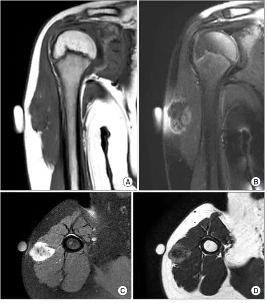
자기공명영상검사에서 상완골 근위부의 삼각근 외측에서 다 방성의 경계가 명확한 종괴를 확인할 수 있었으며 종괴는 삼각근 의 사이에 위치했으며 측정된 크기는 2.1×1.6×3.0 cm이었다. 자 기 공명 영상의 T1 강조영상과 T2 강조영상에서 모두 비균질성의 저-중등도 정도의 신호 강도를 나타냈고 조영제 주사 후에도 비 균질 양상으로 종괴 주위로 조영이 증가되었다(Fig. 2).
접수일 2013년 11월 1일 심사수정일 2013년 11월 30일 게재확정일 2013년 12월 2일
교신저자 전영수
서울시 강동구 동남로 892, 강동경희대학교병원 정형외과 TEL 02-440-6155, FAX 02-440-7498
E-mail [email protected]
미만형 거대세포종은 상대적으로 국소형 거대세포종에 비해 드문 것으로 알려져 있으며 관절외 발생하는 경우는 극히 드물다고 알려져 있다.
이에 저자들은 삼각근 내에 발생한 미만형 거대세포종 1예를 경험하여 이를 문헌고찰과 함께 보고하고자 한다.
색인단어: 삼각근, 미만형 거대세포종
Copyrights © 2013 by The Korean Bone and Joint Tumor Society
“This is an Open Access article distributed under the terms of the Creative Commons Attribution Non-Commercial License (http://creativecommons.org/licenses/by-nc/3.0/) which permits unrestricted non-commercial use, distribution, and reproduction in any medium, provided the original work is properly cited.”
대한골관절종양학회지:제19권 제2호 2013
Figure 1. Anteroposterior simple radiograph of the humerus shows about 3.0 cm sized bulging soft tissue lesion at lateral aspect of right upper arm.
88
전영수·이상훈·이동기 외 3인
Figure 2. These are the magnetic reso- nance findings of the mass. (A) Coronal T1WI shows a mass with lobulations in lateral portion of the deltoid muscle. (B) Coronal T2WI shows low to intermediate signal intensity with heterogeneous enhancement. (C) Axial T1 enhanced image shows high signal intensity with heterogeneous enhancement. (D) Axial T2WI shows low to intermediate signal intensity.
Figure 3. (A) Gross examination reveals an ill-demarcated firm brownish mass. (B) The cut surface is nodular with whitish solid appearance.
전신마취하에 측와위 자세에서 종괴를 촉지 후 종괴 위로 길이 방향으로 절개를 하였고 피하조직을 절개한 후 삼각근을 길이방 향으로 분리한 뒤 종괴를 노출시켰다. 종괴를 근육에서 박리한 후 제거를 하였으며 3.6×2.0×1.6 cm 크기의 갈색의 단단하고 경계 가 분명한 양상을 보였다. 절단면 상에서는 단단한 흰색의 결절 양상임을 확인 할 수 있었다(Fig. 3).
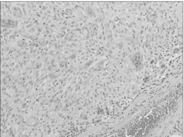
병리 조직검사상 괴사는 관찰되지 않았으며 다핵을 가지는 거 대세포가 미만성으로 분포되어 있었고 유사분열은 고배율에서 3-4개 관찰된 미만형 건막 거대세포종(Diffuse type of tenosyno- vial giant cell tumor)으로 진단 하였다(Fig. 4, 5). 특이 면역 화학 염색 검사 상 SMA, ACTIN (Smooth Muscle): 양성, CD 34: 음성, CD 68, Macrophage: 양성, CK, AE1/AE3: 음성, EMA (Epithelial Membrane Ag): 음성, KI-67 (MIB1): 3% 양성, S-100: 부분양성 소 견을 확인할 수 있었다. 수술 후 1년간 추시 관찰한 결과 재발이나 합병증은 관찰되지 않았다.
고 찰
관절외 색소 융모 결절성 활막염은 국소형 건막 거대세포종과 미 만형 건막 거대세포종으로 분류할 수 있다. 국소형의 거대세포종 은 관절의 활액막이나 건막의 점액낭에서 비교적 자주 관찰 되나 미만형의 거대세포종은 국소형보다 흔히 볼 수 없다. 또한 관찰 된다 하더라도 관절주위의 연부조직에서 발견되는 경우는 있으 나 근육내 또는 피하에서 발견되는 경우는 극히 드물다.2) 미만형 은 세계보건기구에서 활막친화성 단핵구, 다핵 거대세포, 거품세 포(foam cells), 철식세포(siderophage)와 염증세포 등의 파괴적인 증식(destructive proliferation)로 정의하였다. 색소 융모 결절성 활 막염과 관절외 및 미만형의 세분류(varies)는 현재 세계보건기구 에 따라 골과 연부조직 종양 분류에 의해 섬유조직구 종양으로 분
류되어 있다.3) Somerhausen과 Fletcher2)에 의하면 관절외 미만형 거대세포종은 양성종양으로 호발연령은 평균 41세 여자에서 많 으며 호발부위는 손목관절, 슬관절 주위 즉 대퇴골 원위단 및 경 골 근위단에서 잘 발생한다. 국소형 건막 거대세포종의 재발율은 7-23%로 보고하고 있으며, 미만형 건막 거대세포종은 40-50%로 보고하고 있다.1,4)
자기공명영상검사는 거대세포종의 진단에 매우 민감한 검사로 알려져 있다. 일반적으로 T1 강조영상에서는 관절주위의 횡문근 과 동일한 낮은 신호강도를 가지며 T2 강조영상에서는 다양한 신 호강도를 보이는 반면 대개의 연부조직 종양은 T2 강조영상에서 높은 신호강도를 보이므로 T1 및 T2 강조영상 모두에서 낮은 신 호강도를 보이는 연부조직 종양인 경우에는 거대세포종을 의심 할 수 있다.
그러나 본 증례에서는 관절 주위에 발생하지 않고 상완골 근위 부의 근육내에 위치하였다. 또한 전형적인 거대세포종의 자기공 명 영상 소견과는 달리 T1과 T2 강조영상 모두에서 비균질한 양 상의 저-중등도 신호 강도를 확인할 수 있었으며 조영제 주사 후 에도 비균질한 양상으로 조영이 되었고 흔히 발생하지 않는 삼 각근내에 발생하였다. 이에 수술 전 자기 공명 영상을 통한 감 별 진단에서 유건종(desmoids tumor) 또는 근육내 결절성 근막염 (intramuscular nodular fasciitis)을 가장 먼저 의심하였고 횡문근육 종(rhabdomyosarcoma)이나 활막육종(synovial sarcoma), 섬유육 종(fibrosarcoma) 같은 육종 또한 의심할 수 있었다. 종괴의 절제 와 조직생검을 위하여 수술을 시행하였는데, 절개생검(incisional biopsy)이 원칙이나 크기가 작고 경계가 분명하여 절제생검(exci- sional biopsy)을 시행하였다.
Somerhausen과 Fletcher2)은 50명의 미만형 거대세포종 환자 중 27명이 순수한 관절외 형태였고 그 중 5명이 근육 내로 위치 하였 으며 대퇴부 3명, 둔부 1명, 하지 1명이었다고 보고하였다. Sanghvi Figure 4. Extraarticular diffuse-type gaint cell tumor infiltrates irregularly
into fibroadipose tissue (H&E, ×40).
Figure 5. Tumors are composed of multi-nucleated giant cells admixed with round cells and collagen (H&E, ×200).
90
전영수·이상훈·이동기 외 3인
등5)은 대퇴부 피하층에 발생한 증례를 보고하였고, Yoshida 등6)은 슬괵근내 발생한 증례를, Hepp 등7)은 거위발 복합체내에 발생한 증례를 보고 하였다. 이와 같이 관절 외에 발생한 미만형의 거대 세포종은 극히 드문 것으로 알려져 있다. 또한 문헌 고찰 상 삼각 건 내에 발생한 미만형 거대세포종에 대하여 보고한 바가 없어 이 번 증례보고는 의미가 있을 것으로 생각된다.
본 증례에서는 점액낭이나 관절 주위가 아닌 삼각근 내에서 발 생한 미만형 거대세포종으로 자기공명영상 및 조직검사, 또한 특 이 면역 조직 화학 염색을 통해 확진할 수 있었다. 1년간의 추시관 찰 기간 동안 재발이나 합병증은 없었으나 장기간의 추시 관찰이 필요 할 것으로 보인다.
참고문헌
1. Ushijima M, Hashimoto H, Tsuneyoshi M, Enjoji M. Giant cell tumor of the tendon sheath (nodular tenosynovitis). A study of 207 cases to compare the large joint group with the com- mon digit group. Cancer. 1986;57:875-84.
2. Somerhausen NS, Fletcher CD. Diffuse-type giant cell tu- mor: clinicopathologic and immunohistochemical analysis
of 50 cases with extraarticular disease. Am J Surg Pathol.
2000;24:479-92.
3. Fletcher CDM, Unni KK, Mertens F, World Health Organiza- tion, International Agency for Research on Cancer. Pathology and genetics of tumours of soft tissue and bone. Lyon: IARC Press; 2002. 112-4.
4. Lee GW, Lee KS, Song SH, Kim MK, Yun SH. Snow-man shaped nodular tenosynovitis in the knee. case report. J of Ko- rean Arthroscopy Soc. 1999;3:44-7.
5. Sanghvi DA, Purandare NC, Jambhekar NA, Agarwal MG, Agarwal A. Diffuse-type giant cell tumor of the subcutaneous thigh. Skeletal Radiol. 2007;36:327-30.
6. Yoshida T, Sakamoto A, Tanaka K, et al. Intramuscular diffuse- type giant cell tumor within the hamstring muscle. Skeletal Radiol. 2007;36:331-3.
7. Hepp P, Engel T, Marquass B, Aigner T, Josten C, Niederhagen M. Infiltration of the pes anserinus complex by an extraar- ticular diffuse-type giant cell tumor (D-TGCT). Arch Orthop Trauma Surg. 2008;128:155-8.
Diffuse-Type Giant Cell Tumor in Deltoid Muscle
Young Soo Chun, Sang Hoon Lee, Dong Ki Lee, Jung Youn Kim, Jung Suk Kim, and Chung Soo Han
Department of Orthopaedic Surgery, Kyung Hee University Hospital at Gangdong, Seoul, Korea
Diffuse-type giant cell tumor is relatively rare than localized giant cell tumor. Moreover, diffuse type giant cell tumor is common in intraarticular area, rarely occurs at intramuscular or subcutaneous layer. We experienced 1 case of giant cell tumor within the deltoid muscle. So we report this case with review of the literatures.
Key words
: deltoid, diffuse-type giant cell tumorReceived November 1, 2013 Revised November 30, 2013 Accepted December 2, 2013 Correspondence to: Young Soo Chun
Department of Orthopaedic Surgery, Kyung Hee University Hospital at Gangdong, 892, Dongnam-ro, Gangdong-gu, Seoul 134-727, Korea
TEL: +82-2-440-6155 FAX: +82-2-440-7498 E-mail: [email protected]
Copyrights © 2013 by The Korean Bone and Joint Tumor Society
“This is an Open Access article distributed under the terms of the Creative Commons Attribution Non-Commercial License (http://creativecommons.org/licenses/by-nc/3.0/) which permits unrestricted non-commercial use, distribution, and reproduction in any medium, provided the original work is properly cited.”
The Journal of the Korean Bone and Joint Tumor Society Vol. 19, No. 2 (December 2013)