Received:September 24, 2016, Revised:October 10, 2016, Accepted:October 10, 2016
Corresponding to:Yong-Beom Park, Division of Rheumatology, Department of Internal Medicine, Yonsei University College of Medicine, 50-1 Yonsei-ro, Seodaemun-gu, Seoul 03722, Korea. E-mail:[email protected]
pISSN: 2093-940X, eISSN: 2233-4718
Copyright ⓒ 2016 by The Korean College of Rheumatology. All rights reserved.
This is a Free Access article, which permits unrestricted non-commerical use, distribution, and reproduction in any medium, provided the original work is properly cited.
Immunomodulatory Function of Mesenchymal Stem Cells for Rheumatoid Arthritis
Mi Il Kang1, Yong-Beom Park2
1Division of Rheumatology, Department of Internal Medicine, Dankook University Medical College, Cheonan, 2Division of Rheumatology, Department of Internal Medicine, Institute for Immunology and Immunologic Diseases, Brain Korea 21 PLUS Project for Medical Science, Yonsei University College of Medicine, Seoul, Korea
Developments in our comprehension of the autoimmune and inflammation mechanisms in rheumatoid arthritis (RA) have pro- duced targeted therapies that block aberrant immune cells and cytokine networks, and improved treatment of RA patients considerably. Nevertheless, limitations of these treatments include incomplete treatment response, adverse effects requiring drug withdrawal, and refractory cases. Hence, many researchers have redirected efforts towards investigation of other bio- logical aspects of RA, including the mechanisms driving joint tissue repair and balanced immune regulation. This investigation focuses on mesenchymal stem cell (MSC) research, with the ultimate goal of developing interventions for immune modulation and repair of damaged joints. MSCs are multipotent cells capable of differentiating into mesodermal lineage cells. These cells have also attracted interest for their anti-inflammatory and immunomodulatory capacities. They have many distinctive im- munological properties, inhibiting the proliferation and production of cytokines by T, B, natural killer, and dendritic cells.
Indeed, MSCs have the capacity to regulate immunity-induced peripheral tolerance, suggesting they can be used as therapeutic tools in RA. This review discusses properties of MSCs, in vitro studies, animal studies, and clinical trials involving MSCs. Our review discusses the current knowledge of the mechanisms of MSC-mediated immunosuppression and potential therapeutic uses of MSCs in RA. (J Rheum Dis 2016;23:279-287)
Key Words. Inflammation, Mesenchymal stem cells, Rheumatoid arthritis
INTRODUCTION
Rheumatoid arthritis (RA) is a complicated auto- immune disease characterized by joint destruction asso- ciated with production of inflammatory mediators [1].
RA results in significantly reduced ability to perform daily activities and is associated with multiple comorbidities, increased mortality, and socioeconomic loss [2]. The eti- ology of RA is not completely understood. However, nu- merous investigations of the pathogenic mechanisms of inflammation and autoimmunity and our increased un- derstanding of signal mediators implicated in the patho- genesis of RA have led to the development of agents that block tumor necrosis factor-alpha (TNF-α), interleukin
(IL)-6 signaling, the immune activity of T cells by cos- timulation signaling, and, the targeting of pathogenic cells such as B cells and osteoclasts [3]. Based on this re- search, the current care in RA is to apply a treat-to-target (T2T) strategy immediately after diagnosis. The elements of the T2T concept are adjusting the therapy, guided by an assessment of disease activity, with the aim of clinical re- mission [4]. Despite significant development in treat- ments, however, several problems remain unresolved. Up to 30% of RA patients fail to respond to current conven- tional and biologic-disease-modifying therapies [3]. In addition, some RA patients in sustained clinical re- mission presented radiographic progression of joint dam- age [5].
The cause of RA is unknown, but genetic and environ- mental factors are contributory. The pathophysiology of RA is chronic inflammation of the joint synovium, which causes cartilage destruction and bone erosion through in- teractions among infiltrating T cells, B cells, proin- flammatory cytokines and the resident fibroblast-like synoviocytes [6]. Various proinflammatory cytokines, such as TNF-α, IL-6, IL-1β and IL-17, play dominant pathological roles and aberrant T helper cells (Th) 17 and Th1 responses have been connected to pathogenesis of RA [7]. In addition, evidence is increasing that a defect in the number or function of regulatory T cells (Tregs) is cru- cial in the immune imbalance that has a pivotal role in the pathogenesis of RA [8].
MSCs are non-embryonic stromal cells that exist in the bone marrow, peripheral blood, adipose tissue, and synovium. MSCs can be readily isolated from various tis- sue sources, handily expanded in culture, and differ- entiated under appropriate stimulation. These character- istics of MSCs suggest it as an ideal candidate tool for tis- sue engineering efforts aiming to repair damaged structures. In addition to these advantages, MSCs pos- sess multipotent immunomodulatory and anti-in- flammatory effects, through either direct cell-cell inter- action or secretion of various factors. MSC are now widely researched for their immunomodulating and protective qualities based on their regenerative capacities [9].
Several clinical trials showed that the administration of MSC in patients with RA in general well tolerated and the treatment induced a significant remission and a reduction in disease activity score (DAS)-28. In addition, the serum levels of inflammatory cytokines decreased after the MSC therapy [9-11]. This review summarizes our knowledge on the mechanisms underlying MSC properties on im- mune responses and the therapeutic effect of MSCs on RA.
MAIN SUBJECTS
Characteristics of MSC
MSCs are adult stromal cells that they have the ability to differentiate into various mesodermal cell lineages, which are very important in inflammatory arthritis [12].
Despite the many studies on MSCs, there is no uniformly accepted phenotype or surface marker for their isolation.
Currently, MSCs are defined retrospectively based on a grouping of characteristics in vitro, including a combina- tion of phenotypic markers and multi-potential differ-
entiation functional properties. The minimal require- ments for a population of cells to qualify as MSCs are as follows: (1) they must be plastic adherent under standard culture conditions, (2) they should express CD73, CD105, and CD90 and not express CD45, CD34, CD14, CD11b, CD79α, CD19, or human leukocyte antigen (HLA)-DR surface molecules, and (3) they should possess meso- dermal differentiation capability into osteoblasts, chon- drocytes, and adipocytes [13].
MSCs can be expanded ex vivo up to a billion-fold with- out loss of their multipotent properties and are excellent vehicles since they maintain the expression of transfected genes for up to 40 divisions [14]. Even if MSCs isolated from different tissues show similar phenotypic features, it is not distinct whether these are the same MSCs. In ad- dition, MSCs show different dispositions in proliferation and differentiation capacities in response to stimulation with various growth factors. Culture conditions of sur- face, medium, seeding density, and, isolation methods, and the presence of various growth factors influence the expansion, differentiation, and immunogenic properties of MSCs [15].
MSCs are hypoimmunogenic or non-immunogenic and so can easily escape host immune elimination. MSCs ex- press low to intermediate major histocompatibility com- plex (MHC) class I molecules and do not express MHC class II molecules, although an intracellular pool of MHC class II molecules can be stimulated by interferon-gamma (IFN-γ) to be expressed on the cell surface [16]. Because MSCs do not express any costimulatory molecules, in- cluding B7-1 (CD80), B7-2 (CD86), or CD40, they do not activate alloreactive T cells [17]. Even under stimulation, MSCs do not express MHC class II molecules after differ- entiation into adipocytes, osteoblasts, and chondrocytes and remain non-immunogenic [16]. These properties in- dicate that MSCs should be able to be transplanted into an allogeneic host without immune rejection and not elicit a host immune response. An immunoprivileged capacity can be obtained by suppressing alloreactivity through the modulation of most major immune cell activities.
However, the immunoprivileged properties of MSCs seem to be limited. A few mouse studies have reported that allogeneic mismatched MSCs were rejected by the host [18-20].
Immunoregulatory properties of mesenchymal stem cells
Especially for their use in rheumatic diseases, the most
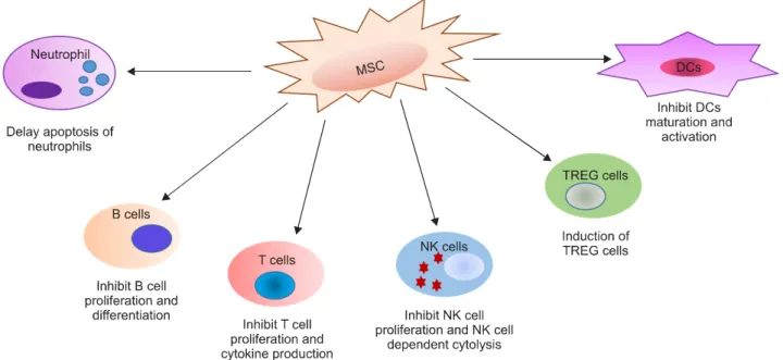
Figure 1. Suppressive effects of MSCs on immune cells. The effects of MSCs on cells of the immune system are anti-inflammatory.
DCs: dendritic cells, MSCs: mesenchymal stem cells, NK cell: natural killer cell, TREG cell: regulatory T cell.
notable and useful features of MSCs, are their potent im- munosuppressive and anti-inflammatory effects. The mechanism of the immunoregulatory activities of MSCs is not completely known, although both direct and in- direct effects have been suggested through either cell to cell interaction or soluble factors. Immunosuppressin of MSCs requires preliminary activation of the MSCs by im- mune cells through the secretion of the proinflammatory cytokine IFN-γ, alone or with TNF-α, IL-1α or IL-1β [21]. A number of studies using bone marrow-derived MSCs have reported that MSC-mediated immunomodu- lation is dependent on IFN-γ [21], and is mainly medi- ated by soluble factors such as indoleamine 2,3-dioxyge- nase (IDO), or prostaglandin E2 (PGE2), which inhibit both T- and B-cell proliferation and function [22]. On stimulation with IFN-γ, MSCs can produce high levels of IDO as a tryptophan-catabolizing enzyme that mediates immune tolerance by limiting the availability of the es- sential amino acid tryptophan and generating toxic me- tabolites for T cells [23]. PGE2 acts as a potent immune suppressant, inhibiting T-cell mitogenesis and IL-2 pro- duction, and is a cofactor of the induction of Th type 2 lymphocyte activity. Production of PGE2 by MSCs is en- hanced by TNF-α or IFN-γ stimulation, and MSC-de- rived PGE2 was shown to act on macrophages by stim- ulating the production of IL-10 and on monocytes by blocking their differentiation to dendritic cells (DCs) [24]. Another MSC-derived factor, IL-6, has been re-
ported to be involved in the inhibition of monocyte differ- entiation to DCs, decreasing their ability to stimulate T cells [25]. Other mediators, such as hepatocyte growth factor, transforming growth factor-beta 1(TGF-β1), leu- kemia inhibitory factor, and heme oxygenase-1, have been found to be produced by activated MSCs [26].
HLA-G5 secreted by MSCs has recently been shown to in- hibit T-cell proliferation, natural killer (NK) cell cytotox- icity, and T-cell cytotoxicity and to promote the gen- eration of Tregs [27]. Cell to cell contact between MSCs and activated T cells induces IL-10 production, which is essential to stimulate the release of soluble HLA-G5.
Finally, MSC-mediated immunoregulation is the result of the cumulative actions of several molecules.
MSCs suppress proliferation of allogeneic lymphocytes and can interfere with and affect cellular differentiation, maturation, and function of immune cells (Figure 1).
Both naïve and memory T cells, as well as CD4+ and CD8+ T-lymphocyte proliferation stimulated by specific antigens, are inhibited by MSCs. Suppression of pro- liferation depends on the arrest of T cells in the G0/G1 phase of the cell cycle, regardless of apoptosis [28]. MSCs change T-cell functions, such as decrease in production of IFN-γ, IL-2, and TNF-α and increase in IL-4 secretion [26]. MSCs can suppress CD8+ cytotoxic T cell-mediated cytolysis [29]. In addition, MSCs have been shown to fa- cilitate the in vivo and in vitro generation of CD4+CD25+
or CD8+ Tregs with functional properties [30]. In addi-
tion to T cells, MSCs suppress proliferation of B cells [31], NK cells [32], and DCs [33]. MSCs inhibit the pro- liferation of B cells, arresting B lymphocytes in the G0/G1 phase of the cell cycle and act their suppressive effect on B-cell terminal differentiation through the secretion of humoral factors [31]. Myeloid DCs as potent anti- gen-presenting cells are vital in the induction of im- munity and tolerance. MSCs inhibit the in vitro matura- tion of monocytes and CD34+ hematopoietic cells into DCs, as shown by a decreased cell surface expression of MHC class II and co-stimulatory molecules, and de- creased production of IL-12 and TNF-α [33]. MSCs in- hibit IL-2-driven or IL-15-driven NK cell proliferation, and, IFN-γ production and cytotoxicity against HLA class I-expressing targets [34]. These effects depend on cell-to-cell contact and on the release of soluble factors, such as TGF-β1 and PGE2, implying the existence of var- ious mechanisms of MSC-mediated NK cell inhibition.
Overall, the effect of MSCs on immune cells is to skew the immune response toward a tolerant and anti-inflam- matory phenotype.
The trafficking and homing properties of MSCs The trafficking and homing properties of MSCs can be useful tools for clinical applications using non-invasive systemic cell administration to treat RA. MSCs have been reported to express diverse chemokines and chemokine receptors and can move to lesions of inflammation by mi- grating towards inflammatory chemokines and cytokines [35-37]. However, the first-line accumulation site of in- travenously administered MSCs is lungs, followed by liv- er and spleen. Several studies showed the intraarticular or intraarterial route of administration were effective in avoiding pulmonary entrapment of MSCs and may im- prove the bioavailability of transplanted MSCs in clin- ically relevant tissues [35-37]. Acting as evidence that host MSCs can migrate in response to inflammation, sys- temically injected MSCs are also often observed within the bone marrow or in damaged tissues [35-37].
Although, data are lacking with regard to the bio- distribution of MSCs, their cellular or molecular target structures, and the mechanisms by which MSCs reach these targets, accumulating data indicate that systemic infusion of MSCs can be used for immunosuppressive treatments [36,37].
Therapeutic potential of MSCs in animal studies Based on their hypoimmunogenicity and immunomodu-
latory abilities, several studies have reported on the ther- apeutic effects of allogenic or xenogenic MSC treatment in collagen-induced arthritis (CIA) mice, a representative animal model of RA [38-43]. However, others have failed to demonstrate such effects [44-46] (Table 1). The first study that utilized an immortalized, allogenic mesen- chymal cell line, administered intravenously in mice, showed that there was no benefit of MSC therapy for the reduction of the pathogenetic development of CIA, de- spite the fact that a potent immunosuppressive activity of the cells was observed in vitro [44]. However, another study showed that a single intraperitoneal injection of al- logeneic MSCs in mice prevented the development of se- vere arthritis [38]. The study showed reduced levels of proinflammatory cytokines, as compared to the levels in controls, and increased levels of IL-10, an immuno- suppressive cytokine produced by Tregs; it also demon- strated that MSC therapy resulted in de novo generation of CD4+CD25+FoxP3+ Tregs specific for type II col- lagen. Further positive results from MSC therapy were re- ported in a later mouse study, in which it was shown that daily intraperitoneal injection of human or murine alloge- neic and syngeneic adipose–derived (AD) MSCs, for 5 days after the onset of disease significantly reduced the severity of arthritis in the CIA model. Intraarticular in- jection of AD-MSCs was less effective than the intra- peritoneal route, adding weight to the argument that the positive effects of MSCs are not simply due to direct tis- sue repair in the joints. The therapeutic efficacy was asso- ciated with decreased antigen-specific Th1/Th17 cell ex- pansion, enhanced secretion of IL-10, and generation of CD4+CD25+FoxP3+ Tregs with the capacity to sup- press self-reactive T-effector responses. The effect de- pended on timing, dose, and route of administration of MSCs [39]. MSCs overexpressing IL-10 have also been shown to attenuate CIA [40]. Results from recent studies [41-43], further support the potential of MSC-based treatment in autoimmune inflammatory arthritis, in that immune modulation and reduction of articular damage following treatment with MSCs have been observed.
These findings suggest that MSC therapy is able to reset the immune system by reducing the deleterious Th1/Th17 response and enhancing the protective Tregs response.
However, other studies have failed to demonstrate any improvement in CIA with MSC treatment [45,46]. One study found that Flk-1+ MSCs exacerbated the arthritis in mice, by promoting the secretion of IL-6 and IL-17
Table 1. The literature describing the effects of mesenchymal stem cells (MSCs) in mouse model of rheumatoid arthritis First author,
year [ref]
Source of MSC
Donor–recipient
MHC match Dose of MSCs Route of administration
Time of treatment
Outcome of treatment Djouad et al.,
2005 [44]
C3 mouse cell line
Allogeneic 1×10⁶, 1×10⁶, 4×10⁶
Intravenous Day 0 or 21, day 0 or 21
UA*
Augello et al., 2007 [38]
Mouse BM Allogeneic 5×10⁶ Intraperitoneal Day 0 or 21 Positive† Gonzalez et al.,
2009 [39]
Adipose tissue:
human, mouse
Xenogeneic, allogeneic
1×10⁶ Intraperitoneal, intraarticular
Once per day for 5 days ADO
Positive† Choi et al.,
2008 [40]
Mouse BM Syngeneic 1×10⁶ Intravenous Day 21, 28, 35 Positive†
Park et al., 2011 [41]
Mouse BM Syngeneic 1×10⁶ Intraperitoneal Week 7 Positive†
Liu et al., 2010 [42]
Human UC Xenogeneic 5×10⁶ Intraperitoneal Day 31 ADO Positive†
Bouffi et al., 2010 [43]
Mouse BM Syngeneic, allogeneic
1×10⁶ Intravenous Day 18, 24, 28, 32
Positive† Chen et al.,
2010 [45]
Mouse BM Syngeneic 1×10⁶,
2×10⁶
Intravenous Day 0 or 21 Negative‡ Schurgers et al.,
2010 [46]
Mouse BM Syngeneic, allogeneic
1×10⁶ Intravenous, intraperitoneal
Day 1, 16, 19, 23, 30
Negative‡
Time of treatment is shown as number of days or weeks before or after induction of arthritis, except where specified as number of days after disease onset (ADO). Ref: reference, MHC: major histocompatibility complex, BM: bone marrow, UC: umbilical cord.
*No effect, indicated by an unaffected disease score. †Positive effect, indicated by a disease score reduction. ‡Negative effect, indicated by a disease score increase.
[45]. The other study was unable to demonstrate any ben- efit from MSC therapy in CIA [46]. Those investigators used both an intravenous route and an intraperitoneal route to administer the MSCs, and also used allogeneic MSCs. None of these changes in MSC therapeutic ap- proaches made any difference to the negative outcome.
Experimental protocols differed between all of these studies (Table 1), which may explain the inconsistencies in results [38-46]. The studies that demonstrated benefi- cial effects of MSC therapy also had a number of differ- ences in their protocols [38-43]. Potential reasons for the discrepancies among these results include the following:
source of MSCs (murine, syngeneic vs. human, alloge- neic), tissue of origin (bone marrow, adipose tissue, cord blood), timing of treatment, number of stem cells in- jected, route of injection (intravenous, intraperitoneal, intraarticular), and treatment regimen (a single injection of MSCs vs. daily injections for 5 consecutive days) [38-47]. They used different administration of routes and used syngeneic, allogeneic, and human sources of MSCs, but the dose of MSCs was fairly consistent between studies. Data showed that human MSCs in completely
MHC-mismatched mice are not rejected by the immune system, even after allogenic or xenogenic transplantation.
MSCs showed similar cell migration and therapeutic ef- fect given by intravenous or intraperitoneal injection [47,48]. A probable factor was the length of culture of the MSCs. Schurgers et al. [46] used MSCs that had been cul- tured for several weeks prior to use in these experiments, and the investigators did not observe any therapeutic ef- fect of MSC administration. Allogeneic MSCs, which were used in the study by Augello et al. [38], were from primary culture of mouse MSCs obtained after the first in vitro passage as opposed to the immortalized cell line that was used in the study by Djouad et al. [44]. In addition, non-measurable variables that may have contributed to study differences and could be relevant might include dif- ferences in the culture medium used for stem cells, lack of standardization of MSC culture conditions, and differ- ences in animal-housing conditions [47].
Recently, a study investigated the therapeutic efficacy of human bone marrow (BM)-, adipose tissue (AD)-, and umbilical cord (UC)-derived MSCs in CIA to find the best condition for clinical application of human MSCs for RA
treatment [49]. The data showed that BM-, AD-, and UC-derived MSCs significantly suppressed joint in- flammation in CIA mice. The cells decreased proin- flammatory cytokines and upregulated anti-inflammatory cytokines and induced Tregs. The results show that the immune modulatory effect of MSCs was associated with Tregs induction, consistent with the increase in anti-in- flammatory cytokine expression in CIA. The BM-MSC-treat- ed mice showed a greater therapeutic efficacy than AD-MSCs or UC-MSCs in CIA in view of the severity of arthritis and histopathological evaluation. Also, Tregs in- duced by BM-MSCs had stronger immunosuppressive ac- tivity than those induced by AD-MSCs or CB-MSCs. As for clinical application, the study investigated the dose-dependent therapeutic effect of MSCs. Although minimal therapeutic effects were seen with 5×10⁵ MSCs, treatment with 5×10⁶ cells was sufficient for the sup- pression of arthritis and the induction of Tregs. The study showed there is no difference in effectiveness between the two administration schedules (daily injection of 1×10⁶ MSCs for 5 consecutive days vs. two injections of 2.5×10⁶ cells over a 3-day interval) [49]. According to this report, BM-MSCs had a greater therapeutic efficacy than the other types of MSCs in CIA.
The ability of MSCs to generate de novo Tregs may be ad- vantageous therapeutically when compared to neutraliz- ing antibodies against single-cytokine signaling, in terms of both safety and efficacy. These data need to be con- firmed in appropriated clinical studies.
The role of MSCs in prevention of bone destruction in RA is still unknown. Recently, one study investigated the effect of AD-MSCs on in vitro formation of bone-resorb- ing osteoclasts and pathological bone loss in a mouse CIA model of RA [50]. The study showed that AD-MSCs con- siderably suppressed receptor activator of NF-B ligand (RANKL)–induced osteoclastogenesis in both a con- tact-dependent and -independent manners. Additionally, AD-MSCs inhibited RANKL-induced osteoclastogenesis in the presence of proinflammatory cytokines such as TNF-α, IL-17, and IL-1β. In addition, treatment with AD-MSCs at the onset of CIA significantly reduced clin- ical symptoms and joint pathology. Moreover, AD-MSCs inhibited autoimmune T cell responses and increased the proportions of peripheral regulatory T and B cells. Thus, the study provides strong evidence that AD-MSCs im- prove inflammation-induced systemic bone destruction in CIA mice by reducing osteoclast precursors and im- proving immune tolerance [50].
Clinical trials with MSCs in patients with rheuma- toid arthritis
A few clinical RA studies have been presented [10,11,51]. In a preliminary report on four RA patients with anti-TNF-failing active RA were intravenous in- jection treated with a single infusion of 1×10⁶ cells per kg of allogeneic BM-MSCs or UC-MSCs. The study showed no significant clinical improvement, though no toxicity was observed. Three of four patients experienced reduc- tion in erythrocyte sedimentation rate, DAS-28, and pain visual analogue scale (VAS) score at 1 and 6 months after transplantation. Two of the patients demonstrated a mod- erate European league against rheumatism (EULAR) re- sponse at 6 months but experienced a relapse at 7 and 23 months, respectively. No patient achieved DAS-28-defined remission in the follow-up period. No adverse events were observed during or immediately after infusions of MSC in any of the four patients. No severe infections, ma- lignancies, or death occurred [10].
Another larger, non-randomized comparative trial in- volving 172 patients with active RA who demonstrated inadequate responses to traditional medication were enrolled to assess the safety and efficacy of human UC-MSCs in the treatment of RA [11]. Patients were div- ided into two treatment groups: disease-modifying an- ti-rheumatic drugs (DMARDs) plus medium without UC-MSCs, or DMARDs plus UC-MSCs (4×10⁷ cells per time) via intravenous injection. Tests of serological mark- ers were conducted to assess safety and disease activity.
Serum levels of inflammatory chemokines/cytokines were measured, and lymphocyte subsets in peripheral blood were analyzed. The serum levels of TNF-α and IL-6 decreased after the first UC-MSC treatment. The percentage of CD4+CD25+ Foxp3+ Tregs in peripheral blood was increased. The treatment induced a significant remission of disease according to the American College of Rheumatology improvement criteria, the DAS-28, and the Health Assessment Questionnaire. The therapeutic effects were maintained for 3∼6 months without con- tinuous administration, correlating with the increased percentage of Tregs in peripheral blood. No serious ad- verse effects were observed during or after infusion, and 4% of the treated patient showed mild adverse effects during the infusion, such as chill and/or mild fever. This study showed that treatment with DMARDs plus UC-MSC can provide safe, significant, and persistent clin- ical benefits for patients with active RA.
The other study presented three RA patients who re-
ceived autologous AD-MSCs [51]. All patients received multiple injections of MSC, intravenous injection in two cases and intraarticular in the third. The first patient re- ceived two intravenous injection of 3×10⁸ AD-MSCs. The second patient received 8×10⁸ AD-MSCs in total, one in- travenous injection of 2×10⁸ AD-MSCs and intraarticular injection of 1×10⁸ AD-MSCs. This patient then received another intravenous injection of 3.5×10⁸ cells and a sec- ond intra-articular injection of 1.5×10⁸ cells. The third patient received four times intravenous injections of 2×10⁸ AD-MSCs at intervals of one month. Clinical ben- efit was seen in all cases without significant toxicity.
Recently clinical trials are ongoing with MSCs in pa- tients with RA [52-54].
Safety of MSCs in human subjects
A recently published article systematically reviewed clinical trials that examined the use of MSCs in order to evaluate their safety [55]. MEDLINE, EMBASE, and the Cochrane Central Register of Controlled Trials (to June 2011) were searched. A total of 1,012 participants with clinical conditions of ischemic stroke, Crohn's disease, cardiomyopathy, myocardial infarction, graft versus host disease, and healthy volunteers were included. Meta-analy- sis of all the randomized controlled trials did not detect any association between acute infusional toxicity, organ systemic complications, infection, death or malignancy, although, there was a significant association between MSC and transient fever [55]. We conclude, based on the current clinical trials, that MSC therapy, seems to be safe.
However, further larger scale controlled clinical trials with radical reporting of adverse events are required to further define the safety profile of MSCs [55].
CONCLUSION
The current data showed that MSCs represent a promis- ing therapeutic tool in the treatment of RA. Encouraging results have been obtained from clinical trials. However, many questions remain to be identified in order to offer better treatment to control inflammation for the benefit of RA patients. This suggests the need for better compre- hension of the underlying mechanisms of immunomo- dulation and satisfaction of safety concerns. Further stud- ies are necessary to ascertain the concept of MSCs in or- der to establish the best treatment strategy for use in RA.
ACKNOWLEDGMENTS
This study was supported by a grant from the Korean Health Technology R&D Project, Ministry of Health and Welfare, Republic of Korea (Grant No. HI13C1270) and Basic Science Research Program through the National Research Foundation of Korea (NRF) funded by the Ministry of Education (NRF-2013R1A1A2058120).
CONFLICT OF INTEREST
No potential conflict of interest relevant to this article was reported.
REFERENCES
1. Firestein GS. Evolving concepts of rheumatoid arthritis.
Nature 2003;423:356-61.
2. Gonzalez A, Maradit Kremers H, Crowson CS, Nicola PJ, Davis JM 3rd, Therneau TM, et al. The widening mortality gap between rheumatoid arthritis patients and the general population. Arthritis Rheum 2007;56:3583-7.
3. McInnes IB, Schett G. The pathogenesis of rheumatoid arthritis. N Engl J Med 2011;365:2205-19.
4. Espinoza F, Fabre S, Pers YM. Remission-induction thera- pies for early rheumatoid arthritis: evidence to date and clinical implications. Ther Adv Musculoskelet Dis 2016;8:
107-18.
5. Cohen G, Gossec L, Dougados M, Cantagrel A, Goupille P, Daures JP, et al. Radiological damage in patients with rheu- matoid arthritis on sustained remission. Ann Rheum Dis 2007;66:358-63.
6. Smolen JS, Aletaha D, Koeller M, Weisman MH, Emery P.
New therapies for treatment of rheumatoid arthritis. Lancet 2007;370:1861-74.
7. Chiang EY, Kolumam GA, Yu X, Francesco M, Ivelja S, Peng I, et al. Targeted depletion of lymphotoxin-alpha-express- ing TH1 and TH17 cells inhibits autoimmune disease. Nat Med 2009;15:766-73.
8. Ehrenstein MR, Evans JG, Singh A, Moore S, Warnes G, Isenberg DA, et al. Compromised function of regulatory T cells in rheumatoid arthritis and reversal by anti-TNFalpha therapy. J Exp Med 2004;200:277-85.
9. Tyndall A, van Laar JM. Stem cells in the treatment of in- flammatory arthritis. Best Pract Res Clin Rheumatol 2010;24:565-74.
10. Liang J, Li X, Zhang H, Wang D, Feng X, Wang H, et al.
Allogeneic mesenchymal stem cells transplantation in pa- tients with refractory RA. Clin Rheumatol 2012;31:157-61.
11. Wang L, Wang L, Cong X, Liu G, Zhou J, Bai B, et al. Human umbilical cord mesenchymal stem cell therapy for patients with active rheumatoid arthritis: safety and efficacy. Stem Cells Dev 2013;22:3192-202.
12. Pittenger MF, Mackay AM, Beck SC, Jaiswal RK, Douglas R, Mosca JD, et al. Multilineage potential of adult human mes- enchymal stem cells. Science 1999;284:143-7.
13. Dominici M, Le Blanc K, Mueller I, Slaper-Cortenbach I, Marini F, Krause D, et al. Minimal criteria for defining mul- tipotent mesenchymal stromal cells. The International Society for Cellular Therapy position statement. Cytothera- py 2006;8:315-7.
14. Tyndall A, LeBlanc K. Stem cells and rheumatology: update on adult stem cell therapy in autoimmune diseases.
Arthritis Rheum 2006;55:521-5.
15. Sotiropoulou PA, Perez SA, Salagianni M, Baxevanis CN, Papamichail M. Characterization of the optimal culture con- ditions for clinical scale production of human mesenchymal stem cells. Stem Cells 2006;24:462-71.
16. Le Blanc K, Tammik C, Rosendahl K, Zetterberg E, Ringdén O. HLA expression and immunologic properties of differ- entiated and undifferentiated mesenchymal stem cells. Exp Hematol 2003;31:890-6.
17. Tse WT, Pendleton JD, Beyer WM, Egalka MC, Guinan EC.
Suppression of allogeneic T-cell proliferation by human marrow stromal cells: implications in transplantation.
Transplantation 2003;75:389-97.
18. Eliopoulos N, Stagg J, Lejeune L, Pommey S, Galipeau J.
Allogeneic marrow stromal cells are immune rejected by MHC class I- and class II-mismatched recipient mice. Blood 2005;106:4057-65.
19. Nauta AJ, Westerhuis G, Kruisselbrink AB, Lurvink EG, Willemze R, Fibbe WE. Donor-derived mesenchymal stem cells are immunogenic in an allogeneic host and stimulate donor graft rejection in a nonmyeloablative setting. Blood 2006;108:2114-20.
20. Prigozhina TB, Khitrin S, Elkin G, Eizik O, Morecki S, Slavin S. Mesenchymal stromal cells lose their immunosuppress- ive potential after allotransplantation. Exp Hematol 2008;
36:1370-6.
21. Ren G, Zhang L, Zhao X, Xu G, Zhang Y, Roberts AI, et al.
Mesenchymal stem cell-mediated immunosuppression oc- curs via concerted action of chemokines and nitric oxide.
Cell Stem Cell 2008;2:141-50.
22. Dazzi F, Krampera M. Mesenchymal stem cells and auto- immune diseases. Best Pract Res Clin Haematol 2011;
24:49-57.
23. Meisel R, Zibert A, Laryea M, Göbel U, Däubener W, Dilloo D. Human bone marrow stromal cells inhibit allogeneic T-cell responses by indoleamine 2,3-dioxygenase-mediated tryptophan degradation. Blood 2004;103:4619-21.
24. Németh K, Leelahavanichkul A, Yuen PS, Mayer B, Parmelee A, Doi K, et al. Bone marrow stromal cells attenu- ate sepsis via prostaglandin E(2)-dependent reprogram- ming of host macrophages to increase their interleukin-10 production. Nat Med 2009;15:42-9.
25. Asari S, Itakura S, Ferreri K, Liu CP, Kuroda Y, Kandeel F, et al. Mesenchymal stem cells suppress B-cell terminal differentiation. Exp Hematol 2009;37:604-15.
26. Aggarwal S, Pittenger MF. Human mesenchymal stem cells modulate allogeneic immune cell responses. Blood 2005;105:1815-22.
27. Selmani Z, Naji A, Zidi I, Favier B, Gaiffe E, Obert L, et al.
Human leukocyte antigen-G5 secretion by human mesen- chymal stem cells is required to suppress T lymphocyte and natural killer function and to induce CD4+CD25highFOXP3+
regulatory T cells. Stem Cells 2008;26:212-22.
28. Glennie S, Soeiro I, Dyson PJ, Lam EW, Dazzi F. Bone mar-
row mesenchymal stem cells induce division arrest anergy of activated T cells. Blood 2005;105:2821-7.
29. Rasmusson I, Ringdén O, Sundberg B, Le Blanc K. Mesen- chymal stem cells inhibit the formation of cytotoxic T lym- phocytes, but not activated cytotoxic T lymphocytes or natu- ral killer cells. Transplantation 2003;76:1208-13.
30. Prevosto C, Zancolli M, Canevali P, Zocchi MR, Poggi A.
Generation of CD4+ or CD8+ regulatory T cells upon mes- enchymal stem cell-lymphocyte interaction. Haematologica 2007;92:881-8.
31. Tabera S, Pérez-Simón JA, Díez-Campelo M, Sánchez- Abarca LI, Blanco B, López A, et al. The effect of mesen- chymal stem cells on the viability, proliferation and differ- entiation of B-lymphocytes. Haematologica 2008;93:1301-9.
32. Spaggiari GM, Capobianco A, Abdelrazik H, Becchetti F, Mingari MC, Moretta L. Mesenchymal stem cells inhibit natural killer-cell proliferation, cytotoxicity, and cytokine production: role of indoleamine 2,3-dioxygenase and pros- taglandin E2. Blood 2008;111:1327-33.
33. Nauta AJ, Kruisselbrink AB, Lurvink E, Willemze R, Fibbe WE. Mesenchymal stem cells inhibit generation and func- tion of both CD34+-derived and monocyte-derived den- dritic cells. J Immunol 2006;177:2080-7.
34. Sotiropoulou PA, Perez SA, Gritzapis AD, Baxevanis CN, Papamichail M. Interactions between human mesenchymal stem cells and natural killer cells. Stem Cells 2006;24:74- 85.
35. Sordi V, Malosio ML, Marchesi F, Mercalli A, Melzi R, Giordano T, et al. Bone marrow mesenchymal stem cells ex- press a restricted set of functionally active chemokine re- ceptors capable of promoting migration to pancreatic islets.
Blood 2005;106:419-27.
36. Stappenbeck TS, Miyoshi H. The role of stromal stem cells in tissue regeneration and wound repair. Science 2009;
324:1666-9.
37. Leibacher J, Henschler R. Biodistribution, migration and homing of systemically applied mesenchymal stem/stromal cells. Stem Cell Res Ther 2016;7:7.
38. Augello A, Tasso R, Negrini SM, Cancedda R, Pennesi G.
Cell therapy using allogeneic bone marrow mesenchymal stem cells prevents tissue damage in collagen-induced arthritis. Arthritis Rheum 2007;56:1175-86.
39. González MA, Gonzalez-Rey E, Rico L, Büscher D, Delgado M. Treatment of experimental arthritis by inducing immune tolerance with human adipose-derived mesenchymal stem cells. Arthritis Rheum 2009;60:1006-19.
40. Choi JJ, Yoo SA, Park SJ, Kang YJ, Kim WU, Oh IH, et al.
Mesenchymal stem cells overexpressing interleukin-10 at- tenuate collagen-induced arthritis in mice. Clin Exp Immunol 2008;153:269-76.
41. Park MJ, Park HS, Cho ML, Oh HJ, Cho YG, Min SY, et al.
Transforming growth factor β-transduced mesenchymal stem cells ameliorate experimental autoimmune arthritis through reciprocal regulation of Treg/Th17 cells and osteoclastogenesis. Arthritis Rheum 2011;63:1668-80.
42. Liu Y, Mu R, Wang S, Long L, Liu X, Li R, et al. Therapeutic potential of human umbilical cord mesenchymal stem cells in the treatment of rheumatoid arthritis. Arthritis Res Ther 2010;12:R210.
43. Bouffi C, Bony C, Courties G, Jorgensen C, Noël D. IL-6-de- pendent PGE2 secretion by mesenchymal stem cells inhibits
local inflammation in experimental arthritis. PLoS One 2010;5:e14247.
44. Djouad F, Fritz V, Apparailly F, Louis-Plence P, Bony C, Sany J, et al. Reversal of the immunosuppressive properties of mesenchymal stem cells by tumor necrosis factor alpha in collagen-induced arthritis. Arthritis Rheum 2005;52:1595- 603.
45. Chen B, Hu J, Liao L, Sun Z, Han Q, Song Z, et al. Flk-1+
mesenchymal stem cells aggravate collagen-induced arthri- tis by up-regulating interleukin-6. Clin Exp Immunol 2010;159:292-302.
46. Schurgers E, Kelchtermans H, Mitera T, Geboes L, Matthys P. Discrepancy between the in vitro and in vivo effects of murine mesenchymal stem cells on T-cell proliferation and collagen-induced arthritis. Arthritis Res Ther 2010;12:R31.
47. MacDonald GI, Augello A, De Bari C. Role of mesenchymal stem cells in reestablishing immunologic tolerance in auto- immune rheumatic diseases. Arthritis Rheum 2011;63:
2547-57.
48. Meyerrose TE, De Ugarte DA, Hofling AA, Herrbrich PE, Cordonnier TD, Shultz LD, et al. In vivo distribution of hu- man adipose-derived mesenchymal stem cells in novel xen- otransplantation models. Stem Cells 2007;25:220-7.
49. Park KH, Mun CH, Kang MI, Lee SW, Lee SK, Park YB.
Treatment of collagen-induced arthritis using immune modulatory properties of human mesenchymal stem cells.
Cell Transplant 2016;25:1057-72.
50. Garimella MG, Kour S, Piprode V, Mittal M, Kumar A, Rani L, et al. Adipose-derived mesenchymal stem cells prevent systemic bone loss in collagen-induced arthritis. J Immunol 2015;195:5136-48.
51. Ra JC, Kang SK, Shin IS, Park HG, Joo SA, Kim JG, et al. Stem cell treatment for patients with autoimmune disease by sys- temic infusion of culture-expanded autologous adipose tis- sue derived mesenchymal stem cells. J Transl Med 2011;
9:181.
52. Safety and efficacy study of human umbilical cord-mesen- chymal stem cells for rheumatoid arthritis. Silver Spring (MD): U.S. Food and Drug Administration; 2016 Jan.
ClinicalTrials.gov Identifier: NCT02643823. Available from: https://clinicaltrials.gov/ct2/show/NCT02643823?
term=NCT02643823&rank=1.
53. Feasibility study of umbilical cord tissue drived mesen- chymal stem cells (UC-MSC) in disease modifying an- ti-rheumatic drugs (DMARD) resistant rheumatoid arthritis. Panama: Panama: Ministry of Health; 2013 Oct.
ClinicalTrials.gov Identifier: NCT01985464. Available from: https://clinicaltrials.gov/ct2/show/NCT01985464?
term=NCT01985464&rank=1.
54. Phase Ib/IIa, Escalating dose, single blind, clinical trial to assess the safety of the i.v administration of allogeneic adi- pose-derived mesenchymal cells (eASCs) to refractory rheumatoid arthritis patients. Madrid: Spanish Agency for Medicines and Health Products; 2011 Aug. ClinicalTrials.
gov Identifier: NCT01663116. Available from: https://
clinicaltrials.gov/ct2/show/NCT01663116?term=NCT01 663116&rank=1.
55. Lalu MM, McIntyre L, Pugliese C, Fergusson D, Winston BW, Marshall JC, et al. Safety of cell therapy with mesen- chymal stromal cells (SafeCell): a systematic review and meta-analysis of clinical trials. PLoS One 2012;7:e47559.
![Table 1. The literature describing the effects of mesenchymal stem cells (MSCs) in mouse model of rheumatoid arthritis First author, year [ref] Source ofMSC Donor–recipient](https://thumb-ap.123doks.com/thumbv2/123dokinfo/5319502.166991/5.892.87.807.162.557/literature-describing-effects-mesenchymal-rheumatoid-arthritis-source-recipient.webp)
