Introduction
Trunk muscles play a major role in the adoption of a sitting posture and in more complex postures, such as reaching, upright standing, or walking. Following a stroke, the control of trunk muscles is often severely impaired, although the loss of muscular strength is greater on the affected than unaffected side (Bohannon, 1995; Davies, 1990; Olney and Martin, 1997). The isometric strength of bilateral trunk mus- cles of stroke patients is also lower than that of healthy individuals (Bertrand and Bourbonnais, 2001;
Bohannon, 1995; Gauthier et al, 1992). Furthermore, hemiplegic patients generally experience controlateral somatosensorial deficiency, which results in dysfunc- tion in the construction of the body scheme (Perennou et al, 1998), hemianopsia, and reorganiza- tion of the sensory collection strategy (Bonan et al, 2004b; Di Fabio and Badke, 1990).
Due to the importance of trunk control in complex postural control, the ability to adopt a correct sitting posture is considered a determinant of the recovery of independent functions after a stroke (Franchignoni et al, 1997; Hsieh et al, 2002; Wade et al, 1983).
Corresponding author: Oh-yun Kwon [email protected]
Effect of Visual Biofeedback Training in Real Time on Buttock Pressure and Pelvic Tilting Angles of Hemiplegic Patients During Sitting
Min-su Cho1, BPT, PT, Kyue-nam Park2, PhD, PT, Sung-dae Choung3, PhD, PT, Oh-yun Kwon4,5, PhD, PT
1Dept. of Physical Therapy, The Graduate School, Yonsei University
2Dept. of Physical Therapy, College of Medical Science, Jeonju University
3Dept. of Physical Therapy, Division of Health Science, Baekseok University
4Dept. of Physical Therapy, College of Health Science, Yonsei University
5Dept. of Ergonomic Therapy, The Graduate School of Health Science, Yonsei University
Abstract
1)Background: After a stroke, the control of the trunk muscle may be severely impaired. Due to the importance of trunk control in complex daily postures, the ability to adopt a correct sitting posture is considered a determinant of the recovery of independent function after a stroke.
Objects: The purposes of this study were to compare differences in buttock pressure between the left and right sides of hemiplegic patients and differences in their pelvic tilting angles (sagittal and coronal planes) after sitting training with visual biofeedback (VBF) in real time.
Methods: Twenty-two individuals with unilateral strokes (11 left-side and 11 right-side hemiplegic stroke patients) participated in this study. Buttock pressure was measured using a pressure mat, and pelvic angles were measured using a palpation meter.
Results: The asymmetry of pressure between the right and left (first and third chamber) sides was significantly decreased after the VBF training. The measurements obtained using the palpation meter revealed a significant decrease in the pelvic angles pre- versus post-intervention.
Conclusion: VBF training may be distribute a patient’s buttock pressure equally while in a sitting posture and increase the length of time a stroke patient can maintain a symmetrical sitting posture. It can also improve pelvic control while sitting in a neutral position.
Key Words: Quiet sitting posture; Visual biofeedback training.
Attempting to change the sitting posture is challenging for several reasons. First, posture is influenced by non- modifiable factors, including genetics and gender, as well as a range of psychosocial and lifestyle factors (Dunk and Callaghan, 2005; O’Sullivan et al, 2011; Seah et al, 2011). Second, the presence of altered proprio- ceptive awareness and changes in body schema may affect attempts to improve the sitting posture (Bray and Moseley, 2011; Brumagne et al, 2000; O’Sullivan et al, 2003; Moseley et al, 2012; Sheeran et al, 2012;). Despite some preliminary evidence that biofeedback may im- prove sitting posture (Horton and Abbott, 2008;
Magnusson et al, 2008; Van Hoof et al, 2011), many studies have demonstrated little benefit from using bio- feedback for non-specific chronic low back pain (NSCLBP) (Bush et al, 1985; Stuckey et al, 1986). Few studies have examined the effect of postural biofeedback on changes in the sitting positions of stroke patients.
Although a correct sitting position is important in the rehabilitation of hemiplegic patients, no specific inter- ventions for ensuring that the patient’s weight is evenly distributed during the sitting posture have been described. A recent study suggested that visual biofeed- back (VBF) on posture in real time may be effective in increasing the awareness of patients with chronic lower back pain of their sitting positions (O’Sullivan et al, 2013). The sufficient trunk muscle control can contribute to trunk stability that reveal the lower variability in buttock pressure and pelvic tilting angle than patients who has poor trunk muscle control during a sitting posture. There have been no studies of the effect of sitting training with VBF in hemiplegic patients.
For investigating whether the sitting training with VBF training has an effect on sitting balance in hemiplegic patients, the purposes of this study were (1) to compare differences in buttock pressure of the left and right sides of hemiplegic patients and (2) to compare differences in pelvic tilting angles (sagittal and coronal planes) in hemiplegic patients after re- al-time VBF sitting training.
We hypothesized that the asymmetric buttock pressure and pelvic tilting angles of the hemiplegic
patients would be reduced after real-time VBF sit- ting training. We also hypothesized that there would be less change in the buttock pressure on the af- fected side than on the less affected side during a sitting posture after real-time VBF sitting training.
Methods
Subjects
A pilot study with five subjects was first conducted. G*power software was used (ver. 3.1.2;
Franz Faul, University of Kiel, Kiel, Germany) to calculate the sample size needed to obtain a power of .80, alpha level of .05, and effect size of .75. The results indicated that a sample size of at least 13 subjects was needed for the study. Twenty-two pa- tients with hemiplegia were recruited. Their mean age was 55.6±9.8 (mean±standard deviation) years, their mean body weight was 62.8±8.8 ㎏, and their mean height was 163.4±7.4 ㎝. The experimental protocols were explained in detail to all the subjects, and all the subjects provided written informed consent. This study protocol was approved by the Yonsei University Wonju Institutional Review Board (approval number: 041849-201511-BM-066-02).
Twenty-two individuals with unilateral strokes (11 left-side and 11 right-side hemiplegic stroke patients) participated in this study. The inclusion criteria were as follows: first-time supratentorial stroke, in- dependent community ambulation, moderate trunk impairment scale (scores between 9 and 17). The Korean version of Trunk Impairment Scale (K-TIS)
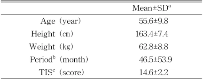
Mean±SDa
Age (year) 55.6±9.8
Height (㎝) 163.4±7.4
Weight (㎏) 62.8±8.8
Periodb (month) 46.5±53.9
TISc (score) 14.6±2.2
astandard deviation, bonset period, ctrunk impairment scale.
Table 1. Demographic data on the participants
scores, on a range from 0 to 23, static and dynamic sitting balance as well as trunk coordination. The ex- clusion criteria were less than a year from stroke on- set, a history of lower back pain or related surgery, or the presence of hemi-neglect, bilateral stroke, visu- al deficits, or comprehension impairment. Demographic data on the participants are presented in Table 1.
Instruments
1. Pressure mat
A pressure mat (Baltube, RELIVE, Gimhae, Korea) was used to measure the changes in pressure while the subject was sitting. The size of the mat was 25×25×7 ㎝, and the mat was divided into four air chambers (Figure 1). The first chamber is the right buttock side, the second chamber is the right thigh side, the third chamber is the left buttock side, and the fourth chamber is the left thigh side.
The “R”, “L”, “A”, and “P” sections of the cham- ber measured the changes in pressure during sitting on the right buttock side, left buttock side, right thigh side, and left thigh side. The subjects were in- structed to place both their buttocks and thighs on the correct chamber. The sensors in the Baltube seats were silicon pressure sensors (TruStability), with series-standard accuracy. These pressure sen- sors estimate the ratiometric analog output for scan- ning pressure values over a full-scale pressure span.
2. Palpation meter
A palpation meter (PALM, Performance Attainment Associates, St. Paul, MN) was used to measure the pel- vic angles in the sagittal and coronal planes (Figure 2).
The palpation meter consisted of an inclinometer, with two caliper arms (Lee and Yoo, 2011). The in- clinometer, which has a semicircular arc, can meas- ure within a range of 0-30˚ in each direction from the middle at 1˚ intervals.
The intratester reliability of the palpation meter was high for both the coronal (intraclass correlation co- efficient (ICC=.84) and sagittal planes (ICC=.98). The
intertester reliability was high for the sagittal plane measurements (ICC=.89) but moderate for the coronal plane measurements (ICC=.65) (Hagins et al, 1998).
In the present study, we utilized a new method, the Baltube, to check the amount of pressure change in a quiet sitting position. The Baltube can also be used to record the time at which the variation in pressure occurs and to check the posture main- tenance time, making it possible to quantify postural changes in a sitting position. We named this method asymmetric occurring time (AOT).
Procedure
Before the subjects sat on the Baltube seat, all the air chambers (first to fourth) of the seat were set at 20 ㎜Hg. The subjects were instructed to sit upright and to place both their buttocks and thighs on each chamber of the pressure mat. They were then asked to place both hands comfortably in their laps.
Figure 2. Palpation meter used to measure the pelvic angles in the sagittal and coronal planes.
Figure 1. Image of the pressure mat measurement device.
1. Preintervention test
The tester placed the subject in a neutral sitting position on the pressure mat. Using the palpation meter, a neutral sitting position was defined as one in which both the anterior superior iliac spine (ASIS) and posterior superior iliac spine (PSIS) were located in the same horizontal plane. The target (a piece of paper stuck on the wall) was placed 150 ㎝ away on the wall in front of the subject at the subject’s eye level. When the tester confirmed that the subject had adopted a neutral sitting posture, the tester clicked the “Start” button on the computer software. The subjects were asked to maintain a neutral sitting po- sition for 5 min. After 5 min, the tester measured the change of pressure in the pressure mat and the amount of pelvic tilting angle.
2. VBF sitting training
After finishing the preintervention test, the sub- jects rested for 2 min to minimize fatigue. A smart- phone screen was then placed on the wall 150 ㎝ away from the subject at the subject’s eye level.
The smartphone was connected and synchronized with the pressure mat using Bluetooth. In this way, the subjects could observe the pressure distribution of both their buttocks and thighs. The tester ex- plained to each subject how the VBF training worked and asked each subject to maintain their center of pressure (COP) in a sitting posture while looking at the smartphone. The smartphone then dis- played the trajectory of the COP while the subject leant forwards, backwards, left, and right while at- tempting to maintain a neutral sitting position. Thus, the subjects were aware when the trunk was in a neutral position and when it was not. When the subjects detected that the trunk was not in a neutral positon (neutral zone) via the smartphone screen, they altered the position of their trunk to move their COP to the neutral zone. The VBF training was performed for 15 min (Figure 3).
During the VBF training, the subjects attempted to keep a real-time COP indicator (1×1 ㎜) inside an
outlined zone (25×25 ㎜) on the smartphone screen located 150 ㎝ away at eye level in front of them.
The size of the zone on the screens was equivalent to an 8×8 ㎜ area on the pressure mat. Leaning the trunk forwards, backwards, left, or right resulted in the indicator moving up, down, left, or right.
3. Postintervention test
To minimize muscle fatigue, each subject rested for 2 min before the postintervention test. After rest- ing, the subjects sat on the pressure mat again and maintained a neutral sitting position, using the same maneuvers for 5 min that they had employed in the preintervention test. After 5 min, the pressure and pelvic angles were reassessed using the pressure mat and palpation meter, respectively.
Data collection
1. Buttock pressure
Buttock pressure data were collected while the subjects performed a target trial for 5 min. The mean pressure value in the initial term was calcu- lated using the data collected at the 1 min. The mean pressure value in the finial term calculated us- ing the data obtained in the following 4 min.
Pressure asymmetry values were calculated by sub- tracting the mean pressure value of the third cham- ber (left side) from the mean pressure value of the first chamber (right side) in the initial term. The pressure asymmetry values were measured in the in- Figure 3. Visual biofeedback sitting training with
Baltube interlocking smartphone application.
itial and final term. For each subject, the change in buttock pressure (from the mean pressure of the ini- tial term to the final term) of the affected and lesser affected sides was calculated.
2. Pelvic angle
The pelvic angles were measured before and after the 5 min target trial in a static sitting position. The pelvic tilting angles were measured using the palpation meter. The investigator palpated the ipsilateral ASIS and PSIS landmarks using their index fingers, insert- ing the tips of both index fingers into the holes of the palpation meter. The tips of the meter were placed on the ASIS and PSIS to measure the angle of pelvic tilting in the sagittal plane. To measure the angle of pelvic tilting in the coronal plane, the two tips of the meter were placed on either side at the top of the iliac crest. Lateral pelvic tilt (coronal plane) toward the left side indicated a minus (-) value, whereas lateral pelvic tilt toward the right side indicated a plus (+) value.
Anterior pelvic tilt indicated a plus (+) value, and pos- terior pelvic tilt indicated a minus (-) value.
Statistical analysis
SPSS, ver. 18.0 software (SPSS Inc., Chicago, IL, USA) was used for statistical analysis. A paired t-test was used to compare the differences between the initial pressure asymmetry values and final pres- sure asymmetry values of the hemiplegic patients.
The significance in terms of intragroup (within) was confirmed. The Komologov-Smirnov analysis was performed to determine whether the data showed a
normal distribution.
To compare the change in pressure values and pelvic tilting angles between the pre- and postintervention trials, a paired t-test was applied. The change in pres- sure was calculated by subtracting the mean of the fi- nal values from the mean of the initial values.
Results
The preintervention pressure asymmetry values and postintervention pressure asymmetry values of the hemiplegic patients were significantly different (p<.05). The initial preintervention asymmetric pres- sure values (difference in pressure values between the left and right sides) and final preintervention values were greater than the postintervention values.
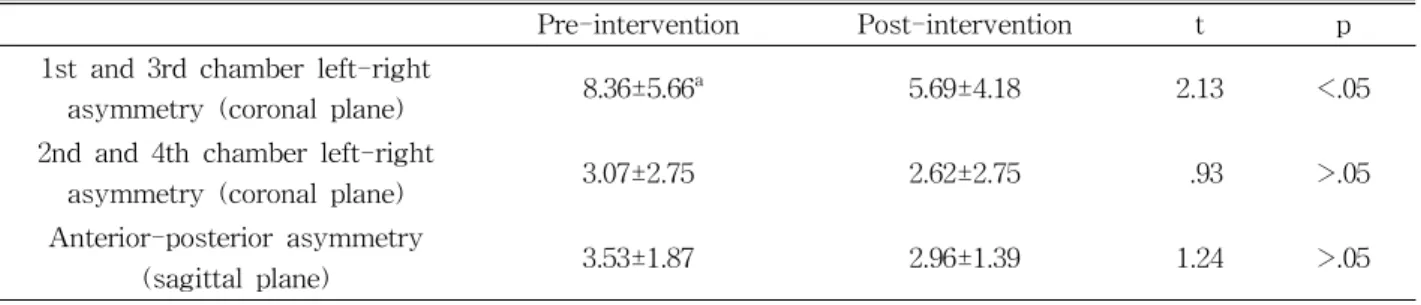
Table 2 shows the difference in the buttock pres- sure values (㎜Hg) of the hemiplegic patients be- tween the pre- and post-intervention periods. The average preintervention buttock pressure value in the coronal plane (first and third chamber) was 8.36 ㎜ Hg, and the average postintervention value was 5.69
㎜Hg. There was a significant difference between the pre- and post-intervention values (p<.05). In contrast, there was no significant difference between the aver- age coronal plane (second and fourth chamber) pre- intervention buttock pressure value (3.07 ㎜Hg) and average postintervention value (2.62 ㎜Hg) (p>.05).
There was also no significant difference between the average sagittal plane (anterior-posterior asymmetry) preintervention and postintervention buttock pressure
Pre-intervention Post-intervention t p
1st and 3rd chamber left-right
asymmetry (coronal plane) 8.36±5.66a 5.69±4.18 2.13 <.05
2nd and 4th chamber left-right
asymmetry (coronal plane) 3.07±2.75 2.62±2.75 .93 >.05
Anterior-posterior asymmetry
(sagittal plane) 3.53±1.87 2.96±1.39 1.24 >.05
amean±standard deviation.
Table 2. Comparison of the buttock pressure values (㎜Hg) of the hemiplegic patients in the pre- and post-
intervention periods (unit: ㎜Hg)
values (3.53 ㎜Hg and 2.96 ㎜Hg, respectively; p>.05).
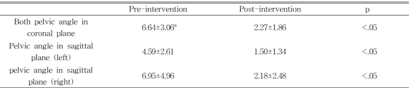
Table 3 shows differences of pelvic angle on the co- ronal and sagittal planes. There was a significant dif- ference in pelvic angle on the coronal plane between the pre- and post-intervention (p<.05). The angle of pelvic tilting variations by 6.64˚ in the preintervention and 2.27˚ in the post-intervention. Also, there was sig- nificant difference in pelvic angle on the sagittal plane between pre- and post-intervention (p<.05). The angle of pelvic tilting variations by left side 4.59˚ and right side 6.95˚ in the preintervention and left side 1.50˚ and right side 2.18˚ in the post intervention, respectively.
Table 4 presents the pre- and post-intervention AOT of 2 ㎜Hg of the hemiplegic patients. In each of the pressure chambers, the average asymmetry between the pre- and post-intervention was AOT of 2 ㎜Hg. All the chambers showed a significant dif- ference in the asymmetry values between the pre- and postintervention, except second chamber.
Discussion
This is the first study to use pressure mat (Baltube) technology in quiet-sitting balance control
training in chronic stroke patients.
The asymmetry of pressure between the right and left (first and third chambers) sides significantly de- creased after the VBF training. There was also a significant decrease in the pelvic tilt angles in the postintervention versus the preintervention, as shown by the measurements obtained using the palpation meter. Visual biofeedback training, using pressure mat technology, would be an alternative way for quiet sitting balance training.
Perlmutter et al. (2010) suggested the use of quantitative methodology to understand the ability to maintain upright during unsupported sitting in chron- ic unilateral stroke patients. They used spatial and temporal analyses to compare the COP trajectories of a stroke group with those of a healthy group. They showed that both groups showed significantly less sway during feedback trials. In their study, when the COP trajectories decreased, the AOT also increased.
Furthermore, when the COP trajectories increased, the AOT was diminished. The results of the present study are similar to those of Perlmutter et al (2010), with a significant increase in the AOT after the VBF training. We concluded that VBF training was an effective method to help stroke patients maintain
Pre-intervention Post-intervention p
Both pelvic angle in
coronal plane 6.64±3.06a 2.27±1.86 <.05
Pelvic angle in sagittal
plane (left) 4.59±2.61 1.50±1.34 <.05
pelvic angle in sagittal
plane (right) 6.95±4.96 2.18±2.48 <.05
amean±standard deviation.
Table 3. Differences in the both pelvic angles on the coronal and sagittal planes (unit: ˚)
Pre-intervention Post-intervention t p
1st Chamber 29.36±30.27a 89.82±109.10 -2.94 <.05
2nd Chamber 102.32±110.47 112.00±113.03 -.35 >.05
3rd Chamber 31.45±44.24 63.68±84.96 -1.93 <.05
4th Chamber 69.36±131.69 142.40±114.48 -2.78 <.05
amean±standard deviation.
Table 4. Comparison of the AOT of 2 ㎜Hg of the hemiplegic patients in the pre- and post-intervention (unit: sec)
an upright-sitting posture.
Previous studies described postural disturbances in stroke patients. Genthon et al (2007) investigated sit- ting balance control among acute stroke subjects sit- ting on a force platform. They tested three con- ditions of quiet sitting: siting with eyes closed, tar- get, and feedback. They detected significantly larger COP displacement and velocity among the stroke pa- tients when compared to the healthy controls. They concluded that postural disturbance occurred in acute stroke patients during sitting. A study by van Nes et al (2008) reported greater postural disturbance in the coronal plane when subjects were seated on an un- stable surface. In the present study, the buttock pres- sure of both right and left sides was significantly re- duced after the VBF training intervention. Genthon et al (2007) reported greater postural disturbance in the sagittal plane, although the stroke subjects in their study had significantly larger maximum sagittal dis- placement than those in the present study. However, decreased in our study occurring time by the pres- sure changes but not significant in the sagittal plane.
In the present study, the pressure values decreased after the intervention, but the level of change was not significant. Previous research with acute hemi- plegic patients used a force plate to measure pres- sure variations. The current study used a pressure mat in chronic hemiplegic patients.
In the pilot study, the pressure variation between the left and right sides was defined as 2 ㎜Hg. In addition, the AOT in each chamber was recorded.
Changing the amount in each chamber changed the COP. After the intervention, an AOT of 2 ㎜Hg emerged as significant. The pressure mat (Baltube) detected the AOT over a 2 ㎜Hg between the pre- and post-interventions for each pressure chamber (AOT above 2 ㎜Hg denoted a nonsignificant differ- ence). There was a significant difference in the pressure variation (2 ㎜Hg) in all the chambers be- tween the pre- and post-interventions, except in chamber two, despite an increase in the time. We attributed this finding to the 2nd, 4th chamber of the
thighs of some tall patients and foot partial weight bearing and is considered a better future consideration.
Previous studies reported that balance training on a force platform and VBF training led to significant improvements in the ability to perform activities of daily living (Chen et al, 2002; Eser et al 2008). Some studies also reported that visual feedback on the COP affected sitting postural control. A number of studies found that visual feedback on the COP in the standing position significantly decreased body sway (Barclay-Goddard et al, 2004; de Haart et al, 2005;
Shumway-Cook et al, 1988). Perlmutter et al. (2010) reported that sitting during visual feedback resulted in decreased trunk sway and maximum displace- ments in both chronic unilateral stroke patients and healthy control groups, as well as significant in- creases in the stroke group. These findings suggest that stroke patients can improve postural efficiency to a level similar to that of controls when provided with an accurate indicator of trunk position in re- al-time. Thus, it is possible that poststroke stability problems in a sitting posture may be related to sen- sory organization of postural trunk control, as is seen in standing balance (Bonan et al, 2004a; Di Fabio et al, 1990, 1991; Marsden et al, 2005).
In the present study, after VBF quiet sitting bal- ance training for 15 min, asymmetry of the buttock pressure and angle of pelvic tilting decreased in the hemiplegic patients. The results suggest that VBF training may be a useful rehabilitation strategy for hemiplegic patients.
Limitations of the study
The results of the pressure mat measurements showed that decreased pressure variation in the co- ronal (chambers two and four) and sagittal planes.
Due to the no significant difficult, we considered that bilateral foot partial weight bearing, and the subjects were asked to place both their hands on their lap.
This resulted in reduced biomechanical constraints.
As a result, the magnitude of the COP was smaller
in the sitting position than it would have been in an upright standing posture (Genthon and Rougier, 2006). Thus, a further quiet sitting study is needed on a controlled unstable surface, with no footplate and both hands in an arm sling.
The pressure mat measurements in the present study revealed a decrease in pressure variation in the coronal (chambers two and four) and sagittal planes and an increase in the time to AOT.
However, due to the absence of any significant dif- ference, it is likely that both feet had partial weight-bearing status.
Conclusion
This study suggested the VBF training can im- prove quiet-sitting balance in chronic hemiplegic patients. Using VBF training, the buttock pressure of the patients was equally distributed, and the time of symmetry sitting increased. In addition, VBF training resulted in improved pelvic control in a neutral position. The results suggest that VBF training could be a new clinical method to improve sitting balance in hemiplegic patients.
References
Barclay-Goddard R, Stevenson T, Poluha, W, et al.
Force platform feedback for standing balance training after stroke. Cochrane Database Syst Rev. 2004;18(4):CD004129.
Bertrand AM, Bourbonnais D. Effects of upper limb unilateral isometric efforts on postural stabiliza- tion in subjects with hemiparesis. Arch Phys Med Rehabil. 2001;82(3):403–411.
Bohannon RW, Smith MB, Larkin PA. Relationship between independent sitting balance and side of hemiparesis. Phys Ther. 1986;66(6):944-945.
Bohannon RW. Recovery and correlates of trunk muscle strength after stroke. Int J Rehabil Res.
1995;18(2):162-167.
Bonan IV, Colle FM, Guichard JP, et al. Reliance on visual information after stroke. Part I: Balance on dynamic posturography. Arch Phys Med Rehabil. 2004a;85(2):268-273.
Bonan IV, Yelnik AP, Colle FM, et al. Reliance on vis- ual information after stroke. Part II: Effectiveness of a balance rehabilitation program with visual cue deprivation after stroke: A randomized controlled trial. Arch Phys Med Rehabil. 2004b;85(2):274-278.
Bray H, Moseley GL. Disrupted working body sche- ma of the trunk in people with back pain. Br J Sports Med. 2011;45(3):168-173. https://do- i.org/173.10.1136/bjsm.2009.061978
Brumagne S, Cordo P, Lysens R, et al. The role of paraspinal muscle spindles in lumbosacral position sense in individuals with and without low back pain. Spine (Phila pa 1976). 2000;25(8):989-994.
Bush C, Ditto B, Feuerstein M. A controlled evalua- tion of paraspinal EMG biofeedback in the treatment of chronic low back pain. Health Psychol. 1985;4(4):307-321.
Chen IC, Cheng PT, Chen CL, et al. Effect of bal- ance training on hemiplegic stroke patients.
Chang Gung Med J. 2002;25(9):583-590.
Davies PM. Right in the middle-selective trunk ac- tivity in the treatment of adult hemiplegia. 1st ed. Berlin. Springer-Verlag, 1990;16-20.
Di Fabio RP, Badke MB. Relationship of sensory or- ganization to balance function in patients with hemiplegia. Phys Ther. 1990;70(9):542-548.
Di Fabio RP, Badke MB. Stance duration under sensory conflict conditions in patients with hemiplegia.
Arch Phys Med Rehabil. 1991;72(5):292-295.
de Haart M, Guerts AC, Dault MC, et al. Restoration of weight-shifting capacity in patients with postacute stroke: A rehabilitation cohort study.
Arch Phys Med Rehabil. 2005;86(4):755-762.
Dunk NM, Callaghan JP. Gender-based differences in postural responses to seated exposures. Clin Biomech. 2005;20(10):1101-1110.
Eser F, Yavuzer G, Karakus D, et al. The effect of
balance training on motor recovery and ambula- tion after stroke: A randomized controlled trial.
Eur J Phys Rehabil Med. 2008;44(1):19-25.
Franchignoni FP, Tesio L, Ricupero C, et al. Trunk control test as an early predictor of stroke re- habilitation outcome. Stroke. 1997;28:1382-1385.
Genthon N, Vuillerme N, Monnet JP, et al.
Biomechanical assessment of the sitting posture maintenance in patients with stroke. Clin Biomech (Bristol, Avon). 2007;22(9):1024-1029.
Genthon N, Rougier P. Does the capacity to appro- priately stabilize trunk movements facilitate the control of upright standing? Motor Control.
2006;10(3):232-243.
Gauthier J, Bourbonnais D, Filiatrault J, et al.
Characterization of contralateral torques during static hip efforts in healthy subjects and subjects with hemiparesis. Brain. 1992;115(Pt 4):1193-1207.
Hagins M, Brown M, Cook C, et al. Intratester and intertester reliability of the palpation meter (palm) in measuring pelvic position. J Man Manip Ther. 1998;6(3):130-136.
Horton S, Abbott J. A novel approach to managing graduated return to spinal loading in patients with low back pain using the spine-angel de- vice: A case series report. NZ Journal of Physiotherapy. 2008;36(1):22-28.
Hsieh CL, Sheu CF, Hsueh IP, et al. Trunk control as an early predictor of comprehensive activities of daily living function in stroke patients. Stroke 2002;33(11):2626-2630.
Lee JH, Yoo WG. Changes in gluteal pressure and pelvic inclination angles after continuous cross-legged sitting. Work. 2011;40(2):247-252.
https://doi.org/10.3233/WOR-2011-1225
Magnusson ML, Chow DH, Diamandopoulos Z, et al.
Motor control learning in chronic low back pain.
Spine (Phila Pa 1976). 2008;33(16):E532-E538.
https://doi.org/10.1097/BRS.0b013e31817dfd9a Marsden JF, Playford DE, Day BL. The vestibular
control of balance after stroke. J Neurol Neurosurg Psychiatry. 2005;76(5):670-678.
Moseley GL, Gallagher L, Gallace A. Neglect-like tactile dysfunction in chronic back pain.
Neurology. 2012; 79(4):327-332. https://doi.org/10.1212/
WNL.0b013e318260cba2
Olney SJ, Martin CS, Rehabilitation: Physical therapy for stroke. In: Welch KMA, Caplan LR, Reiss DJ. Eds. Primer on Cerebrovascular Diseases. 1 st ed. San Diego, CA, Academic Press. 1997.
O’Sullivan K, O’Sullivan L, O’Sullivan P, et al.
Investigating the effect of real-time spinal pos- tural biofeedback on seated discomfort in people with non-specific chronic low back pain.
Ergonomics. 2013;56(8):1315-1325. https://do- i.org/10.1080/00140139.2013.812750
O’Sullivan PB, Burnett A, Floyd AN, et al. Lumbar repositioning deficit in a specific low back pain population. Spine (Phila Pa 1976). 2003;28(10):
1074-1079.
O’Sullivan PB, Smith AJ, Beales DJ, et al.
Association of biopsychosocial factors with de- gree of slump in sitting posture and self-report of back pain in adolescents: A cross-sectional study.
Phys Ther. 2011;91(4):470-483. https://doi.org/10.2522/
ptj.20100160
Perennou DA, Amblard B, Leblond C, et al. Biased postural vertical in humans with hemispheric cerebral lesions. Neurosci Lett. 1998;252(2):75-78.
Perlmutter S, Lin F, Makhsous M. Quantitative anal- ysis of static sitting posture in chronic stroke. Gait Posture. 2010;32(1):53-56. https://doi.org/10.1016/
j.gaitpost.2010.03.005
Seah SH, Briggs AM, O’Sullivan PB, et al. An ex- ploration of familial associations in spinal pos- ture defined using a clinical grouping method.
Man Ther. 2011;16(5);501-509. https://doi.org/10.1016/
j.math.2011.05.002
Sheeran L, Sparkes V, Caterson B, et al. Spinal posi- tion sense and trunk muscle activity during sitting and standing in non-specific chronic low back pain: classification analysis. Spine. 2012;37(8):
E486-E495.
Shumway-Cook A, Anson D, Haller S. Postural
sway biofeedback: Its effect on reestablishing stance stability in hemiplegic patients. Arch Phys Med Rehabil. 1988;69(6):395-400.
Stuckey S, Jacobs A, Goldfarb J. EMG biofeedback training, relaxation training, and placebo for the relief of chronic back pain. Percept Mot Skills.
1996;63(3):1023-1036.
Van Hoof W, Volkaerts K, O’Sullivan K, et al.
Cognitive functional therapy intervention includ- ing biofeedback for lbp during cycling. a single case study. Sport Geneeskunde. 2011;44(4):20-26.
van Nes IJ, Nienhuis B, Latour H, et al. Posturographic assessment of sitting balance recovery in the sub-
acute phase of stroke. Gait Posture. 2008;28(3):
507-512. https://doi.org/10.1016/j.gaitpost.2008.03.004 Wade DT, Skilbeck CE, Hewer RL. Predicting Barthel ADL score at 6 months after an acute stroke.
Arch Phys Med Rehabil. 1983;64(1):24-28.
This article was received January 19, 2017, was reviewed January 19, 2017, and was accepted May 10, 2017.