CASE REPORT
J Korean Surg Soc 2011;81:S12-16
http://dx.doi.org/10.4174/jkss.2011.81.Suppl1.S12
JKSS
Journal of the Korean Surgical Society pISSN 2233-7903ㆍeISSN 2093-0488
Received February 9, 2011, Revised August 5, 2011, Accepted August 16, 2011 Correspondence to: Sun Hyoung Shin
Department of Surgery, Chonnam National University Medical School, 42 Jebong-ro, Dong-gu, Gwangju 501-757, Korea Tel: +82-62-220-6356, Fax: +82-62-227-1635, E-mail: [email protected]
cc Journal of the Korean Surgical Society is an Open Access Journal. All articles are distributed under the terms of the Creative Commons Attribution Non-Commercial License (http://creativecommons.org/licenses/by-nc/3.0/) which permits unrestricted non-commercial use, distribution, and reproduction in any medium, provided the original work is properly cited.
Primary papillary carcinoma originated from a branchial cleft cyst
Jin Seong Cho, Sun Hyoung Shin, Hee Kyung Kim
1, Ji Shin Lee
2, Min Ho Park, Jung Han Yoon, Young Jong Jegal
Departments of Surgery, 1Internal Medicine and 2Pathology, Chonnam National University Medical School, Gwangju, Korea
Although branchial cleft cysts are common, papillary carcinomas arising from them are rare. Here we report a 41-year-old woman with papillary carcinoma originating from a right lateral branchial cleft cyst without any evidence of a papillary car- cinoma in the thyroid gland. The patient underwent right lateral neck dissection followed by total thyroidectomy. We then confirmed papillary carcinoma arising from the branchial cleft cyst through microscopic and immunohistochemical staining with thyroglobulin (TG), thyroid-associated transcription factor -1 (TTF-1) and p63. It is the 10th case worldwide describing papillary carcinoma in a branchial cleft cyst with a review of the literature on the features of the disease and discussion of the role of immunohistochemical staining with TG, TTF -1 and p63. In conclusion, it should be emphasized that the surgeon must be cautioned of the possibility of primary papillary carcinoma in the branchial cleft cyst.
Key Words: Branchioma, Thyroid neoplasms, Papillary carcinoma
INTRODUCTION
Although branchial cleft cysts (BCCs) are common, squ- amous cell carcinomas of BCC are rare [1], but papillary carcinomas arising from them are extremely rare. Here we report a patient with papillary carcinoma originated from right lateral BCC without any evidence of a papillary car- cinoma in thyroid gland. Papillary carcinoma of BCC may be clinically indistinguishable from benign BCC or meta- static papillary carcinoma, and the diagnosis in most cases is incidental after surgical resection. We report the 10th case for papillary carcinoma in a BCC, and review the liter- ature on the features of the disease and discuss the role of
immunohistochemical staining with thyroglobulin (TG), thyroid-associated transcription factor-1 (TTF-1) and p63.
CASE REPORT
A 41-year-old woman presented to our hospital with a 2-year history of palpable mass in the right lateral aspect of the neck. There was no past history of irradiation or trau- ma in the head and neck. On physical examination, a firm and non-tender mass was palpable in the right lateral neck measuring 3.0 cm at the level of the cricoid cartilage with- out associated cervical lymphadenopathy. No abnormal
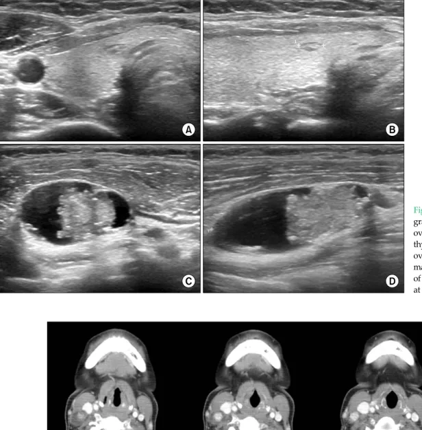
Fig. 1. Neck ultrasono- graphy shows a flat ovoid 0.5 cm mass in the thyroid (A, B) and an oval 3.0 cm size solid mass on the cystic wall of the right lateral neck at level III (C, D).
Fig. 2. Axial computed tomography demonstrating a uni-loculated cystic lesion measuring about 3.0 cm in diameter, just lateral to right inter- nal jugular vein at the level of cricoid cartilage. Note the high-density area within the cystic lesion consistent with focal calcification. The mass abuts the right internal jugular vein medially and the sternocleidomastoid muscle posteriorly.
laboratory findings were seen in routine laboratory inves- tigations and euthyroid state in thyroid function test.
Neck ultrasonography revealed a solitary nodule meas- uring 0.5 cm in the right thyroid gland, and a 3.0 cm mixed solid and cystic mass in the right lateral aspect of the neck at level III (Fig. 1). Computed tomography also showed
mixed solid and cystic lesion associated with punctate cal- cification (Fig. 2).
Examination of fine needle aspiration cytology (FNA) on thyroid gland confirmed follicular cell proliferative lesion. But, the right lateral neck mass was suggestive of metastatic papillary carcinoma. Therefore, the patient was
Fig. 3. Papillary carci- noma originated from a branchial cleft cyst. In- tracystic papillary pro- jections are observed (A, H&E, ×20). The cyst was lined by squamous epithelium (B, H&E,
×200). Squamous epi- thelium was positive for p63 (C, ×200) and ne- gative for thyroglobulin and thyroid-associated transcription factor-1 (D, ×200).
diagnosed with presumed metastatic papillary thyroid carcinoma (PTC) in the right lateral neck and occult papil- lary thyroid carcinoma in the right thyroid gland.
The patient underwent right lateral neck dissection fol- lowed by total thyroidectomy. It must be emphasized that papillary carcinoma arising in a right lateral BCC was not in the differential diagnoses preoperatively. The intra- operative findings included a 3.0 cm dark brown fluid fil- led cystic mass lateral to the internal jugular vein without cyst rupture at the time of the operation, and revealed PTC on the frozen section biopsy. Therefore, a total thyroi- dectomy was followed.
Histopathologic examination of the 3.0 cm cystic mass was confirmed papillary carcinoma in a BCC (Fig. 3A). The cyst was partially lined by squamous epithelium sur- rounded by fibrous tissues associated with chronic in- flammation (Fig. 3B). But there was no normal thyroid tis- sue adjacent to the focus of papillary carcinoma within the cyst wall (Fig. 3A).
Histopathologic examination of the thyroid gland re- vealed nodular hyperplasia of the right lobe without evi-
dence of malignancy. Repeated serial sectioning of the whole thyroid and even in postoperative positron emis- sion tomography does not reveal any foci of carcinoma.
Other harvested right lateral lymph nodes were examined but there were no evidence of any other abnormal findings.
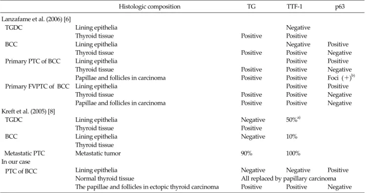
It is known that immunohistochemical staining through TG, TTF-1 and p63 could help in the differential diagnosis in thyroglossal duct cyst, BCC, and papillary carcinoma in BCC (Table 1). In our case, the squamous epithelium was positive for p63 and negative for TG and TTF-1 (Fig. 3C, D). On the other hand, papillary carcinoma in BCC was positive on TG and TTF-1, but negative on p63. This issue for immunohistochemical staining will be explained later on discussion.
The patient has remained disease free having been fol- lowed up for 28 months. She is being managed with post-operative levothyroxine replacement without radio- active iodine therapy.
Histologic composition TG TTF-1 p63 Lanzafame et al. (2006) [6]
TGDC Lining epithelia Negative
Thyroid tissue Positive Positive
BCC Lining epithelia Negative Positive
Thyroid tissue Positive Positive Negative
Primary PTC of BCC Lining epithelia Positive Positive
Thyroid tissue Positive Positive Negative
Papillae and follicles in carcinoma Positive Positive Foci (+)b)
Primary FVPTC of BCC Lining epithelia Positive Positive
Thyroid tissue Positive Positive Negative
Papillae and follicles in carcinoma Positive Positive Negative
Kreft et al. (2005) [8]
TGDC Lining epithelia Negative 50%a)
Thyroid tissue Positive
BCC Lining epithelia Negative 10%
Thyroid tissue
Metastatic PTC Metastatic tumor 90% 100%
In our case
PTC of BCC Lining epithelia Negative Negative Positive
Normal thyroid tissue All replaced by papillary carcinoma
The papillae and follicles in ectopic thyroid carcinoma Positive Positive Negative TGDC, thyroglossal duct cyst; BCC, branchial cleft cyst; PTC, papillary thyroid carcinoma; FVPTC, follicular variant papillary thyroid carcinoma; TG, thyroglobulin; TTF-1, thyroid-associated transcription factor-1.
a)Positive on only basal part of lining epithelium of TGDC. b)Staining was focal rather than extensive or confluent and the extent of staining varied from multiple foci to scattered or rare foci and observed in only 1 PTC of BCC.
Table 1. Differential diagnosis based on immunohistochemical staining
DISCUSSION
Ectopic thyroids are rarely found in the region of the sublingual, submandibular, intratracheal, mediastinum, esophagus, lung, heart, aorta, and even in abdomen. The most frequent site is the thyroglossal remnants or BCC. In our knowledge, about 215 cases papillary carcinoma of thyroglossal remnants were reported, but only nine cases of BCC have been reported by 2010 [2-4].
Because metastatic papillary carcinoma may become cystic [5], all lining cells of the cyst must be carefully reviewed. And it is important to differentiate a metastatic derivation with a missed primary tumor or represents papillary thyroid carcinoma arising in ectopic thyroid tis- sue in a BCC [5-7].
Sidhu’s criteria for the papillary carcinoma in BCC [3]
was suggested as 1) an epithelial lining layer, subepithelial lymphoid tissue collection, 2) normal thyroid tissue ad- jacent to the focus of papillary carcinoma within the wall, 3) and no evidence of papillary carcinoma in the thyroid or
other area.
In our case, there was no normal thyroid tissue adjacent to the papillary carcinoma within the wall. It may be as- sumed as 1) papillary carcinoma was initiated in the nor- mal thyroid tissues in BCC, 2) and all normal thyroid tis- sues within the wall were replaced by papillary carcino- ma. But there was epithelial lining layer - not in cystic change of metastatic carcinoma -, and there was no evi- dence of papillary carcinoma within the thyroid gland even in the repeated serial section and immunohisto- chemical staining.
In immunohistochemical staining, TTF-1 cannot dis- tinguish between primary and metastatic tumors of BCC [8]. The p63 is a potentially oncogenic protein that would contribute to the onset of papillary carcinoma. It was sug- gested that he detection of p63 in papillary carcinomas of BCC could distinguish from primary BCC or metastatic origin [6]. Unfortunately, p63 staining on papillary area of carcinoma of BCC in our case did not display any positive foci. As reviewed by Lanzafame et al [6], the involvement
of p63 immunoreactive cells in the tumorigenic process re- mains to be proven because they may simply reflect squ- amous differentiation of papillary thyroid carcinoma. In addition, staining of p63 was focal rather than extensive or confluent, and the extent of staining varied from multiple foci to scattered or rare foci. It was also in only one papil- lary type carcinoma in BCC case, and follicular variant car- cinoma of BCC in his study showed negativity on p63 [6].
We need some more research for this debate on positivity on p63 in PTC of BCC.
In our case, there was no evidence of papillary carcino- ma found within the thyroid gland when meticulous his- tological examination including immunohistochemical staining on all thyroid section. Also, diagnostic radio- active iodine scan and positron emission tomography does not reveal the any focus of malignancy. Taken togeth- er, these findings confirmed papillary carcinoma arising from the BCC at level III of the right neck even though ab- sence of normal thyroid tissue within the BCC.
In heterotopic thyroid tissue in thyroglossal duct cyst and BCC, it is important to evaluate if the case has a meta- static of occult thyroid cancer. So FNA should be per- formed under ultrasound guidance in order to best sam- ple the solid part of the lesion and any thyroid nodule in any size. This can help to reduce false-negative rate from dilution by the cystic contents. and TG levels in FNA fluids can aids the adequate diagnosis and decision of operation extent [4]. Papillary carcinoma arising from branchial remnants has the ability to metastasize to regional lymph nodes. Neck node metastases are found in 20% [9,10], but distant metastases were not reported.
In conclusion, it should be emphasized that the surgeon must be cautioned that the possibility of primary papillary carcinoma in the branchial cleft cyst.
CONFLICTS OF INTEREST
No potential conflict of interest relevant to this article was reported.
REFERENCES
1. Kim DS, Kim CB, Min JS. Carcinoma arising in a branchial cleft cyst. J Korean Surg Soc 1984;26:278-80.
2. Balasubramaniam GS, Stillwell RG, Kennedy JT. Papillary carcinoma arising in ectopic thyroid tissue within a bran- chial cyst. Pathology 1992;24:214-6.
3. Sidhu S, Lioe TF, Clements B. Thyroid papillary carcinoma in lateral neck cyst: missed primary tumour or ectopic thy- roid carcinoma within a branchial cyst? J Laryngol Otol 2000;114:716-8.
4. Park J, Kwon SY, Kim NH, Baik SH, Choi DS. Papillary thy- roid carcinoma arising in a branchial cleft cyst. Thyroid 2010;20:347-9.
5. Nuttall FQ. Cystic metastases from papillary ad- enocarcinoma of the thyroid with comments concerning carcinoma associated with thyroglossal remnants. Am J Surg 1965;109:500-5.
6. Lanzafame S, Caltabiano R, Puzzo L, Cappellani A.
Thyroid transcription factor 1 (TTF-1) and p63 expression in two primary thyroid papillary carcinomas of branchial cleft cysts. Virchows Arch 2006;449:129-33.
7. Seven H, Gurkan A, Cinar U, Vural C, Turgut S. Incidence of occult thyroid carcinoma metastases in lateral cervical cysts. Am J Otolaryngol 2004;25:11-7.
8. Kreft A, Hansen T, Kirkpatrick CJ. Thyroid transcription factor 1 expression in cystic lesions of the neck: an im- munohistochemical investigation of thyroglossal duct cysts, branchial cleft cysts and metastatic papillary thyroid cancer. Virchows Arch 2005;447:9-11.
9. Matsumoto K, Watanabe Y, Asano G. Thyroid papillary carcinoma arising in ectopic thyroid tissue within a bran- chial cleft cyst. Pathol Int 1999;49:444-6.
10. Mehmood RK, Basha SI, Ghareeb E. A case of papillary car- cinoma arising in ectopic thyroid tissue within a branchial cyst with neck node metastasis. Ear Nose Throat J 2006;
85:675-6.