Letter to the Editor
Vol. 26 No. 3, 2014 399
Ann Dermatol Vol. 26, No. 3, 2014 http://dx.doi.org/10.5021/ad.2014.26.3.399
LETTER TO THE EDITOR
Received June 20, 2012, Revised April 26, 2013, Accepted for publication May 29, 2013
Corresponding author: Jong Keun Seo, Department of Dermatology, Busan Paik Hospital, Inje University College of Medicine, 75 Bokji-ro, Busanjin-gu, Busan 614-735, Korea. Tel: 82-51-890-6135, Fax: 82-51-897-6391, E-mail: [email protected]
This is an Open Access article distributed under the terms of the Creative Commons Attribution Non-Commercial License (http://
creativecommons.org/licenses/by-nc/3.0) which permits unrestricted non-commercial use, distribution, and reproduction in any medium, provided the original work is properly cited.
Fig. 1. Multiple nonfollicular pustules with surround erythema on the trunk.
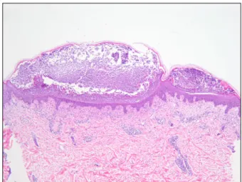
Fig. 2. Histopathologic findings shows subcorneal pustules in the epidermis, perivascular and interstitial inflammatory cells infil- tration in the dermis. The inflammatory cells are comprised of lymphohistiocytes, neutrophils and a few eosinophils (H&E,
×40).
Acute Generalized Exanthematous Pustulosis Induced by Parvovirus B19 Infection
Deborah Lee
1, Jeong Nan Kang, Sung Hwan Hwang, Young Suk Lee, Hyojin Kim, Jong Keun Seo, Ho Suk Sung
Department of Dermatology, Busan Paik Hospital, Inje University College of Medicine, Busan,
1Sinsa Theme Dermatology Clinic, Seoul, Korea
Dear Editor:
A 49-year-old Korean woman presented to dermatologic department with asymptomatic multiple pustules with surrounding erythema on whole body. Four days ago, the patient developed a high fever (>40oC) with common cold symptoms. Two days later, erythematous patches occurred on whole body. To evaluate the cause of fever and skin rash, she was admitted in the department of internal medicine and started to treat with empirical antibiotics, 3rd cephalosporin. On the 2nd day of admi- ssion, more than 100 non-follicular pustules occurred on the previous erythematous patches. Before the skin eru-
ption occurred, there were no history of skin disease.
Physical examination demonstrated multiple small non- follicular pustules with surrounding erythema on whole body (Fig. 1). Laboratory finding showed leukocytosis with neutrophilia (42.52×10⁹/L, 96.1%), elevated erythrocyte sedimentation rate (89 mm/h) and parvovirus B19 immunoglobulin M (IgM) and IgG were both positive. And on the echocardiography and chest computed tomogra- phy, dilated cardiomyopathy with myocarditis was obser-
Letter to the Editor
400 Ann Dermatol
ved. A culture of the blood, sputum, urin and pustule did not produce any growth of organisms. The other labo- ratory studies were unremarkable. A skin biopsy was performed on pustular lesion of trunk. The histopathologic findings showed subcorneal pustules in the epidermis and perivascular, interstitial infiltration of lymphohistiocytes, neutrophils and a few eosinophils in the dermis (Fig. 2).
The patient was dermatologically treated with topical de- sonide. Despite the ongoing treatment with empirical antibiotics, the pustules resolved within 7 days leaving desquamation and pigmentation.
Acute generalized exanthematous pustulosis (AGEP) is an uncommon cutaneous drug reaction characterized by the acute onset of generalized, non-follicular and sterile pustu- les on the erythematous background. The eruption can be accompanied by fever and neutrophilc leukocytosis. Alth- ough AGEP is induced by drug in over 90% of cases, it can develop without preceding medication history1. Mer- cury exposure, spider bites, lacquer have been reported to the causes of AGEP, in addition, viral infections may serve as occasional causes.
Parvovirus B19 is a single-stranded nonenveloped DNA virus that can cause several diseases. Its clinical manife- stations exist in various forms including arthritis, hemato- logic disorders, myocarditis and dermatologic involve- ment such as erythema infectiosum, gloves and socks syndrome2. However, there were only 2 cases of AGEP in- duced by parvovirus B19 infection in the Englinsh lite- rature3,4. The pathogenesis of AGEP induced by parvovirus B19 is uncertain, it has been thought that viral infection may induce inflammatory cytokines, interleukin 8 or gra- nulocyte-microphage colony stimulating factor and trigger AGEP.
The differential diagnosis of this case is AGEP, generalized pustular psoriasis, subcorneal pustular dermatosis and sub- corneal IgA dermatosis. The findings of abscense of psori- asiform acanthosis, tortuous and dilated blood vessels and neutrophils migrate through the epidermis to aggregate beneath the stratum corneum can be differentiated from generalized pustular psoriasis5. Subcorneal pustular der- matosis and subcorneal IgA dermatosis are characterized by the subacute development of larger pustules than present case5. In our patient, the clinical course and histopathologic finding corresponded with AGEP, the cause was parvovirus B19 infection confirmed by se- rologic test. To our knowledge, AGEP induced parvovirus B19 is a rare entity, we should keep in mind that par- vovirus B19 is one of the causative agent of AGEP.
REFERENCES
1. Sidoroff A, Dunant A, Viboud C, Halevy S, Bavinck JN, Nal- di L, et al. Risk factors for acute generalized exanthematous pustulosis (AGEP)-results of a multinational case-control study (EuroSCAR). Br J Dermatol 2007;157:989-996.
2. Vafaie J, Schwartz RA. Parvovirus B19 infections. Int J Der- matol 2004;43:747-749.
3. Perceau G, Derancourt C, Salmon-Ehr V, Durlach A, Raclot P, Bredard V, et al. Acute generalized exanthematous pustu- losis in hypercalcemia. Ann Dermatol Venereol 2000;127:
1090-1093.
4. Ofuji S, Yamamoto O. Acute generalized exanthematous pu- stulosis associated with a human parvovirus B19 infection. J Dermatol 2007;34:121-123.
5. Fernando SL. Acute generalised exanthematous pustulosis.
Australas J Dermatol 2012;53:87-92.