Letter to the Editor
340 Ann Dermatol
Received May 30, 2014, Revised June 25, 2014, Accepted for publication July 10, 2014
Corresponding author: Hee Young Kang, Department of Dermatology, Ajou University School of Medicine, 164 World Cup-ro, Yeongtong-gu, Suwon 443-380, Korea. Tel: 82-31-219-5190, Fax: 82-31-219-5189, E-mail: [email protected]
This is an Open Access article distributed under the terms of the Creative Commons Attribution Non-Commercial License (http://
creativecommons.org/licenses/by-nc/4.0) which permits unrestricted non-commercial use, distribution, and reproduction in any medium, provided the original work is properly cited.
http://dx.doi.org/10.5021/ad.2015.27.3.340
Changes in Melanin and Melanocytes in Mottled Hypopigmentation after Low-Fluence 1,064-nm Q-Switched Nd:YAG Laser Treatment for Melasma
Yong Hyun Jang, Ji-Youn Park1, Young Joon Park1, Hee Young Kang1
Department of Dermatology, Kyungpook National University School of Medicine, Daegu, 1Department of Dermatology, Ajou University School of Medicine, Suwon, Korea
Dear Editor:
Melasma is a common acquired hyperpigmentary disorder of the face, but its treatment remains challenging. A proce- dure called “laser toning” that uses a low-energy 1064-nm Q-switched Nd:YAG laser was recently introduced for the treatment of melasma, demonstrating good results1-3. However, the possibility of mottled hypopigmentation is a major concern with this treatment, because it rarely recov- ers spontaneously4. The underlying mechanisms of mot- tled hypopigmentation are not completely understood.
Whether the hypopigmentation is caused by reduced pig- mentation with intact melanocytes or a decrease or ab- sence of melanocytes remains to be elucidated.
Therefore, this study investigated the changes in melanin and melanocytes in hypopigmented lesions that develop after low-fluence 1,064-nm Q-switched Nd:YAG laser treatment for melasma. This study was approved by the in- stitutional review board of Ajou University Hospital (AJIRB-MED-KSP-12-374).
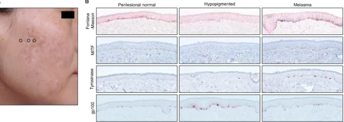
Patient 1, a 41-year-old woman with Fitzpatrick skin type IV, presented with typical mottled hypopigmentation after treatment with 1,064-nm Q-switched Nd:YAG laser for melasma (Fig. 1A). The duration of hypopigmentation was
7 years. Skin biopsies were obtained from the hypopig- mented, hyperpigmented (i.e., melasma), and adjacent perilesional normal skin areas. Hematoxylin and eosin and Fontana-Masson staining as well as immunohisto- chemical staining using melanocyte-specific markers in- cluding monoclonal antibodies against human gp100 (NKI/
beteb; Monosan, Uden, the Netherlands), tyrosinase (Thermo Scientific, Fremont, CA, USA), and microphthalmia- asso- ciated transcription factor (MITF; Leica Biosystems, Newcastle, UK) were performed. The numbers of MITF+
melanocytes per 1-mm length of rete ridge were counted.
The expression levels of gp100 and tyrosinase were meas- ured as the ratio of stained area to measured epidermal area.
The general histological findings of the hypopigmented skin were unremarkable. There was no collagen remodel- ing or scarring. Fontana-Masson staining demonstrated the almost complete absence of melanin pigment in hypo- pigmented skin (Fig. 1B). The number of melanocytes, as determined by MITF expression, was not substantially dif- ferent in lesional skin (n=4) compared to perilesional nor- mal skin (n=5) or melasma skin (n=3). Consistently, ty- rosinase level was not lower in lesional skin than perile- sional normal skin but was higher than that in melasma skin as expected. Interestingly, gp100 expression was higher in the hypopigmented lesion than perilesional nor- mal skin and even melasma skin. Similar findings were ob- served in patient 2, a 49-year-old woman with Fitzpatrick skin type III who presented with typical mottled hypo- pigmentation after laser toning for 1 year (Fig. 2A).
Lesional skin biopsy showed the complete absence of melanin pigment in hypopigmented skin. However, there was no reduction in the number of melanocytes, and gp100 expression was elevated (Fig. 2B).
Letter to the Editor
Vol. 27, No. 3, 2015 341 Fig. 1. (A) Mottled hypopigmentation developing after laser toning for melasma treatment in a 41-year-old woman with Fitzpatrick skin type IV. Hypopigmented, hyperpigmented (i.e., melasma), and adjacent perilesional normal skin were evaluated. Circles indicate biopsy sites of perilesional normal (left), hypopigmented (middle), and melasma (right) skin. (B) Histopathologic examination showed melanin pigmentation was markedly reduced in the basal layer of lesional skin (Fontana-Masson staining). The number of melanocytes, determined according to microphthalmia-associated transcription factor (MITF) expression, did not differ much between lesional skin and perilesional normal skin or melasma skin. The expression of gp100 was higher in hypopigmented lesional skin than perilesional normal skin or even melasma skin (×200).
Fig. 2. (A) A 49-year-old woman with Fitzpatrick skin type III presented with typical mottled hypopigmentation after laser toning for 1 year. Circle indicates biopsy sites of the hypopigmented lesion. (B) Lesional skin biopsy showed the complete absence of melanin pigment in hypopigmented skin. However, there was no reduction in the number of melanocytes and rather increased gp100 expression (×200). MITF: microphthalmia-associated transcription factor.
The results of the present study clearly demonstrate that the histologic features of laser toning-induced hypo- pigmentation are characterized by almost destroyed mela- nosome pigments and a preserved the number of melanocytes. However, it is unclear whether the number of melanocytes is reduced in the hypopigmented skin.
One study indicates hypopigmentation might be due to melanocytopenia5. However, in that study, lesional skin was not compared with adjacent perilesional normal skin.
The other studies suggest the number of melanocytes in hypopigmented skin is normal6,7. In the present study,
there was no difference in the mean numbers of MITF- stained melanocytes between lesional skin and perile- sional normal skin. Interestingly, we noticed that the lev- els of the structural protein gp100 were preserved and rather elevated in lesional skin compared to perilesional normal skin. Moreover, in patient 1, gp100 levels of hypo- pigmented skin were clearly elevated compared to that in hyperpigmented skin. Although the reason for this result is unclear, these findings nonetheless suggest the melano- genic activity in the melanocytes was impaired and the cells failed to produce fully matured melanosomes.
Letter to the Editor
342 Ann Dermatol
Considering that the proposed action mechanism of laser toning is subcellular selective photothermolysis of mela- nosomes and not melanocytes, it is speculated that mela- nocytes survived but were functionally downregulated such that they did not produce fully matured melanosomes.
The cumulative dose of repetitive laser treatment may af- fect melanocyte function, resulting in the development of hypopigmentation. Therefore, treatment activating or stim- ulating melanogenesis in the melanocytes would be required. To this end, treatment with focused narrowband ultraviolet B therapy has been used with some success8. In conclusion, laser toning-induced hypopigmentation is characterized by almost destroyed melanosome pigments and a preserved number of melanocytes, which seem to be functionally downregulated not to produce fully ma- tured melanosomes. Thus, early intervention aiming to re- store melanocyte function would be required.
REFERENCES
1. Jeong SY, Chang SE, Bak H, Choi JH, Kim IH. New melasma treatment by collimated low fluence Q-switched Nd : YAG laser. Korean J Dermatol 2008;46:1163-1170.
2. Polnikorn N. Treatment of refractory dermal melasma with the MedLite C6 Q-switched Nd:YAG laser: two case reports.
J Cosmet Laser Ther 2008;10:167-173.
3. Cho SB, Kim JS, Kim MJ. Melasma treatment in Korean women using a 1064-nm Q-switched Nd:YAG laser with low pulse energy. Clin Exp Dermatol 2009;34:e847-e850.
4. Kim MJ, Kim JS, Cho SB. Punctate leucoderma after melasma treatment using 1064-nm Q-switched Nd:YAG laser with low pulse energy. J Eur Acad Dermatol Venereol 2009;
23:960-962.
5. Kim T, Cho SB, Oh SH. Punctate leucoderma after 1,064-nm Q-switched neodymium-doped yttrium aluminum garnet laser with low-fluence therapy: is it melanocytopenic or melanopenic? Dermatol Surg 2010;36:1790-1791.
6. Ryu HJ, Kim J. A case of mottled hypopigmentation after low-fluence 1,064-nm Q-switched neodymium-doped yttrium aluminum garnet laser therapy. J Cosmet Laser Ther 2013;15:290-292.
7. Chan NP, Ho SG, Shek SY, Yeung CK, Chan HH. A case series of facial depigmentation associated with low fluence Q-switched 1,064 nm Nd:YAG laser for skin rejuvenation and melasma. Lasers Surg Med 2010;42:712-719.
8. Kim HS, Jung HD, Kim HO, Lee JY, Park YM. Punctate leucoderma after low-fluence 1,064-nm quality-switched neodymium-doped yttrium aluminum garnet laser therapy successfully managed using a 308-nm excimer laser.
Dermatol Surg 2012;38:821-823.