2020 Korean Society for Surgery of the Hand, Ko- rean Society for Microsurgery, and Korean Society for Surgery of the Peripheral Nerve. All Rights re- served.
This is an open-access article distributed under the terms of the Creative Commons Attribution Non-Commercial license (http://creativecommons.
org/licenses/by-nc/4.0/), which permits unrestricted non-commercial use, distribution, and reproduction in any medium, provided the original work is prop- erly cited.
INTRODUCTION
Severe traumatic injuries of the hand involving various soft tissue loss, are cata- strophic events. It is a challenge to reconstruct the affected part and restore its functions. Reconstructive surgery often requires multiple procedures that can prolong hospital stay and delay hand rehabilitation. Hence, one-stage reconstruc- tive surgery is recommended to overcome these problems [1]. Chimeric flap con- sisting of free fasciocutaneous anterolateral thigh flap with fascia lata and free vascularized osteomyocutaneous iliac bone graft with isolated pedicled from sin- gle vascular origin is a promising option for one-stage reconstruction of complex tissue defects [2-4].
CASE REPORT
The patient, who is a 20-year-old female student with right-hand dominance, suffered near-amputated left-hand degloving injury due to a road traffic accident.
In that accident, she was a backseat passenger and thrown out from the car after a collision with a high-speed motorcycle. She was brought to a local district hospi- Hand injuries, involving extensor tendon with carpal bone loss are catastrophic events.
Reconstructive surgeons typically face difficulties in minimizing the number of opera- tions, recovery period and restoring its functions with an acceptable aesthetic out- come. However, chimeric flap, which consists of multiple composite flaps from one of the sources of main vascular system is a promising option for reconstruction. This pa- per presents a case of near-amputated degloving injury over the extensor tendon with carpal bone loss on left hand due to road traffic accidents. The patient had a hand re- construction with chimeric flap consisted of free fasciocutaneous anterolateral thigh flap and vascularized osteomyocutaneous iliac bone graft with tensor fascia lata from the lateral circumflex femoral vascular system. As a result, the hand functioned well and appeared aesthetically acceptable. Thus, this surgery is an effective option in re- covering and reconstructing a complex and traumatic hand injury that involved multi- ple composite tissue defects.
Keywords: Hand injuries, Carpal bones, Tendon, Microsurgical free flap Sains Malaysia, Kubang Kerian, Kelantan, Malaysia
3Plastic and Reconstructive Unit, MSU Medical Center, Shah Alam, Selangor, Malaysia Arch Hand Microsurg 2020;25(2):156-160
https://doi.org/10.12790/ahm.20.0007
Received: January 31, 2020 Revised: April 15, 2020 Accepted: April 21, 2020 Corresponding author:
Wan Azman Wan Sulaiman
Reconstructive Sciences Unit, School of Medical Sciences, Universiti Sains Malaysia, Kubang Kerian, Kelantan, Malaysia
Tel: +60129085060 Fax: +6097676909 E-mail: [email protected] ORCID:
https://orcid.org/0000-0003-4120-4918
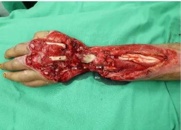
tal with no plastic and reconstructive surgery expertise. She sustained an extensive hand injury that consisted of deep de- gloving injury over extensor surface zone five to eight, extensor tendon loss over all six compartments, all carpal bone losses ex- cept trapezium and pisiform, bone loss over distal end radius and ulna, flexor carpi ulnaris (FCU) and flexor carpi radialis (FCR) total cut with tendon loss, total cut of the flexor digitorum super- ficialis (FDS) and flexor digitorum profundus (FDP) of the little finger, total cut of the FDP of the middle and ring fingers, 20%
cut of the FDS of the middle finger, 80% cut of the FDS of the ring finger, total cut of the FDS of the index finger, as well as 80%
cut of the FDP of the index finger, radial nerve and ulnar nerve cut (Fig. 1A, 1B).
In an early stage, she had wound debridement and external fixator performed by an orthopaedic team in that hospital. Af- ter 1 week of post trauma, she was transferred to our centre for further management. In view of extensive injury including loss of all extensor compartments with metacarpal bone beyond re- pair, the plan was to reconstruct each of the defects accordingly, using an appropriate composite flap that would be able to cover the defects in an operation. Angiogram was performed to as- sess the vasculature of the injured hand and showed a compro- mise of the ulnar artery.
Three weeks later, she was scheduled for operation as planned, involving orthopaedic and reconstructive team. Fol- lowing meticulous wound debridement, defect size over the dorsum of the hand was found to be 12 cm×20 cm. Proximal and distal stumps of extensor tendons were identified for ten- don reconstruction, these include extensor pollicis longus (EPL), extensor carpi radialis (ECR), extensor digitorum (ED), and extensor digiti minimi (EDM) (Fig. 2).
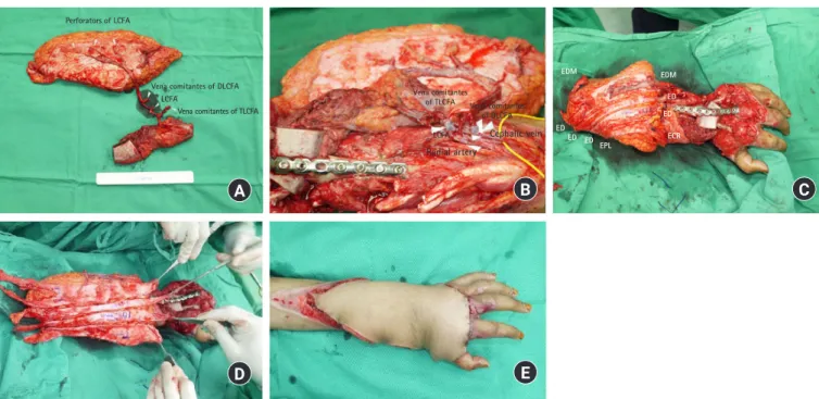
Flap was harvested as a chimeric flap that consisted of free fasciocutaneous anterolateral thigh (ALT) flap with fascia lata supply by descending branch of the lateral circumflex femoral artery (DLCFA) and free vascularized osteomyocutaneous iliac
bone graft supply by transverse branch of lateral circumflex femoral artery (TLCFA) which both flaps vascular pedicle orig- inate from lateral circumflex femoral artery (LCFA) (Fig. 3A).
Vessel anastomoses were done by the end-to-side anastomosis via LCFA to radial artery of recipient and end to side anasto- mosis between vena comitantes of TLCFA and vena comitantes of DLCFA to cephalic vein (Fig. 3B).
Vascularized iliac bone graft was inserted to cover the carpal bone defect and K-wire was inserted from the metacarpal bone through iliac bone graft. Locking plate was applied across the metacarpal bone, iliac bone graft and distal radius representing wrist joint arthrodesis (Fig. 3C). Fascia lata was laid down on top of bone as vascularised tendon grafts and slit into small strips to cover extensor tendon defects for each digit. Fascia lata was used to repair the thumb by connecting EPL tendon stump at the forearm and ECR tendon stump at the hand. For the in- dex and middle fingers, fascia lata was used by connecting ED tendon stump at the forearm to ED tendon stump at the index and middle fingers, respectively. Similar technique was also used for the ring and little fingers which the EDM tendon stump at the forearm was connected to ED tendon stump of the ring finger and also EDM tendon stump of the little finger by fascia lata (Fig. 3D). The flap skin component was used to resurface remaining tissue loss at dorsum of hand (Fig. 3E).
The remaining defects and donor area were covered with split thickness skin graft 3 weeks later after the flap was stable.
Postoperative course was uneventful, as the flap was viable with satisfactory outcome. Wound over donor site of harvesting ALT flap healed satisfactorily without impairment of lower limb function. Patient started on early hand rehabilitation. Lat- Fig. 1. (A) Left forearm wound and (B) X-ray of left forearm,
lateral view.
A B
Fig. 2. Defects wound after debridement. ED, extensor digitorum;
EDM, extensor digiti minimi; ECU, extensor carpi ulnaris.
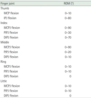
er, she had additional flap debulking surgery and tenolysis for the following 5 months (Fig. 4). During this operation, the vas- cularized tendon graft was found to be viable. After 1 year, the reconstructed left hand showed reasonable hand functions with stiffness of ring and little fingers even after tenolysis was per- formed (Fig. 5A-5C). The range of motion of each digit was as- sessed and measured (Table 1). Despite limited range of mo- tion, she was satisfied with the outcomes, as she was able to perform most of the coarse hand functions for daily activities such as riding bicycle, motorbike, holding an umbrella and plates. However, the fine hand movement still require further improvement.
Written informed consent was obtained from the patient.
DISCUSSION
Composite tissue defects including bony involvement in hand injury are a challenge for reconstructive surgeons. As the case was presented with degloving injury over dorsum of the hand involving extensor tendon and carpal bone loss, it is man- datory to reconstruct the defect accordingly to maximize the hand function. Reconstruction of such extensive degloving in-
jury defects usually required multiple stages of reconstruction.
However, with advancement in microsurgical techniques and deeper understanding of vascular anatomy in harvesting flap, one-stage reconstruction using chimeric flap is able to serve as an alternative method in soft tissue coverage [2-5]. Composite flaps that include multiple and different vascularized tissue from various components from a single source of vascular sys- Fig. 3. (A) Chimeric flap of free fasciocutaneous anterolateral thigh flap with fascia lata and vascularized osteomyocutaneous iliac bone graft. (B) Vascular anastomoses between recipient and donor vessels. (C) Vascularized iliac bone graft was inserted to cover the carpal bone defect and K-wire was inserted from the metacarpal bone through iliac bone graft. Locking plate was applied across the metacarpal bone. (D) Fascia lata slit into small strips and laid down on top of bone as vascularised tendon grafts for the extensor tendon defects. (E) Immediate result after flap inset. LCFA, lateral circumflex femoral artery; TLCFA, transverse branch of LCFA; DLCFA, descending branch of LCFA.
Fig. 4. Flap debulking and tenolysis surgery.
D E
tem can be raised in a single setting. This will not only reduce the number of operations, but also hastens the recovery period.
Here, the flap was harvested as chimeric flap that consisted of free fasciocutaneous anterolateral thigh flap with fascia lata supply by DLCFA and free vascularized osteomyocutaneus iliac bone crest supply by TLCFA via tensor fascia lata muscle, in which both flap vascular pedicle origin from LCFA. Multiple tissue components in this flap allowed reconstruction of the missing multiple structures in this traumatic case such as the
Fig. 5. (A) Dorsum part of the left forearm in full extensor. (B) Stiffness of ring and little fingers of left hand. (C) X-ray of left forearm after 1 year, anteroposterior view.
A B C
Table 1. ROM of left hand digits after 1 year postoperative
Finger joint ROM (°)
Thumb
MCP flexion 0–10
IPJ flexion 0–80
Index
MCPJ flexion 0–90
PIPJ flexion 0–30
DIPJ flexion 0–70
Middle
MCPJ flexion 0–90
PIPJ flexion 0–20
DIPJ flexion 0–10
Ring
MCPJ flexion 0–10
PIPJ flexion 0–10
DIPJ flexion 0
Little
MCPJ flexion 0–10
PIPJ flexion 0–10
DIPJ flexion 0
ROM, range of motion; MCP, metacarpophalangeal joint; IPJ, interphalangeal joint; PIP, proximal interphalangeal joint; DIP, distal interphalangeal joint.
bone, the tendons and the soft tissue coverage. Several authors have described relevant techniques to incorporate multiple tis- sue components, such as chimeric flap in this type of extensive injury. The ALT flap with fascia lata which previously described in the literature was used to provide gliding surface over the dorsum area of the hand [6,7]. In this case, the fascia lata was slit into several strips and functioned as a bridge interconnect- ing the distal and proximal part of extensor tendon loss and bulk of tissue. Also, this was able to obliterate the dead space.
Meanwhile, the vascularized iliac bone graft which nourished by the attachment of periosteal layer, supplied by the TLCFA was used as carpal bone reconstruction. The abundance tissue components from ALT and its variability in harvesting chime- ric flap are adequate to cover the large tissue defects over the dorsum part of hand [4,6].
Considering the promising outcomes of this case, salvaging a large defect did not permit full recovery of the hand function.
However, the patient reported an overall satisfaction despite only three functioning fingers as evident of functional and aes- thetic outcome [8]. Hence, the ability of chimeric flap based on LCFA vascular system including ALT flap and vascularized ili- ac bone graft is a reliable one-stage microsurgical reconstruc- tion in managing complex hand injury.
CONFLICTS OF INTEREST
The authors have nothing to disclose.
REFERENCES
1. Koul AR, Patil RK, Philip V. Complex extensor tendon inju- ries: early active motion following single-stage reconstruction.
J Hand Surg Eur Vol. 2008;33:753-9.
metacarpal defects with free chimeric iliac osteocutaneous flaps. J Plast Surg Hand Surg. 2017;51:143-8.
5. Huang WC, Chen HC, Wei FC, Cheng MH, Schnur DP. Chi- meric flap in clinical use. Clin Plast Surg. 2003;30:457-67.
of the forearm and hand with osteocutaneous free flaps. Plast Reconstr Surg. 2006;118:443-56.