51세 여성 환자가 파노라마방사선촬영 및 콘빔형전산화 단층촬영 (CBCT) 검사시 오른쪽 상악 및 상악동 부위에 병 적 소견이 우연히 발견되어 개인의원으로부터 의뢰되어 본 원에 내원하였다. 초진시, 환자는 코에서 반짝반짝한 누런 물이 나오기도 하였으나 현재 그 누런 물의 색은 짙어졌다 고 하였다. 또한 상악 우측 대구치 부위는 종창이 관찰되 었고 상악 제2대구치 원심부에서 배농이 되었다. 환자는 미약한 부갑상선기능항진증을 제외하고는 기타 뚜렷한 전 신질환은 있지 않다고 하였다.
초진 파노라마방사선영상에서 상악 우측 제3대구치로 보 이는 매복된 치아는 치관이 상방으로 향한 채 상악 제1대 구치 치근단부위까지 변위되었고 우측 상악동의 후벽은 좌 측에 비하여 비박, 팽창되어 있었으며 상악 제2대구치의 원 심부 치조정 피질골은 소실된 양상이었다 (Fig. 1). Waters 영상에서도 측벽을 비박시키며 치관이 포함된 피질골성 변 연을 보이는 병소가 상부 일부를 제외하고 상악동을 채우 고 있는 소견이었다 (Fig. 2).
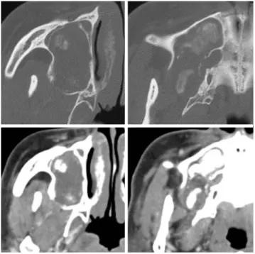
CBCT 영상에서 낭성병소가 우측 상악동을 거의 채우면
서 매복 치아의 치관부를 포함하였으며 병소의 전하방 및 상악동의 후벽측에서 석회화물이 관찰되었다(Fig. 3). CBCT 검사 후 5일 뒤 촬영한 조영증강 전산화단층영상에서 병
─ 99 ─ 접수일 (2010년 5월 15일), 수정일 (2010년 5월 27일), 채택일 (2010년 5월 31일)
Correspondence to : Prof. Kyung-Hoe Huh
Department of Oral and Maxillofacial Radiology and Dental Research Institute, School of Dentistry, Seoul National University, Chang-kyeong-gung-ro 62-1, Jongno-gu, Seoul 110-768, Korea
Tel) 82-2-2072-0211, Fax) 82-2-744-3919, E-mail) [email protected]
대한구강악안면방사선학회지2010; 40 : 99-101
상악에서 발생한 석회화를 동반한 치성각화낭
서울대학교 치의학대학원 구강악안면방사선학교실
*서울대학교 치의학대학원 구강악안면방사선학교실, 치학연구소
김태영∙허경회*
Odontogenic keratocyst with dystrophic calcifications in the maxilla
Tae-Young Kim, Kyung-Hoe Huh*
Department of Oral and Maxillofacial Radiology, School of Dentistry, Seoul National University
*Department of Oral and Maxillofacial Radiology and Dental Research Institute, School of Dentistry, Seoul National University ABSTRACT
Odontogenic keratocyst (OKC) with secondary inflammation involving the maxillary sinus was presented. Radiolo- gical diagnosis of this case was made based on the various findings from the cone-beam computed tomography, com- puted tomography and magnetic resonance images. There were calcified materials and impacted tooth within the lumen of the lesion, which is not uncommon in OKC. Histopathologic findings confirmed this lesion as OKC with secondary inflammation. (Korean J Oral Maxillofac Radiol 2010; 40 : 99-101)
KEY WORDS : Odontogenic Cysts; Pathologic Calcification; Diagnostic Imaging
Fig. 1.Cropped panoramic radiograph shows that the posterior wall of the right maxillary sinus was thinned and expanded and the cortex of alveolar crest distal to the right upper second molar was resorbed. In addition, an impacted molar was visible within the sinus around the periapical region of the first molar.
소 내부는 조영증강이 되지 않는 낭액으로 채워져 있으면 서 석회화물이 비교적 크게 관찰되었으며 병소의 피질골 성 변연은 부분적으로 소실되고 희미해져 병소가 2차적으 로 염증에 이환된 양상이었다(Fig. 4).
초진 12일 뒤 촬영한 환자의 MR (magnetic resonance) 영 상에서 병소는 T2 강조영상에서 높은 신호 강도 (Fig. 5), T1 강조영상에서 낮은 신호강도를 보였으며 조영증강 T1 강 조영상에서 변연이 조영 증강 신호를 나타내었고 상악동의 후벽에서는 비박과 팽윤상이 관찰되었다 (Fig. 6). 또한, MR 영상에서 낮은 신호의 매복 치아 치관이 병소에 포함되어 있었다.
환자는 전신마취하에 낭의 적출수술을 받았으며, 조직병 리검사 결과 염증이 심한 치성각화낭으로 판명되었다.
Ng 등은 시간이 오래 경과된 치성각화낭에서 낭의 결합 조직 벽에서 이형성 석회화물이 발견될 수 있다고 보고하
였다.1Sun 등도 치성각화낭에서 이형성 석회화물이 드물
지 않다고 하였으며 주로 이 석회화물은 병소의 결합조직 낭벽에서 주로 나타나는 것이 보통이지만, 낭내부에서도 낮 은 빈도로 관찰된다고 보고하고, 구강-상악동 누공 등에 의 하여 구강내 체액으로부터 오랫동안 무기물 (mineral)이 유
─ 100 ─ 상악에서 발생한 석회화를 동반한 치성각화낭
Fig. 3.CBCT images show a tooth crown and patch-like calcified materials inside the lumen of the lesion which occupies and ex- pands almost completely the right maxillary sinus.
Fig. 2.Cropped Waters’ view reveals the corticated margin of the lesion and the thinning of the lateral wall of the right maxillary sinus.
Fig. 4.Contrast enhanced CT images show non-enhancing fluid attenuation with corticated margin representing the cystic charac- teristics of this lesion. Patch-like calcifications are noted inside the lumen of the lesion. Also, the diffuse corticated margin suggests the secondary infection of the lesion.
Fig. 5. T2-weighted MR images show a high signal intensity of the cystic lesion with a low signal intensity of the impacted tooth (arrow).
Fig. 6.Contrast enhanced T1-weighted MR images show an inter- nal low signal intensity and enhancing rim of the lesion. These are typical findings of the cyst.
래되면 치성각화낭에 이형성 석회화가 나타날 수 있을 것 으로 추정하였다.2
본 증례의 치성각화낭에서 발견된 이형성 석회화물은
Sun 등2의 주장처럼 배농로를 통하여 유입된 구강내 체액
으로부터 기원된 것일 수도 있는 반면 코와 입안에 배농 이 될 정도의 심한 2차 염증이 동반되었다는 점으로 미루 어 염증으로 인하여 생긴 석회화물일 가능성도 충분히 있
을 것으로 생각된다.
참 고 문 헌
1. Ng KH, Siar CH. Odontogenic keratocyst with dentinoid formation.
Oral Surg Oral Med Oral Pathol Oral Radiol Endod 2003; 95 : 601-6.
2. Sun ZJ, Liu B, Zhao YF. Radiopacity in syndrome keratocystic odon- togenic tumour. Dentomaxillofac Radiol 2008; 37 : 175-8.
─ 101 ─
김태영 외