서 론
매복 정중치란 상악 전치 부위에 매복된 과잉치로 치과 의사가 가장 흔히 만날 수 있는 과잉치이다. 이러한 매복
정중치의 유병률은 연구마다 약간의 차이를 보이지만 보통 0.15-3.8% 정도의 범위로 알려져 있고 관련된 합병증으로 는 주변 치아의 맹출방해나 변위, 정중이개, 치근흡수, 낭 형성 등이 있다.1-12방사선영상은 매복 정중치의 위치나 합 병증 여부를 확인하고 평가하는데 필수적인데 일반적으로 파노라마방사선영상과 치근단영상이 사용된다.7,9,10,13-15
파 노라마방사선영상이나 치근단영상으로는 매복 정중치의 근 원심 위치를 파악할 수 있으며 협설측 위치는 치근단영상
접수일 (2010년 4월 22일), 수정일 (2010년 5월 20일), 채택일 (2010년 5월 27일) Correspondence to : Prof. Sam-Sun Lee
Department of Oral and Maxillofacial Radiology, School of Dentistry, Seoul National University, Chang-kyeong-gung-ro 62-1, Jongno-gu, Seoul 110-768, Korea Tel) 82-2-2072-3978, Fax) 82-2-744-3919, E-mail) [email protected]
매복 정중치의 진단영상분석
서울대학교 치의학대학원 치의학과
*서울대학교 치의학대학원 구강악안면방사선학교실
**서울대학교 치의학대학원 구강악안면방사선학교실 및 치학연구소
***서울대학교 치의학대학원 구강악안면방사선학교실, 치학연구소 및 BK 치의학생명과학사업단
노정준∙최보람*∙정환석*∙허경회**∙이원진***∙허민석**∙이삼선***∙최순철**
Diagnostic imaging analysis of the impacted mesiodens
Jeong-Jun Noh, Bo-Ram Choi*, Hwan-Seok Jeong*, Kyung-Hoe Huh**, Won-Jin Yi***, Min-Suk Heo**, Sam-Sun Lee***, Soon-Chul Choi**
Department of Dentistry, School of Dentistry, Seoul National University
*Department of Oral and Maxillofacial Radiology, School of Dentistry, Seoul National University
**Department of Oral and Maxillofacial Radiology and Dental Research Institute, School of Dentistry, Seoul National University
***Department of Oral and Maxillofacial Radiology, Dental Research Institute and BK21 Craniomaxillofacial Life Science, School of Dentistry, Seoul National University
ABSTRACT
Purpose : The research was performed to predict the three dimensional relationship between the impacted mesiodens and the maxillary central incisors and the proximity with the anatomic structures by comparing their panoramic images with the CT images.
Materials and Methods : Among the patients visiting Seoul National University Dental Hospital from April 2003 to July 2007, those with mesiodens were selected (154 mesiodens of 120 patients). The numbers, shapes, orientation and positional relationship of mesiodens with maxillary central incisors were investigated in the panoramic images.
The proximity with the anatomical structures and complications were investigated in the CT images as well.
Results : The sex ratio (M : F) was 2.28 : 1 and the mean number of mesiodens per one patient was 1.28. Conical shape was 84.4% and inverted orientation was 51.9%. There were more cases of anatomical structures encroachment, especially on the nasal floor and nasopalatine duct, when the mesiodens was not superimposed with the central inci- sor. There were, however, many cases of the nasopalatine duct encroachment when the mesiodens was superimpoised with the apical 1/3 of central incisor (52.6%). Delayed eruption (55.6%), crown rotation (66.7%) and crown resorp- tion (100%) were observed when the mesiodens was superimposed with the crown of the central incisor.
Conclusion : It is possible to predict three dimensional relationship between the impacted mesiodens and the maxil- lary central incisors in the panoramic images, but more details should be confirmed by the CT images when neces- sary. (Korean J Oral Maxillofac Radiol 2010; 40 : 69-74)
KEY WORDS : Tooth, Impacted; Panoramic Radiography, Computed Tomography, X-ray
의 관구이동법으로 파악할 수 있다.
파노라마방사선영상이나 치근단영상만으로는 매복 정중 치의 3차원적 위치에 대한 충분한 정보와 주변 해부학적 구조물이나 주변 치아와의 정확한 위치관계를 얻기 어려 운 경우가 있다.13,14이 때 CT영상을 이용하여 매복 정중 치의 3차원 위치에 대한 보다 정확하고 자세한 정보를 얻 을 수 있지만 상대적으로 비용이 많이 들고 방사선노출량 이 증가하게 된다.13그러나 CT영상을 사용하지 않고 일반 방사선영상만 사용하는 경우 매복 정중치와 인접치 간의 정확한 협설 거리와 인접 해부학적 구조물과의 정확한 위 치 관계를 파악하지 못한 상태로 발치를 시행하게 되므로 인접치나 주변 해부학적 구조물을 손상시킬 위험성이 증가 된다. 따라서 CT영상을 사용하지 않을 경우 일반 방사선영 상에서 매복 정중치와 인접 치아나 구조물과의 위치 관계 에 대한 최대의 정보를 얻을 필요가 있다.13,14
이 논문에서는 파노라마방사선영상으로 매복 정중치와 상악 중절치와의 3차원적 위치 관계나 주위 구조물과의 근 접도를 어느 정도 나타내줄 수 있는 지를 CT영상과 비교 하여 알아보고자 하였다.
대상 및 방법
1. 연구대상
2003년 4월부터 2007년 7월까지 서울대학교 치과병원에 내원하여 파노라마방사선영상과 CT영상을 모두 촬영한 환자 중에서 매복 정중치가 관찰된 환자를 대상으로 하였 다. CT 촬영기는 Somatom Sensation 10 (Siemens AG, For- chheim, Germany)을 사용하였고 파노라마 촬영기는 Ortho- pantomograph OP-100 (Instrumentarium Corp., Tuusula, Fin- land)을 사용하였으며 모든 파노라마방사선영상은 PSP 영 상판을 이용하여 FCR system (Fuji Computed Radiography 5000R, Fuji Photo Film Co. Ltd., Düsseldorf, Germany)으로 CR 영상을 얻었다. 사용된 모니터는 일반 LCD monitor (Nexview 1830, Dicon Co. Ltd., Seoul, Korea)였으며 화면 해상도는 1024×768 픽셀이고 색 품질은 32 bit이었고 π- View STAR (Infinitt Co., Seoul, Korea) PACS프로그램을 사 용하여 관찰하였다. 모든 영상 자료는 서울대학교 치과병 원 PACS를 통해 얻었으며 모두 120명의 환자에서 남성은 82명, 여성은 38명이었고 총 154개의 정중치를 대상으로 하였다. 평균연령은 남성이 18.8세였고 여성이 18.9세였다.
2. 연구방법
1) 매복 정중치의 개수, 모양, 매복양상
매복 정중치의 개수를 조사하였으며, 모양을 원추모양, 절치모양, 결절모양, 소구치모양으로 나누어 조사하였다. 매 복 양상의 분류 기준은 치관이 치근보다 위에 있고 치축과
시상 정중선이 이루는 예각이 30도 보다 작을 때를 역위매 복 (inverted), 30도보다 크고 60도보다 작을 때를 경사진 역위매복 (obliquely inverted), 60도보다 크고 90도보다 작 을 때를 수평매복 (horizontal)이라고 하였으며, 이 때 치관 이 근심 쪽으로 기울어져 있으면 근심수평매복 (horizontal- m), 치관이 원심 쪽으로 기울어져 있을 때를 원심수평매복 (horizontal-d)이라고 하였다. 치관이 치근보다 아래에 있고 치축과 시상 정중선이 이루는 예각이 30도보다 작을 때를 수직매복 (vertical), 30도보다 크고 60도보다 작을 때를 경 사진 수직매복 (obliquely vertical), 60도보다 크고 90도보다 작을 때를 마찬가지로 수평 매복 (horizontal)이라고 하였다.
또한 매복 정중치가 협설 쪽으로 길게 위치했을 때를 횡단 매복 (transverse)이라고 하였다.
2) 매복 정중치와 상악 중절치와의 관계
파노라마방사선영상에서 매복 정중치와 상악 중절치가 겹치는 경우와 겹치지 않는 경우로 분류하였으며, 겹치는 경우는 다시 치관과 겹치는 경우, 치근의 치경부 1/3 부위 와 겹치는 경우, 중간 1/3 부위와 겹치는 경우, 치근단 1/3 부위와 겹치는 경우로 나누었다. 이 때 판정은 매복 정중 치의 치관이 상악 중절치의 치관, 치근의 치경부 1/3, 중간 1/3, 치근단 1/3 부위 중에서 가장 많이 겹치는 부분을 선 택하여 분류하였다. 겹치지 않는 경우에는 매복 정중치가 상악 중절치 치관 사이에 있는 경우, 치근 사이에 있는 경 우, 치근단 상방에 있는 경우로 나누었다.
3) 매복 정중치와 인접 해부학적 구조물과의 근접도 평가 CT영상에서 매복 정중치와 비강저, 비구개관, 순측 피질 골, 중절치 치근단과의 근접도를 근접하지 않음, 근접, 침 범, 관통, 사라짐의 5가지로 분류하였다. 이 때 근접한 경우 는 매복 정중치와 해부학적 구조물의 경계와의 거리가 1 mm 이하인 경우, 침범한 경우는 매복 정중치가 구조물의 피질골을 침범한 경우, 관통한 경우는 완전히 구조물을 뚫 은 경우, 사라진 경우는 그 해부학적 구조물이 완전히 사라 진 경우로 정의하였다.
4) 매복 정중치로 인한 합병증 평가
매복 정중치로 인해 생기는 합병증을 매복 정중치의 위 치 관계와 매복 양상에 따라 조사하였다. 합병증은 맹출이 지연된 경우, 정중이개가 있는 경우, 상악 중절치의 치관이 회전된 경우, 낭이 있는 경우, 상악 중절치의 치관이 흡수 된 경우, 상악중절치의 치근이 흡수된 경우, 상악 중절치가 앞쪽으로 전진되었을 경우로 나누었다.
결 과 1. 매복 정중치의 개수, 모양, 매복 양상
환자 당 정중치 개수는 1개인 경우가 88명 (73.3%), 2개
인 경우가 30명 (25.0%), 3개인 경우가 2명 (1.7%)으로 한 환자 당 평균 1.28개의 정중치를 가지고 있었다.
정중치의 모양은 총 154개의 정중치 중 원추모양 (coni- cal)이 130개 (84.4%), 절치모양 (incisor-like)이 4개 (2.6%), 결절모양 (tuberculate)이 11개 (7.1%), 소구치모양 (premolar- like)은 9개 (5.8%)로 원추모양이 가장 많았다 (Table 1). 또 한, 매복양상은 역위매복 (inverted)이 80개 (51.9%), 횡단매 복 (transverse)이 25개 (16.2%), 근심수평매복 (horizontal-m) 이 6개 (3.9%), 원심수평매복 (horizontal-d)이 9개(5.8%), 경 사진 역위매복 (obliquely inverted)이 20개 (13.0%), 경사진 수직매복 (obliquely vertical)이 4개 (2.6%), 수직매복 (verti- cal)이 10개 (6.5%)로 역위매복 (inverted)이 가장 빈도가 높
게 나왔다 (Table 2).
2. 매복 정중치와 상악 중절치와의 관계
파노라마방사선영상에서 매복 정중치가 상악 중절치와 겹치는 경우가 78증례 (50.6%), 겹치지 않는 경우가 76증 례 (49.4%)로 비슷한 빈도를 보였다. 겹치는 경우에는 중절 치의 치근단 1/3과 겹치는 경우가 49증례 (62.8%)로 가장 높은 빈도를 보였고 치관과 겹치는 경우가 16증례 (20.5
%), 치근의 중간 1/3과 겹치는 경우가 9증례 (11.5%), 치경 부 1/3과 겹치는 경우가 4증례 (5.1%)였다 (Table 3). 겹치지 않는 경우에는 매복 정중치가 상악 중절치의 치근단 상방 에 있는 경우가 64증례 (84.2%)로 가장 높은 빈도를 보였 Table 1.Distribution of mesiodenses according to shape
Shape Conical Incisor-like Tuberculate Premolar-like Total
Number 130 4 11 9 154
Table 2.Distribution of mesiodenses according to orientation
Orientation Inverted Transverse Horizontal-m Horizontal-d Obliquely inverted Obliquely vertical Vertical Total
Number 80 25 6 9 20 4 10 154
Table 3.Location of superimposition between the impacted mesiodens and the cental incisor
Location of superimposition Crown of central incisor Cervical 1/3 of root Middle 1/3 of root Apical 1/3 of root Total
Number of cases 16 4 9 49 78
Table 4.Location of mesiodenses in case of not superimposed with upper incisors
Location of mesiodens Between the crowns Between the roots Between the apex Over the root apex Total
Number of cases 1 7 4 64 76
Table 5.Proximity of mesiodens with other anatomical structures according to the location of superimposition
Nasal floor Nasopalatine duct Labial cortex Incisor root apex
1 2 3 4 5 1 2 3 4 5 1 2 3 4 5 1 2 3 4 5
43 5 1 0 0 11 9 29 0 0 48 0 0 1 0 49 0 0 0 0
8 0 1 0 0 3 4 2 0 0 9 0 0 0 0 9 0 0 0 0
3 0 1 0 0 1 1 2 0 0 3 0 1 0 0 4 0 0 0 0
13 0 3 0 0 6 2 8 0 0 15 1 0 0 0 16 0 0 0 0
67 5 6 0 0 21 16 41 0 0 75 1 1 1 0 78 0 0 0 0
1 0 0 0 0 0 0 1 0 0 1 0 0 0 0 1 0 0 0 0
2 2 3 0 0 2 0 5 0 0 6 0 0 1 0 7 0 0 0 0
2 0 2 0 0 1 1 2 0 0 4 0 0 0 0 4 0 0 0 0
21 14 29 0 0 26 7 28 0 3 59 4 0 1 0 62 2 0 0 0
26 16 34 0 0 29 8 36 0 3 70 4 0 2 0 74 2 0 0 0
1: No proximity, 2: Proximity, 3: Encroachment, 4: Perforation, 5: Disappearance S: Superimposition
Position Cases
Apical 1/3 49
Middle 1/3 9
S Cervical 1/3 4
Crown 16
Total 78
Between crowns 1 Between roots 7 No S Between apex 4
Over apex 64
Total 76
고 중절치 치근 사이에 있는 경우가 7증례 (9.2%), 치근단 사이에 있는 경우가 4증례 (5.2%), 치관 사이에 위치한 경 우가 1증례 (1.3%)였다 (Table 4).
3. 매복 정중치와 인접 해부학적 구조물과의 근접도 평가
1) 파노라마방사선영상에서 매복 정중치와 상악 중절치의 중첩에 따른 근접도 분류
매복 정중치가 상악 중절치와 겹치는 78증례 중 비구개 관과 근접도 3 이상인 증례는 41증례 (52.6%)로 가장 높은
빈도를 보였다. 또한 이 41증례 중 중첩된 위치가 치근단 1/3 부위인 경우가 29증례 (70.7%)를 보였다. 매복 정중치 가 상악 중절치와 겹치지 않는 76증례 중에서도 비강저와 비구개관과 근접도 3 이상인 증례는 각각 34증례 (44.7%), 39증례 (51.3%)로 역시 높은 빈도를 보였다 (Table 5).
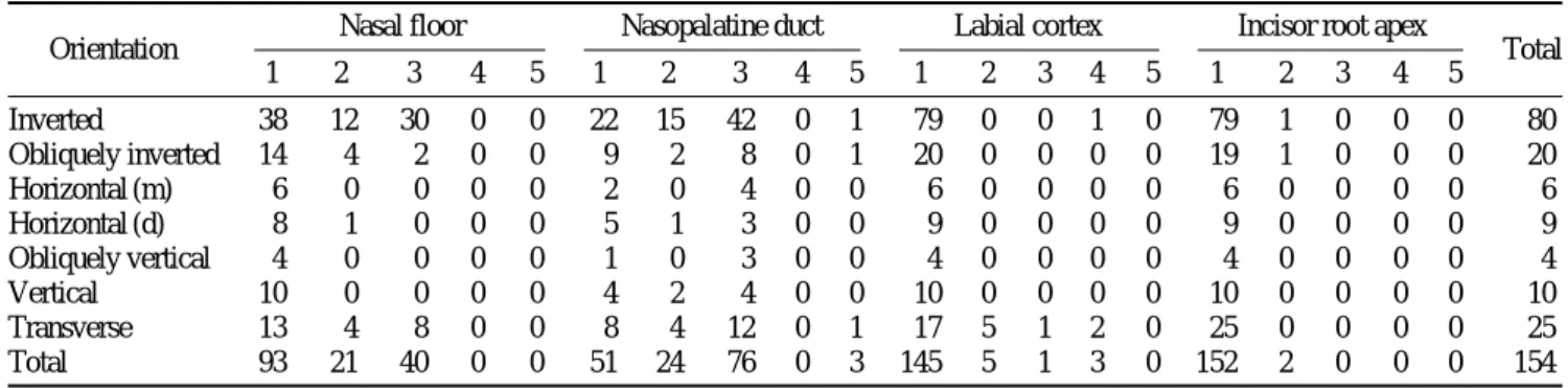
2) 매복 정중치의 매복 양상에 따른 근접도 분류
매복 양상에 따른 근접도 분포는 근접도 4 이상의 총 6 증례 중 3증례 (50.0%)가 횡단매복인 경우에서 나타났다 (Table 6).
Table 6.Proximity of mesiodens with other anatomical structures according to the orientation of the impaction
Nasal floor Nasopalatine duct Labial cortex Incisor root apex
Total
2 3 4 5 1 2 3 4 5 1 2 3 4 5 1 2 3 4 5
12 30 0 0 22 15 42 0 1 79 0 0 1 0 79 1 0 0 0 80
4 2 0 0 9 2 8 0 1 20 0 0 0 0 19 1 0 0 0 20
0 0 0 0 2 0 4 0 0 6 0 0 0 0 6 0 0 0 0 6
1 0 0 0 5 1 3 0 0 9 0 0 0 0 9 0 0 0 0 9
0 0 0 0 1 0 3 0 0 4 0 0 0 0 4 0 0 0 0 4
0 0 0 0 4 2 4 0 0 10 0 0 0 0 10 0 0 0 0 10
4 8 0 0 8 4 12 0 1 17 5 1 2 0 25 0 0 0 0 25
21 40 0 0 51 24 76 0 3 145 5 1 3 0 152 2 0 0 0 154
Orientation 1
Inverted 38
Obliquely inverted 14 Horizontal (m) 6 Horizontal (d) 8 Obliquely vertical 4
Vertical 10
Transverse 13
Total 93
Table 7.Complication due to mesiodens according to superimposition Complication No Delayed
Diastema Crown
Cyst Crown Root Protrusion of
Total eruption rotation resorption resorption central incisor
Apical 1/3 37 2 4 1 5 0 0 0 49
Super Middle 1/3 3 1 1 1 2 0 0 0 8
imposition Cervical 1/3 2 1 1 1 0 0 0 0 5
Crown 5 5 2 6 0 3 0 0 21
Total 47 9 8 9 7 3 0 0 83
Between crowns 1 0 0 0 0 0 0 0 1
No super Between roots 0 2 0 2 2 0 0 0 6
imposition Between apex 4 0 0 0 0 0 0 0 4
Over apex 40 1 4 7 11 0 1 1 65
Total 45 3 4 9 13 0 1 1 76
Table 8.Complication due to mesiodens according to orientation Complication No Delayed
Diastema Crown
Cyst Crown Root Protrusion of
Total eruption rotation resorption resorption central incisor
Inverted 49 6 7 8 9 2 0 0 81
Obliquely inverted 11 1 1 2 3 0 0 1 19
Horizontal (m) 3 1 0 0 1 0 0 0 5
Horizontal (d) 5 0 1 1 3 0 1 0 11
Obliquely vertical 3 1 0 0 0 0 0 0 4
Vertical 8 0 1 1 0 1 0 0 11
Transverse 13 3 2 6 4 0 0 0 28
Total 92 12 12 18 20 3 1 1 159
4. 매복 정중치로 인한 합병증 평가
1) 매복 정중치와 상악 중절치의 중첩에 따른 합병증의 분포 매복 정중치가 상악 중절치의 치관과 겹치는 경우, 맹출 지연 9증례 중 5증례 (55.6%), 치관회전 9증례 중 6증 례 (66.7%), 치관흡수 3증례 중 3증례 (100.0%)를 보였다.
정중치가 상악 중절치와 겹치지 않는 경우에는 중절치의 치근 사이에 정중치가 위치할 때 맹출지연 3증례 중 2증 례 (66.7%)가 나타났다 (Table 7).
2) 매복 정중치의 매복 양상에 따른 합병증의 분포
횡단 매복양상의 경우 치관회전 합병증을 일으킨 비율 이 전체 18증례 중 6증례로 33.3%를 보였다 (Table 8).
고 찰
Kim과 Lee10의 매복 정중치에 대한 연구에 따르면 국내
정중치의 유병률은 0.15-1%이고 전 세계적으로는 연구마 다 차이가 있지만 유병률이 0.15-3.8% 범위 정도로 국내의 정중치 유병률과 큰 차이를 보이지 않는다.1-5,8,10국내 매복 정중치의 성비는 4 : 1이고 Liu 등13의 연구에서는 중국의 매복 정중치의 성비가 2.64 : 1, 코카시안의 경우에는 1.3 : 1-2.5 : 1, 인도의 경우는 1.5 : 1로 보고하고 있는데10,12,13 이번 연구에서는 2.28 : 1로 가장 증례가 많은 중국의 연구 결과와 비슷했고 코카시안이나 인도의 경우보다는 차이가 있었다. 이러한 차이는 인종적 차이나 표본추출의 차이에 의한 것으로 보인다.
매복 정중치 모양은 원추모양이 84.4%로 가장 많이 나 와 이전의 연구결과와 유사하였고1,3,5,7-10,15,16
특히 83.5%라 고 보고한 Liu 등13의 연구와 매우 비슷한 결과를 보였다.
매복 양상 또한 역위매복이 51.9%로 가장 많은 결과를 보 였고 이 결과 역시 다른 연구들의 결과와 유사하였 다.7,10,12,13,16
파노라마방사선영상에서 매복 정중치가 상악 중절치와 겹치는 것과 관계없이 두 경우 모두에서 비구개관과 근접 도 3 (침범) 이상을 보이는 경우가 50% 이상이었으나 겹치 지 않는 경우에는 비강저에 근접한 경우도 많았다. 매복양 상에 따른 근접도 분포는 횡단 매복양상인 경우 주변 해부 학적 구조물과의 근접도가 높았다.
파노라마방사선영상에서의 매복 정중치의 위치에 따른 합병증의 분포를 분석해 보면, 맹출지연, 치관회전, 치관흡 수는 상악 중절치의 치관과 정중치의 치관이 가까이 있을 경우에 많았다. 매복양상에 따른 합병증 분포를 보면, 횡단 매복의 경우 전체 165증례 (중복허용) 중 28증례로 17.0%
이지만 치관회전 합병증을 일으킨 비율은 전체 18증례 중 6증례로 33.3%를 보이며 상대적으로 다른 매복 양상에 비 해 높은 빈도를 보였다.
이 연구에서는 우리나라 120명의 환자, 154증례의 매복
정중치 증례에 대해 파노라마방사선영상에서 얻을 수 있 는 정보로 CT영상으로부터 얻을 수 있는 실제 위치에 대 한 정보를 유추하기 위한 시도를 하였다. 이 연구가 다른 CT영상을 이용한 매복치 연구와 다른 점은 파노라마방사 선영상에서의 위치 정보와 CT영상을 이용한 위치 정보를 연관시키고 전자로부터 후자를 유추해보려는 노력을 했다 는 점이다. 기존 연구에서는 파노라마방사선영상이나 치근 단방사선영상을 기반으로 한 매복 정중치의 통계나 CT영 상을 기반으로 정중치의 3차원적 위치에 대한 통계처리가 대부분이었지만 이 연구는 그러한 3차원적 위치 정보를 파노라마방사선영상에서 유추할 수 있는 근거를 찾아보았 다. 그 결과 파노라마방사선영상을 통해 실제 정중치의 위 치에 대한 정보를 유추할 수 있는 경우도 있었으나 정중 치의 삼차원적 위치에 대해 유추할 수 있는 정도에 한계가 있었다. 따라서 매복 정중치 발치 시 정중치와 인접 구조물 과의 위치 정보를 좀 더 자세하게 얻기 위해서는 CT영상 이 필요하다.
참 고 문 헌
1. Gabris K, Fabian G, Kaan M, Rozsa N, Tarjan I. Prevalence of hypo- dontia and hyperdontia in paedodontic and orthodontic patients in Budapest. Community Dent Health 2006; 23 : 80-2.
2. Alberti G, Mondani PM, Parodi V. Eruption of supernumerary perma- nent teeth in a sample of urban primary school population in Genoa, Italy. Eur J Paediatr Dent 2006; 7 : 89-92.
3. Costa Pinho TM, Figueiredo Pollmann MC. Study of the frequency and the features of supranumerary teeth found in one Portuguese popu- lation. Bull Group Int Rech Sci Stomatol Odontol 2004; 46 : 52-62.
4. Ray D, Bhattacharya B, Sarkar S, Das G. Erupted maxillary conical mesiodens in deciduous dentition in a Bengali girl - A case report. J Indian Soc Pedod Prev Dent 2005; 23 : 153-5.
5. Russell KA, Folwarczna MA. Mesiodens-diagnosis and management of a common supernumerary tooth. J Can Dent Assoc 2003; 69 : 362-6.
6. Zmener O. Root resorption associated with an impacted mesiodens: a surgical and endodontic approach to treatment. Dent Traumatol 2006;
22 : 279-82.
7. Asaumi JI, Shibata Y, Yanagi Y, Hisatomi M, Matsuzaki H, Konou- chi H, et al. Radiographic examination of mesiodens and their asso- ciated complications. Dentomaxillofac Radiol 2004; 33 : 125-7.
8. Fernández Montenegro P, Valmaseda Castellón E, Berini Aytés L, Gay Escoda C. Retrospective study of 145 supernumerary teeth. Med Oral Patol Oral Cir Bucal 2006; 11 : E339-44.
9. Huang WH, Tsai TP, Su HL. Mesiodens in the primary dentition stage:
a radiographic study. ASDC J Dent Child 1992; 59 : 186-9.
10. Kim SG, Lee SH. Mesiodens: a clinical and radiographic study. J Dent Child 2003; 70 : 58-60.
11. Lustmann J, Bodner L. Dentigerous cysts associated with supernumer- ary teeth. Int J Oral Maxillofac Surg 1988; 17 : 100-2.
12. Roychoudhury A, Gupta Y, Parkash H. Mesiodens: a retrospective study of fifty teeth. J Indian Soc Pedod Prev Dent 2000; 18 : 144-6.
13. Liu DG, Zhang WL, Zhang ZY, Wu YT, Ma XC. Three dimensional evaluations of supernumerary teeth using cone-beam computed tomo- graphy for 487 cases. Oral Surg Oral Med Oral Pathol Oral Radiol
Endod 2007; 103 : 403-11.
14. Liu DG, Zhang WL, Zhang ZY, Wu YT, Ma XC. Localization of im- pacted maxillary canines and observation of adjacent incisor resorption with cone-beam computed tomography. Oral Surg Oral Med Oral Pa- thol Oral Radiol Endod 2008; 105 : 91-8.
15. von Arx T. Anterior maxillary supernumerary teeth: a clinical and radiographic study. Aust Dent J 1992; 37 : 189-95.
16. Stellzig A, Basdra EK, Komposch G. Mesiodentes: incidence, morpho- logy, etiology. J Orofac Orthop 1997; 58 : 144-53.