Alteration of Lipid Metabolism Related Proteins in Liver of High-Fat Fed Obese Mice
Eunhui Seo, Ying Han, So-Young Park, Hyongjong Koh and Hye-Jeong Lee*
Department of Pharmacology, Medical Science Research Center, Dong-A University College of Medicine, Busan 602-714, Korea
Received May 13, 2010 /Accepted May 24, 2010Obesity and being overweight are strongly associated with the development of metabolic disease such as diabetes, hypertension, dyslipidemia. High-fat diet (HFD) is one of the most important factors which cause obesity. In this study, C57BL/6 mice were treated with a HFD for 22 weeks in order to induce obesity and hyperglycemia. Twenty-two weeks later, body weight and plasma glucose level of the HFD group were significantly increased, compared with the normal diet (ND) group. Intra-peri- toneal glucose tolerance test (IPGTT) showed glucose intolerance in the HFD group compared with the ND group. These results confirmed that a HFD induced obesity and hyperglycemia in C57BL/6 mice. Plasma levels of triglyceride (TG) and total cholesterol (TC) were increased in the HFD group compared with the ND group. Hepatic levels of TG and TC were also increased by a HFD. To inves- tigate the alteration of lipid metabolism in liver, proteins which are related to lipid metabolism were observed. Among lipid synthesis related enzymes, fatty acid synthase (FAS) and glycerol phosphate acyl transferase (GPAT) were significantly increased in the HFD group. Apolipoprotein B (apoB) and microsomal triglyceride transport protein (MTP), which are related to lipid transport, were sig- nificantly increased in the HFD group. Interestingly, protein level and phosphorylation of AMP-acti- vated protein kinase (AMPK), which is known as a metabolic regulator, were significantly increased in the HFD group compared with the ND group. In the present study we suggest that HFD may physiologically increase the proteins which are related with lipid synthesis and lipid transport, but that HFD may paradoxically induce the activation of AMPK.
Key words : High fat diet, lipid metabolism, triglyceride
*Corresponding author
*Tel:+82-51-240-2859, Fax:+82-51-241-0778
*E-mail : [email protected]
Introduction
High-fat diet (HFD) has contributed to the accumulation of adipose tissue, resulting in obesity, which is one of the most metabolic disorders in developing and developed countries. Particularly, a shift from a low-fat diet to a high-fat diet in developing countries, including South Korea, markedly increased in the prevalence of obesity in the last two decades [12,19]. Weight gain and obesity are major risk factors for conditions and diseases ranging from insulin re- sistance and type 2 diabetes mellitus (T2D) to atherosclerosis and the sequelae of nonalcoholic fatty liver disease [30].
Additionally, obesity is one of the most important risk fac- tors for complex and chronic liver disorder [11]. These liver disorders begin as steatosis and may progress to steatohepa- titis, cirrhosis, liver failure, and hepatocellular carcinoma [2,20]. It has been previously reported that insulin-resistant T2D and hyperlipidemia occur in most liver disease cases
[32]. It means that obesity, insulin resistance and liver fail- ure, all of three, are closely connected. The liver plays a cen- tral role in the control of glucose and lipid metabolism.
When a high-carbohydrate meal is ingested, several metabol- ic events aimed at decreasing endogenous glucose pro- duction by the liver (glycogenolysis and gluconeogenesis) and increasing glucose uptake (glycolysis) and storage in the form of glycogen in the liver are turned on. In addition, when glucose is delivered into the portal vein in large quan- tities and once hepatic glycogen concentrations are restored, glucose can be converted in the liver into TG through de novo lipogenesis. In fact, TG represents the principal energy storage fuel in mammals. Lipids are essential for energy ho- meostasis, reproductive and organ physiology, and numer- ous aspects of cellular biology. They are also linked to many pathological processes, such as obesity, diabetes, heart dis- ease, and inflammation [5]. In particular, glucose and fatty acids are able to regulate hepatic gene expression in a tran- scriptional manner.
The synthesis of TG in liver is nutritionally regulated, and
its formation from simple carbohydrates requires multiple
metabolic pathways, including glycolysis and pyruvate oxi- dation to generate acetyl-CoA for fatty acid synthesis, NADPH generation to supply the reductive power, pack- aging of fatty acids into a glycerophosphate backbone, and finally, lipoprotein packaging to export TG. Hepatic TG is produced by several key enzyme, including (i) ATP citrate lyase [6], ACC [15] and FAS [16] for lipogenesis; (ii) long-chain elongase (LCE) (also known as ELOVL6) [13] and stearoyl-CoA desaturase 1 (SCD1) [22] synthesizing for fatty acid elongation and desaturation steps; and finally (iii), mi- tochondrial GPAT and diacylglycerol acyltransferase (DGAT) for TG synthesis [29]. It is widely accepted that hep- atic lipid availability is obligatory for apoB-containing lip- oprotein assembly within the liver, a finding supported by studies demonstrating the necessity of TG [7,27] and phos- pholipid [35]. The hepatic production of apoB-containing lip- oproteins is regulated largely, with nascent apoB molecules being secreted or degraded intracellularly [36]. The assembly of apoB with lipid to form a secretion-competent particle is a complex process [7,36]. The availabilities of cholesterol and cholesterol esters, synthesized by the HMG-CoA reduc- tase (HMGCR) pathway, have also been shown to be im- portant determinants of the rate of hepatic apoB secretion [3,4,34]. The MTP also plays a key role in apoB secretion by catalyzing the transfer of lipids to the nascent apoB mole- cule as it is cotranslationally translocated across the endo- plasmic reticulum membrane [14,33].
The aims of the present study were as follows: 1) to devel- op an obesity mouse model fed with HFD, 2) to investigate the alteration of hepatic proteins which are associated with the lipid metabolism under the normal or high-fat diet condition. The results of this study might be basic in- formation for understanding the connection of obesity, dia- betes and lipid disorder, and searching for therapeutic tar- gets of the metabolic disorder.
Materials and Methods Animal treatment
Male, 6 weeks old C57BL/6 inbred mice weighing 22-25 g were supplied by Samtako Bio Korea (Kyoung-Ki, South Korea). All mice were maintained in room under controlled temperature (22
oC), humidity (50%), and air-flow condition, with a fixed 12 hr light-dark cycle. Mice were provided with food and water in ad libitum during 2 weeks for acclimatization. Mice were classified into two groups and
fed with the normal (ND) and high-fat diet (HFD) (protein 14.3%, carbohydrate 20.1%, and fat 64.4% (kcal %)) for up to 22 weeks. All period of experiment, body weight and food intake are measured daily, except weekend. Tissue samples were frozen immediately using liquid nitrogen and were stored at -70
oC until assayed.
Intraperitoneal glucose tolerance test (IPGTT)
After 22 weeks ND or HFD feeding, the mice were fasted for 6 hr following intraperitoneal injection with glucose (1 mg/g body weight), and blood glucose levels were meas- ured at 0, 5, 10, 30, 60 and 120 min post injection.
Measurements of blood glucose, plasma lipids and GOT/GPT
Blood samples were collected to measure glucose, total cholesterol (TC), and triglyceride (TG) levels. The sampling line was filled with 4.5% EDTA to prevent blood clotting.
Samples were kept on ice, and plasma was isolated using centrifuagation and stored at -20°C until analysis. Glucose levels were measured with a glucose analyzer, GlucoDR (Allmedicus, An-Yang, South Korea). TC and TG levels were measured with the ASAN Set Total-cholesterol Reagent and Triglyceride-S Reagent (ASAN PHARM., Seoul, South Korea), respectively. Glutamic oxaloacetic transaminase (GOT)/ glutamic-pyruvate transaminase (GPT) were meas- ured with the ASAN set GOT·GPT Reagent by Reitman- Frankel method (ASAN PHARM.).
Analysis of hepatic lipids
Liver samples were lysed in ethanolic KOH (2 parts EtOH: 1 part 30% KOH) and incubated overnight at 55°C.
The lysates were mixed with H
2O:EtOH (1:1) and centri- fuged for 5 min. The supernatant was mixed with 1 M MgCl
2, incubated for 10 min on ice and centrifuged for 5 min. The resulting supernatant was used to measure TG or TC.
Immunoblotting
Total proteins were isolated form liver sample using ra- dioimmunoprecipitation assay (RIPA) buffer containing 50 mM Tris-HCl, pH 8.0, with 150 mM sodium chloride, 1.0%
Igapal CA-630 (NP-40), 0.5% sodium deoxycholide, 0.1% so- dium dodesyl sulfate, protease Inhibitor Cocktail (P8340;
Sigma, St Louis, MO, USA) and Phosphatase Inhibitor
Cocktails (P2850 and P5726, Sigma). The lysates were centri-
fuged at 13,000 rpm for 30 min to remove insoluble debris.
Supernatants were collected and assayed for protein content prior to storage at -70
oC. Protein concentration was de- termined in triplicate using the bicinchoninic acid solution (B9643, Sigma) with cooper (Ⅱ) surfate solution (C2284, Sigma).
Protein sample were mixed Laemmli Sample Buffer (LSB) and placed in a boiling water bath for 5 min. Proteins were resolved by 6 or 10% SDS-polyacrylamide gel electro- phoresis (SDS-PAGE; each loaded with same μg of total pro- tein per lane), and transferred to nitrocellulose membranes.
Blots were probed with antibodies to ACC Antibody (#3662;
280 kDa) and Phospho-ACC (Ser79) Antibody (#3661; 280 kDa) (Cell Signaling Technology), Actin (I-19) (sc-1616; 43 kDa), HMGCR (C-18) (sc-27578; 43 kDa), SCD1 (E-15) (sc-14720; 37 kDa), MTP (N-17) (sc-33116; 97 kDA), Fatty Acid Synthase (H-300) (sc-20140; 270 kDa) and DGAT2 (H-70) (sc-66859; 44 kDa) (Santa Cruz Biotechnology Inc., Santa Cruz, CA), Rabbit polyclonal to apoB (ab20737; 549 kDa; Abcam, Cambridge, MA) and AMPK alpha1 antibody (A300-507A; 62 kDa; Bethyl Laboratories, Inc., Montgomery, TX). Membrane were developed using horseradish perox- idase-conjugated donkey anti-rabbit (NA934; GE Healthcare, Chalfont, St Giles, UK), and donkey anti-goat IgG (sc-2056;
Santa Cruz Biotechnology Inc.), followed by incubation with ECL PlusTM Western Blotting Detection Reagents (RPN2135; GE Healthcare, Chalfont, St Giles, UK), and sig- nals were detected using a Fuji LAS 3000 Imaging station (Fuji Photo Film, Fuji, Japan).
Statistical analysis
Results are expressed as means±SEM. Differences be- tween groups were analyzed by paired or non-paired Student’s t test. Differences were considered significant when the p value was less than 0.05.
Results
Obese and hyperglycemic mice model by feeding with HFD
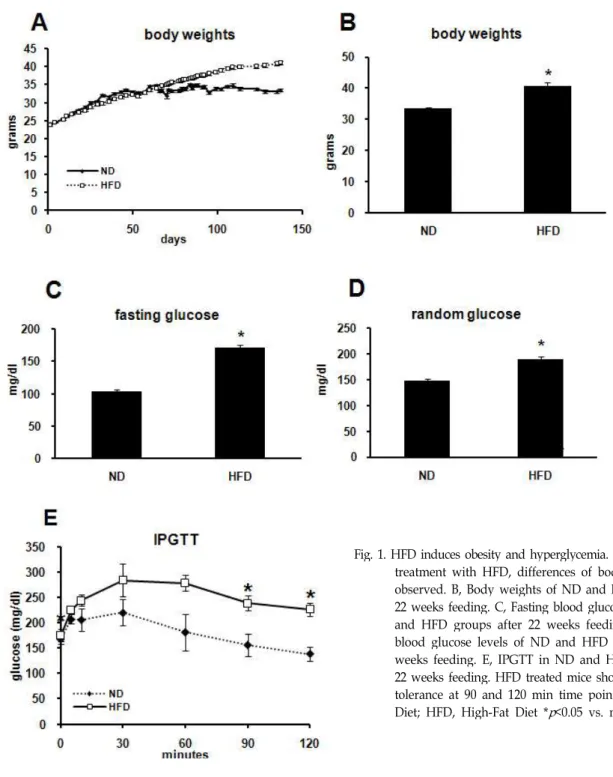
For inducing obesity and hyperglycemia, animals were treated with HFD for 22 weeks. Body weight was measured daily, except weekend. After 9 weeks of treatment with HFD, a difference in body weight was observed (Fig. 1A).
Twenty-two weeks later with ND or HFD, body weight of ND group was 33.58±0.38 g. On the other hand, that of HFD
group was 40.83±0.89 g; differences were significant (Fig.
1B). Blood glucose levels of both fasting and random glucose increased in HFD group (Fig. 1C and 1D). Fasting glucose level was 103.20±3.30 mg/dl in ND group, and 171.69±4.02 mg/dl in HFD group (Fig. 5C). In the same manner, random glucose level was 147.15±23.86 mg/dl in ND group, and 188.72±5.43 mg/dl in HFD group (Fig. 5D). Analysis of IPGTT also showed significant differences between ND and HFD groups. Especially, later at 90 and 120 min following glucose injection, plasma glucose was significantly higher in HFD group than in ND group (Fig. 1E).
GOT/GPT measurement for verification of hepatic toxicity by HFD
To examine that HFD itself causes cytotoxicity, ver- ification of toxicity of the liver and several other organs was necessary. GOT is raised in acute liver damage. It is also present in red blood cells, cardiac muscle, skeletal muscle, kidney and brain tissue, and may be elevated due to damage to those sources as well. GPT is commonly measured clin- ically as a part of diagnostic liver function testing for deter- mination of liver health, because, compared with GOT, GPT is located in liver more than in other organs. The normal range for GOT is 8-40 karmen/ml, and GPT is 5-30 kar- men/ml. The HFD itself induced a significantly increase of both GOT and GPT levels. Plasma level of GOT was 9.70±3.57 karmen/ml in ND group, but 41.88±6.79 kar- men/ml in HFD group. Plasma level of GPT was 4.46±1.68 karmen/ml in ND group, but 10.61±2.75 karmen/ml in HFD group (Fig. 2). However, the increment induced by HFD was within normal range. These results suggest that hepatic tox- icity may not be induced significantly by HFD.
Changes of lipid parameters by HFD
To determine the alteration of plasma and hepatic lipids by HFD, lipid profile analysis performed. The HFD group showed a high plasma levels of TG and TC compared with the ND group. Plasma level of TG was 46.3±2.1 mg/dl in ND group, but 60.6±4.9 mg/dl in HFD group (Fig. 3).
Plasma level of TC was 106.6±4.7 mg/dl in ND group, but
158.5±7.8 mg/dl in HFD group (Fig. 3). Hepatic lipids were
also higher at HFD group than ND group. Hepatic level of
TG was 89.2±5.8 mg/g in ND group, but 191.4±20.7 mg/g
in HFD group (Fig. 3). Hepatic level of TC was 26.4±4.6
mg/g in ND group, but 59.7±8.9 mg/g in HFD group con-
dition (Fig. 3). These results indicated that HFD induced hy-
Fig. 1. HFD induces obesity and hyperglycemia. A, 9 weeks after treatment with HFD, differences of body weight were observed. B, Body weights of ND and HFD groups for 22 weeks feeding. C, Fasting blood glucose levels of ND and HFD groups after 22 weeks feeding. D, Random blood glucose levels of ND and HFD groups after 22 weeks feeding. E, IPGTT in ND and HFD group after 22 weeks feeding. HFD treated mice showed glucose in- tolerance at 90 and 120 min time points. ND, Normal Diet; HFD, High-Fat Diet *
p
<0.05 vs. normal diet.perlilpidemia and fatty liver.
Changes of lipid metabolism-related proteins by HFD
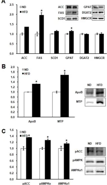
To verify protein expression associated with lipid metabo- lism, we performed western blotting with liver tissues of both ND and HFD groups (Fig. 4). Proteins involved in lipid synthesis, ACC, FAS, SCD1, GPAT, HMGCR and DGAT2 were checked (Fig. 4A). Protein levels of FAS and GPAT were higher in HFD than ND group. Protein levels of ACC
and SCD1 showed tendency of increase, but statistically
non-significant (P=0.051, P=0.093, respectively) Protein lev-
els of apoB and MTP, which are associated with lipid exocy-
tosis, were significantly increased in HFD compared with
ND group (Fig. 4B). Protein levels of AMPK known as a
metabolic regulator were unexpectedly increased and the
phosphorylation of AMPK activated in HFD group com-
pared to ND group. ACC which is a major substrate of
AMPK was also phosphorylated in HFD group more than
ND group.
Fig. 3. Measurement of plasma and hepatic lipid levels. Plasma and hepatic concentrations of TG and was increased in HFD group compared with ND group after 22 weeks feeding. *
p
<0.05 vs. normal diet.Fig. 2. Analysis of hepatic toxicity. GOT/GPT levels were in- creased in HFD group, but within normal limit. *
p
<0.05 vs. normal diet.Discussion
Weight gain and obesity are major risk factors for con- ditions and diseases ranging from insulin resistance and T2D to atherosclerosis [30]. The prevalence of liver disorders is high in obese people, and in patients who have T2D, insulin
resistance is one of the key elements leading to liver disease in obese people [21]. Obesity is continuous with T2D [23,25,28].
The present study investigated the alteration of lipid me- tabolism related proteins in obese C57BL/6 mice induced by HFD. To confirm that HFD was successfully induced obe- sity and T2D, the body weight and blood glucose level were measured (Fig. 1A-D). Difference of Body weight was ob- served after 9 weeks of treatment with HFD (Fig. 1A).
Fasting and random blood glucose were also higher in HFD group than ND group (Fig. 1C, D). The result from IPGTT confirmed glucose intolerance in HFD group (Fig. 1E). These results mean that feeding of HFD during 22 weeks induced obesity and T2D. Levels of plasma and hepatic lipids were also significantly higher in HFD than ND group (Fig. 3). It means that as already known HFD induced not only obesity and hyperglycemia, but also hyperlipidemia and fatty liver in this study.
Because HFD induced obesity and fatty liver, plasma lev-
els of GOT/GPT were measured for knowing whether HFD
itself may cause cytotoxicity. The levels of GOT/GPT in HFD group were significantly higher than ND group, al- though levels of GOT/GPT stayed within normal range in both ND and HFD groups. It means that HFD itself does not trigger cytotoxicity even though the numerical value was slightly increased in HFD group.
Hepatic TG synthesis is conducted in the order of ACC-FAS-SCD1-GPAT-DGAT2 [26]. The present study shows the comparison of the protein levels related with hep- atic TG synthesis between ND and HFD. The protein levels of FAS and GPAT were increased in HFD group (Fig. 4A).
FAS is the enzyme catalyzing the terminal steps in the de novo biosynthesis of long-chain fatty acids. FAS activity has been shown to significantly influence energy expenditure rates, fat mass, insulin sensitivity and cancer risk [36]. GPAT, which catalyzes the initial and committing step in glycer- olipid biosynthesis, is predicted to play a pivotal role in the regulation of cellular triacylglycerol and phospholipid levels [17]. This result is corresponded with present of hyper- lipidemia and fatty liver in this study.
ApoB and MTP are necessary for lipoprotein assembly and exocytosis [8]. Lipid transfer activity of MTP is required for the assembly of lipoproteins. This activity renders nas- cent apoB secretion-competent and may be involved in the import of triglycerides into the lumen of endoplasmic retic- ulum [1,8,24]. In this study, protein levels of apoB and MTP were increased by HFD (Fig. 4B). Therefore, increased pro- tein levels of apoB and MTP by HFD is thought to induce exocytosis of lipoprotein. This hypothesis harmonized with our results, increase of both hepatic and plasma lipid (Fig. 3).
AMPK is thought to act as a cellular ‘fuel gauge’ or ‘meta- bolic switch’ and has been suggested to play an important role in glucose and lipid metabolism [9,10]. The expression and activity of AMPK are generally suppressed with HFD for short-term period [37]. In this study, AMPK protein level and activity were increased by HFD (Fig. 4C). This is un- expected result and the discrepancy might be caused by the feeding duration of HFD. HFD experiment is commonly at- tempted for 5-10 weeks while this study performed it for 22 weeks. The long period HFD induced high plasma and hepatic lipids. It is thought to a compensatory action to re- verse the increased concentration of plasma and hepatic lipids. The enzyme might serve as a metabolic switch, de- crease fatty acid synthesis and increase fatty acid oxidation.
In summary, C57BL/6 mice fed HFD during 22 weeks induce obesity, hyperglycemia, hyperlipidemia and fatty
Fig. 4. Analysis of immunoblots with proteins associated with lipid metabolism by HFD. A, Protein levels related with lipid synthesis. Protein levels of FAS and GPAT were increased by HFD. B, Protein levels related with lipid transfer. Protein levels of ApoB and MTP were also in- creased by HFD. C, Protein levels of AMPK and phos- phorylation of AMPK and ACC. AMPK , pAMPK and pACC were increased by high-fat diet. *
p
<0.05 vs. nor- mal diet.liver. The increase of hepatic lipid synthesis and exocytosis
by HFD is considered that these phenomena might be
caused excessive supplementation of high fat. To rectify
these metabolic phenomena, AMPK protein level and activ-
ity might be increased. Therefore, we suggest that early acti-
vation of AMPK might be better targets for amelioration of
high-fat diet induced obesity. These differential expression
of lipid metabolism related proteins may be applied to the
development of therapeutic targets for obesity and hyper- glycemia treatment.
Acknowledgement
This paper was supported by Dong-A University Research Fund in 2007.
References
1. Berriot-Varoqueaux, N. L. P., A. M. Samson-Bouma, and J. R. Wetterau. 2000. The role of the microsomal triglygeride transfer protein in abetalipoproteinemia.
Annu. Rev. Nutr.
20, 663-697.
2. Bugianesi, E., P. Manzini, S. D'Antico, E. Vanni, F. Longo, N. Leone, P. Massarenti, A. Piga, G. Marchesini, and M.
Rizzetto. 2004. Relative contribution of iron burden, HFE mutations and insulin resistance to fibrosis in non alcoholic fatty liver.
Hepatology
39, 179-187.3. Burnett, J. R., L. J. Wilcox, D. E. Telford, S. J. Kleinstiver, P. H. R. Barrett, R. S. Newton, and M. W. Huff. 1999.
Inhibition of ACAT by avasimibe decreases both VLDL and LDL apolipoprotein B production in miniature pigs.
J. Lipid Res.
40, 1317-1327.4. Cianflone, K., Z. Yasruel, M. A. Rodriguez, D. Vas, and A.
D. Sniderman. 1990. Regulation of apoB secretion from HepG2 cells: evidence for a critical role for cholesteryl ester synthesis in the response to a fatty acid challenge.
J. Lipid Res.
31, 2045-2055.5. Dentin, R., P. D. Denechaud, F. Benhamed, J. Girard, and C. Postic. 2006. Hepatic gene regulation by glucose and pol- yunsaturated fatty acids: a role for ChREBP.
J. Nut.
136, 1145-1149.6. Elshourbagy, N. A., J. C. Near, P. J. Kmetz, G. M. Sathe, C. Southan, J. E. Strickler, M. Gross, J. F. Young, T. N. Wells, and P. H. Groot. 1990. Rat ATP citrate-lyase. Molecular cloning and sequence analysis of a full-length cDNA and mRNA abundance as a function of diet, organ, and age.
J. Biol. Chem.
265, 1430-1435.7. Ginsberg, H. N. 1997. Role of lipid synthesis, chaperone proteins and proteasomes in the assembly and secretion of apoprotein Bcontaining lipoproteins from cultured liver cells.
Clin. Exp. Pharmacol. Physiol.
24, A29-A32.8. Gordon, D. A. and H. Jamil. 2000. Progress towards under- standing the role of microsomal triglyceride transfer protein in apolipoprotein-B lipoprotein assembly.
Biochim. Biophys.
Acta.
1486, 72-83.9. Hardie, D. G. 2004. AMP-activated protein kinase: a master switch in glucose and lipid metabolism.
Rev. Endocr. Metab.
Disord.
5, 119-125.10. Hardie, D. G., J. W. Scott, D. A. Pan, and E. R. Hudson.
2003. Management of cellular energy by the AMP-activated protein kinase system.
FEBS Lett
546, 113-120.11. Haynes, P., S. Liangpunsakul, and N. Chalasani. 2004.
Nonalcoholic fatty liver disease in individuals with severe obesity.
Clin. Liver Dis.
8, 535-547.12. Hedley, A. A., C. L. Ogden, C. L. Johnson, M. D. Carroll, L. R. Curtin, and K. M. Flegal. 2004. Prevalence of over- weight and obesity among US children, adolescents, and adult, 1999-2002.
JAMA
291, 2847-285013. Jakobsson, A., R. Westerberg, and A. Jacobsson. 2006. Fatty acid elongases in mammals: their regulation and roles in metabolism.
Prog. Lipid Res.
45, 237-249.14. Jamil, H., J. K. Dickson, Jr., C. Chu, M. W. Lago, J. K.
Rinehart, S. A. Biller, R. E. Gregg, and J. R. Wetterau. 1995.
Microsomal triglyceride transfer protein: specificity of lipid binding and transport.
J. Biol. Chem.
270, 6549-6554.15. Katsurada, A., N. Iritani, H. Fukuda, Y. Matsumura, N.
Nishimoto, T. Noguchi, and T. Tanaka. 1990. Effects of nu- trients and hormones on transcriptional and post-transcrip- tional regulation of acetyl-CoA carboxylase in rat liver.
Eur.
J. Biochem.
190, 435-441.16. Katsurada, A., N. Iritani, H. Fukuda, Y. Matsumura, T.
Noguchi, and T. Tanaka. 1989. Effects of nutrients and in- sulin on transcriptional and post-transcriptional regulation of glucose-6-phosphate dehydrogenase synthesis in rat liver.
Biochim. Biophys. Acta.
1006, 104-110.17. Lewin, T. M., S. Wang, C. A. Nagle, C. G. Van Horn, and R. A. Coleman. 2005. Mitochondrial glycerol-3-phosphate acyltransferase-1 directs the metabolic fate of exogenous fat- ty acids in hepatocytes.
Am. J. Physiol. Endocrinol. Metab.
288, E835-E844.18. Menendez, J. A., A. Vazquez-Martin, F. J. Ortega, and J.
M. Fernandez-Real. 2990. Fatty Acid Synthase: Association with Insulin Resistance, Type 2 Diabetes, and Cancer.
Clin.
Chem.
55, 425-438.19. Mokdad, A. H., E. S. Ford, and B. A. Bowman, W. H. Dietz, F. Vinicor, V. S. Bales, J. S. Marks. 2003. Prevalence of obe- sity, diabetes, and obesity-related health risk factors.
JAMA
289, 76-79.20. Mori, S., T. Yamasaki, I. Sakaida, T. Takami, E. Sakaguchi, T. Kimura, F. Kurokawa, S. Maeyama, and K. Okita. 2004.
Hepatocellular carcinoma with nonalcoholic steatohepatitis.
J. Gastroenterol.
39, 391-396.21. Nicol, C. J., M. Adachi, T. E. Akiyama, and F. J. Gonzalez.
2005. ARgamma in endothelial cells influences high fat di- et-inducedhypertension.
Am. J. Hypertens
18, 549-556.22. Ntambi, J. M. 1992. Dietary regulation of stearoyl-CoA desa- turase 1 gene expression in mouse liver.
J. Biol. Chem.
267, 10925-10930.23. Olefsky, J. M., O. G. Kolterman, and A. Scarlett. 1982.
Insulin action and resistance in obesity and non- insulin-dependent type II diabetes mellitus.
Am. J. Physiol.
Endocrinol. Metab.
243, E15-E30.24. Olofsson, S. O., L. Asp, and J. Boren. 1990. The assembly and secretion of apolipoprotein B-containing lipoproteins.
Curr. Opin. Lipido.
10, 341-346.25. Pinkney, J. 2002. Prevention and cure of type 2 diabetes,
BMJ
325, 232-233.26. Postic, C. and J. Girard. 2008. Contribution of de novo fatty acid synthesis to hepatic steatosis and insulin resistance: les-
sons from genetically engineered mice.
J. Clin. Invest
. 118, 829-838.27. Pullinger, C. R., J. D. North, B. B. Teng, V. A. Rifici, A.
E. Ronhild de Brito, and J. Scott. 1989. The apolipoprotein B gene is constitutively expressed in HepG2 cells: regulation of secretion by oleic acid, albumin, and insulin, and meas- urement of the mRNA halflife.
J. Lipid Res.
30, 1065-1077.28. Reaven, G. M. 1988. Banting lecture. Role of insulin resist- ance in human disease.
Diabetes
37, 1595-1607.29. Saha, A. K., P. R. Avilucea, J. M. Ye, M. M. Assifi, E. W.
Kraegen, and N. B. Ruderman. 2004 Pioglitazone treatment activates AMP-activated protein kinase in rat liver and adi- pose tissue
in vivo
.Biochem. Biophys. Res. Commun.
314, 580-585.30. Shoelson, S. E., L. Herrero, and A. Naaz. 2007. Obesity, in- flammation, and insulin resistance.
Gastroenterology
132, 2169-2180.31. Tao, R., F. Ye, Y. He, J. Tian, G. Liu, T. Ji, and Y. Su. 2009.
Improvement of high-fat-diet-induced metabolic syndrome by a compound from Balanophora polyandra Griff in mice.
Eur. J. Pharmacol.
616, 328-333.32. Van Hoek, B. 2004. Non-alcoholic fatty liver disease: a brief
초록:고지방식이 비만쥐의 지방관련 단백질의 변화 서은희․한영․박소영․고형종․이혜정*
(동아대학교 의과대학 약리학교실)
과체중과 비만은 당뇨병, 고혈압, 고지혈증과 같은 신진 대사 질환의 발병과 강력하게 연관되어 있다. 비만의 원인은 여러가지가 있겠지만, 고지방식이는 비만의 원인 중 가장 중요한 요소 중 하나이다. 본 연구에서는 C57BL/6 생쥐에게 22주간의 고지방 식이를 주었으며, 이를 통해 비만과 고혈당을 유도하였다. 22주 후에 고지방 식이를 한 생쥐들에서 체중과 혈장 포도당 수준이 정상 식이를 한 생쥐들에 비해 크게 증가함을 관찰하였다. 복 막 내 당 부하 검사(IPGTT)에서도 고지방 식이를 한 생쥐들은 정상 식이를 한 생쥐들에 비해서 당 내성 이상 반응을 보여주었다. 이러한 결과들은 고지방식이가 C57BL/6 생쥐에서 비만 및 고혈당을 유도한다는 사실을 확 인시켜 주었다. 고지방식이군 생쥐들에서는 정상식이군의 생쥐들에 비해 혈장의 중성지방과 총 콜레스테롤의 양 이 증가됨이 관찰되었다. 간에서의 중성지방 및 총 콜레스테롤의 수준도 역시 증가하였다. 따라서, 간에서의 지질 대사가 어떻게 변하였는지를 알기 위해, 지질대사에 관련된 단백질들의 변화를 관찰하였다. 지방 합성과 관련된 효소들 중 FAS와 GPAT가 고지방식이 군에서 의미있게 증가 되어있었으며, 지방 수송에 관련하는 단백질 중에서 도 ApoB 및 MTP의 큰 증가가 고지방식이군에서 관찰되었다. 흥미롭게도, 대사 조절 인자로 알려진 AMPK의 단백질의 양과 인산화 정도는 정상식이군에 비해 고지방식이군에서 의미있게 증가되었음이 관찰되었다. 결론적 으로, 본 연구에서 우리는 고지방식이가 지질 합성과 지질 수송과정을 생리학적으로 증가시키지만, 역설적으로 AMPK의 활성화를 유발한다는 것을 확인하였다.
review.
Scand J. Gastroenterol.
241, 56-5933. Wetterau, J. R., R. E. Gregg, T. W. Harrity, C. Arbeeny, M.
Cap, F. Connolly, C. H. Chu, R. J. George, D. A. Gordon, H. Jamil, K. G. Jolibois, L. K. Kunselman, S. J. Lan, T. J.
Maccagnan, B. Ricci, M. Yan, D. Young, Y. Chen, O. M.
Fryszman, J. V. M. Logan, C. L. Musial, M. A. Poss, J. A.
Robl, L. M. Simpkins, W. A. Slusarchyk, R. Sulsky, P.
Taunk, D. A. Magnin, J. A. Tino, R. M. Lawrence, J. K.
Dickson, Jr., and S. A. Biller. 1998. An MTP inhibitor that normalizes atherogenic lipoprotein levels in WHHL rabbits.
Science
282, 751-754.34. Wilcox, L. J., P. H. R. Barrett, R. S. Newton, and M. W.
Huff. 1999. ApoB100 secretion from HepG2 cells is de- creased by the ACAT inhibitor CI-1011: an effect associated with enhanced intracellular degradation of apoB.
Arterioscler.
Thromb. Vasc. Biol.
19, 939-949.35. Yao, Z. and D. E. Vance. 1988. The active synthesis of phos- phatidylcholine is required for very low density lipoprotein secretion from rat hepatocytes.
J. Biol. Chem.
263, 2998-3004.36. Yao, Z., K. Tran, and R. S. McLeod. 1997. Intracellular deg- radation of newly synthesized apolipoprotein B.
J. Lipid Res.
38, 1937-1953.