Biomedical Science Letters 2018, 24(4): 341~348 https://doi.org/10.15616/BSL.2018.24.4.341 eISSN : 2288-7415
Effects of Gyeongshingangjeehwan 18 on Pancreatic Fibroinflammation in High-Fat Diet-Fed Obese C57BL/6J Mice
Joonseong Jang
§,*, Younghyun Park
§,*and Michung Yoon
†,* * Department of Biomedical Engineering, Mokwon University, Daejeon 35349, KoreaThe polyherbal drug Gyeongshingangjeehwan 18 (GGEx18) from Rheum palmatum L. (Polygonaceae), Laminaria japonica Aresch (Laminariaceae), and Ephedra sinica Stapf (Ephedraceae) has traditionally been used as an antiobesity drug in Korean local clinics. This study investigates the effects of GGEx18 on pancreatic fibroinflammation in high-fat diet (HFD)-fed obese C57BL/6J mice and the molecular mechanism involved in this process. After HFD-fed obese C57BL/6J mice were treated with GGEx18 (125, 250, and 500 mg/kg) for 12 weeks, variables and determinants of obesity, pancreatic inflammation, and fibrosis were measured using histology, immunohistochemistry, and real-time polymerase chain reaction. Administration of GGEx18 at 500 mg/kg/day to obese mice decreased body weight gain, mesenteric adipose tissue mass, and adipocyte size. GGEx18 treatment not only reduced mast cells and CD68-immunoreactive cells, but also decreased collagen levels and α-smooth muscle actin-positive cells in the pancreas of HFD-fed mice.
Concomitantly, GGEx18 decreased the expression of genes for inflammation (i.e., CD68 and tumor necrosis factor α) and fibrosis (i.e., collagen α1 and transforming growth factor β) in the pancreas of obese mice. These results suggest that GGEx18 may inhibit visceral obesity and related pancreatic fibroinflammation in HFD-fed obese mice.
Key Words: Rheum palmatum, Laminaria japonica, Ephedra sinica, Pancreas, Inflammation, Fibrosis, Visceral obesity
INTRODUCTION
Visceral obesity is a marker of ectopic fat accumulation that can occur in the liver, heart, skeletal muscle, and pan- creas (van Herpen and Schrauwen-Hinderling, 2008; Smith, 2015). Pancreatic steatosis or nonalcoholic fatty pancreatic disease is characterized by increased cytokine secretion and a heavier pancreas, which is due to more pancreatic fat con- sisting of a high composition of triglycerides and free fatty acids (Mathur et al., 2007; Fernandes-Santos et al., 2009).
It has been reported that fatty pancreas in obese individuals
exaggerates pancreatic inflammation, resulting in pancreatitis (Acharya et al., 2014; Noel et al., 2016). Macrophages also play an important role in the development of pancreatic fibrosis, as evidenced by the observation that inhibition of macrophage infiltration and inflammatory factor secretion ameliorates pancreatic fibrosis (Duan et al., 2017).
Gyeongshingangjeehwan 18 (GGEx18), a polyherbal drug derived from Rheum palmatum L. (Polygonaceae), Lami- naria japonica Aresch (Laminariaceae), and Ephedra sinica Stapf (Ephedraceae), is already being used as an antiobesity drug in local clinics of Korea. The three medicinal plants in GGEx18 are known to be quite useful in treating colds,
Original Article
Received: September 18, 2018 / Accepted: December 10, 2018
*Graduate student, **Professor.
§These authors contributed equally: Joonseong Jang, Younghyun Park.
†Corresponding author: Michung Yoon. Department of Biomedical Engineering, Mokwon University, Daejeon 35349, Korea.
Tel: +82-42-829-7581, Fax: +82-42-829-7590, e-mail: [email protected]
○CThe Korean Society for Biomedical Laboratory Sciences. All rights reserved.
○CCThis is an Open Access article distributed under the terms of the Creative Commons Attribution Non-Commercial License (http://creativecommons.org/licenses/by-nc/3.0/) which permits unrestricted non-commercial use, distribution, and reproduction in any medium, provided the original work is properly cited.
asthma, edema, diabetes, steatosis, gastrointestinal disease, and chronic renal failure (Chen, 1928; Yen, 1996; Greenway et al., 2004; Wang et al., 2009; Yang et al., 2016). Our pre- vious study showed that GGEx18 was more effective than individual herbs with respect to antiobesity effects (Yoon et al., 2010). We recently reported that GGEx18 inhibited obesity, hepatic steatosis, and hepatic fibroinflammation in high-fat diet (HFD)-fed obese mice (Lim et al., 2018). Accor- dingly, we hypothesized that pancreatic fibroinflammation may be effectively regulated by GGEx18.
In this study, we treated HFD-induced obese mice with GGEx18 for 12 weeks and assessed the effects on body weight gain, visceral mesenteric adipose tissue mass, adipo- cyte size, pancreatic inflammation and fibrosis, and expres- sion of pancreatic genes involved in inflammation and fibrosis. Our results suggest that GGEx18 inhibits pan- creatic fibroinflammation by reducing inflammatory cell infiltration, collagen accumulation, and pancreatic expression of fibroinflammation-related genes.
MATERIALS AND METHODS
Preparation of GGEx18GGEx18 was prepared from food-grade aqueous extracts of the three herbs: Laminaria japonica, Ephedra sinica, and Rheum palmatum (Hwalim, Busan, Korea). The composition of GGEx18 was described previously (Lim et al., 2018). The percent composition of each ingredient is equal to that cur- rently used to treat human patients. Voucher specimens for Ephedra sinica (FOS-05-04), Laminaria japonica (FOS-05- 05), and Rheum palmatum (FOS-05-06) were deposited at the Department of Formula Sciences, Dongeui University.
Briefly, three dried herbs with their contents weighted were boiled together in distilled water for 22 h at 95℃. The aqueous extracts were then filtered and freeze-dried under vacuum for the production of GGEx18. The yield of GGEx18 was 27.74%.
Animal treatments
Eight-week-old male wild-type C57BL/6J mice (n=8/
group) were purchased from Central Lab Animal (Seoul, Korea) and randomly divided into five groups. Mice were
fed a low-fat diet (LFD; 13% kcal fat, CJ, Incheon, Korea), a HFD (45% kcal fat, Research Diets, New Brunswick, NJ, USA) or the HFD supplemented with one of three doses of GGEx18 (HFD-GGEx18; 125, 250, or 500 mg/kg/day) for 12 weeks. Body weights were measured daily by a person blinded to each treatment group. At the end of the study, mice were sacrificed under diethyl ether anesthesia. Tissues were harvested, weighed, snap-frozen in liquid nitrogen, and stored at -80℃. Additional sections of pancreas and mes- enteric adipose tissues were prepared for histological analyses.
All animal experiments were approved by the Institutional Animal Care and Use Committee of Dongeui University and followed National Research Council Guidelines.
Histological analysis
Tissue specimens were fixed in 10% phosphate-buffered formalin for 1 day and processed in a routine manner for paraffin sectioning. To quantify adipocyte size, mesenteric adipose tissue sections (5 μm) were cut and stained with hematoxylin and eosin. To analyze pancreatic mast cells and collagen, pancreas sections were stained with toluidine blue and Masson's trichrome, respectively. Stained prepar- ations were examined using an image analysis system (Media cybernetics, Bethesda, MD).
Immunohistochemistry
Pancreatic inflammation and fibrosis were also detected using a monoclonal mouse anti-CD68 (ab955, Abcam, Cambridge, UK) and a monoclonal mouse anti-α-smooth muscle actin (α-SMA; ab7817, Abcam) antibodies, respec- tively. Irradiated pancreas sections were incubated with CD68 (1:200 dilution) and α-SMA (1:150 dilution) primary antibodies and an anti-mouse IgG biotinylated secondary antibody (Vector Laboratories, Burlingame, CA, USA), and diaminobenzidine (Vector Laboratories) as a color substrate.
Quantitative real-time polymerase chain reaction (PCR)
Total cellular RNA from pancreas tissues was prepared using Trizol reagent (Gibco-BRL, Grand Island, NY, USA).
Total cellular RNA (2 μg) was reverse transcribed to gene- rate an antisense cDNA template. The genes of interest were amplified from the synthesized cDNA using AccuPower®
GreenStarTM qPCR PreMix (Bioneer, Deajeon, Korea) on an ExcyclerTM 96 Real Time Quantitative Thermal Block machine (Bioneer). The PCR primers used for gene expres- sion analysis are shown in Table 1. PCR was performed using the following conditions: denaturing at 95℃ for 5 min fol- lowed by 45 cycles of 95℃ for 10 s, 60℃ for 40 s and 72℃
for 10 s. Transcript concentrations were calculated as copies per μL using a standard curve, and the relative expression levels were calculated as the ratio of target gene cDNA to β-actin cDNA.
Statistical analysis
All values are expressed as the mean ± standard deviation (SD). Statistical analysis was performed by ANOVA fol- lowed by either Tukey's multiple comparison or Dunnett's post hoc tests. A P value <0.05 was considered statistically significant.
RESULTS
Body weight gain, mesenteric adipose tissue mass, and adipocyte size
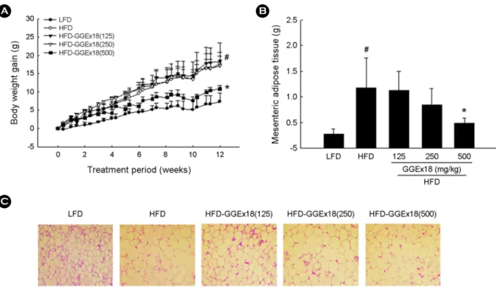
Body weight gain and mesenteric adipose tissue mass were significantly increased by 128% and 337%, respectively, in HFD-fed obese mice compared with lean LFD-fed mice (P<0.05; Figs. 1A and B). In contrast, HFD-fed obese mice that were supplemented with GGEx18 (500 mg/kg/day) [HFD-GGEx18(500)] exhibited significant reductions in
body weight gain and mesenteric adipose tissue weight of 41% and 57%, respectively, compared with untreated HFD- fed mice (P<0.05). Similarly, histological analysis showed that the mesenteric adipocyte size was markedly lower in HFD-GGEx18(500)-treated HFD-fed mice than in untreated HFD-fed mice (Fig. 1C). Treatment with GGEx18(500) sig- nificantly decreased the size of adipocytes by 22% in HFD- fed mice (data not shown).
Infiltrated mast cells and collagen accumulation in the pancreas
The effect of GGEx18 on pancreatic inflammation, in- vestigated by examining mast cells in pancreas sections stained with toluidine blue, was characterized by a higher number of mast cells in the pancreas of HFD-fed mice than LFD-fed mice (Fig. 2A). However, HFD-fed mice treated with GGEx18 had fewer infiltrating inflammatory cells than untreated HFD-fed mice, with the highest effect observed at the dose of 500 mg/kg/day. The inhibitory effects of GGEx- 18 on pancreatic inflammation were assessed using pancreas sections stained with an antibody against CD68. The CD68- positive cells were almost completely eliminated in HFD- GGEx18 (500) mice compared with untreated HFD-fed mice (Fig. 2B).
Collagen levels were assessed using pancreas sections stained with Masson's trichrome. HFD-fed mice had much higher collagen levels than LFD mice (Fig. 3A). However, GGEx18 reduced pancreatic collagen levels in HFD-fed Table 1. Sequences of primers used for quantitative real-time PCR assays
Genes Gene bank Primer sequences
CD68 NM_001291058.1 Forward : 5'- AACAGGACCTACATCAGAGC -3'
Reverse : 5'- CTGTAGCCTTAGAGAGAGCA -3'
Collagen α1 NM_007742.4 Forward : 5'- GCCCGAACCCCAAGGAAAAGAAGC -3'
Reverse : 5'-CTGGGAGGCTCGGTGGACATTAG -3'
TGFβ NM_011577.2 Forward : 5'- ACCGCAACAACGCCATCTAT -3'
Reverse : 5'- GTAACGCCAGGAATTGTTGC -3'
TNFα NM_001278601.1 Forward : 5'- GGCAGGTCTACTTTGGAGTCATTGC -3'
Reverse : 5'- ACATTCGAGGCTCCAGTGAATTCGG-3'
β-actin NM_007393.5 Forward : 5'- TGGAATCCTGTGGCATCCATGAAA -3'
Reverse : 5'- TAAAACGCAGCTCAGTAACAGTCCG -3'
Fig. 1. Effects of GGEx18 on body weight gain, mesenteric adipose tissue mass, and adipocyte size in HFD-induced obese mice.
Adult male C57BL/6J mice were fed a LFD, a HFD, or a HFD supplemented with 125, 250, or 500 mg/kg/day GGEx18 for 12 weeks. (A) Modulation of body weight gain by GGEx18. Body weights at the end of the treatment period are significantly different between the LFD and HFD groups (#P<0.05) and between the HFD and HFD-GGEx18(500) groups (*P<0.05). (B) Regulation of mesenteric adipose tissue mass by GGEx18. (C) Representative hematoxylin and eosin-stained mesenteric adipose tissue sections (original magnification, ×100). All values are expressed as the mean ± SD (n = 8 per group). #P<0.05 vs. LFD, *P<0.05 vs. HFD.
Fig. 2. Effects of GGEx18 on pancreatic inflammation in HFD-induced obese mice. Adult male C57BL/6J mice (n = 6 per group) were fed a LFD, a HFD, or a HFD supplemented with 125, 250, or 500 mg/kg/day GGEx18 for 12 weeks. (A) Representative toluidine blue-stained pancreas sections (original magnification, ×400). (B) Representative CD68-immunostained pancreas sections (original magnification, ×200).
Fig. 3. Effects of GGEx18 on pancreatic fibrosis in HFD-induced obese mice. Adult male C57BL/6J mice (n = 6 per group) were fed a LFD, a HFD, or a HFD supplemented with 125, 250, or 500 mg/kg/day GGEx18 for 12 weeks. (A) Representative Masson's trichrome- stained pancreas sections (original magnification, ×100). (B) Representative α-SMA-immunostained pancreas sections (original magnifi- cation, ×200).
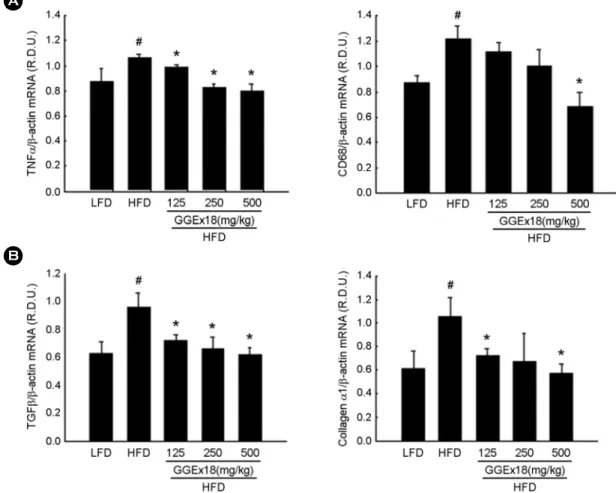
Fig. 4. Effects of GGEx18 on the expression of genes involved in (A) inflammation and (B) fibrosis in the pancreas of HFD-induced obese mice. Adult male C57BL/6J mice were fed a LFD, a HFD, or a HFD supplemented with 125, 250, or 500 mg/kg/day GGEx18 for 12 weeks. All values are expressed as the mean ± SD (n = 6 per group). #P<0.05 vs. LFD, *P<0.05 vs. HFD.
mice compared with untreated HFD-fed mice. Similarly, α- SMA-positive cells were also decreased in GGEx18-treated HFD-fed mice compared with untreated HFD-fed mice (Fig. 3B).
Pancreatic expression of genes involved in inflammation and fibrosis
Consistent with the histological and immunohistochemical data, HFD-fed mice exhibited significant increases in the expression of inflammatory genes, such as tumor necrosis factor α (TNFα) and CD68 (P<0.05; Fig. 4A). However, GGEx18 treatment of HFD-fed obese mice decreased the higher mRNA levels of TNFα and CD68 present in the pan- creas of HFD-fed obese mice (P<0.05). Similarly, mRNA levels of fibrogenic genes, such as transforming growth factor β (TGFβ) and collagen α1 mRNA, were more highly expressed in the pancreas of HFD-fed mice than in LFD-fed mice (P<0.05; Fig. 4B). In this case, GGEx18 treatment reduced the elevated mRNA levels of collagen α1 and TGFβ in HFD-fed mice compared with untreated HFD-fed mice (P<0.05).
DISCUSSION
Our previous study suggested that GGEx18 curtails im- paired glucose metabolism and pancreatic fat accumulation by regulating pancreatic genes that govern lipid metabolism and by improving insulin sensitivity (submitted). Based on report that showed an association between visceral obesity and ectopic fat accumulation in different organs (e.g., liver, pancreas, skeletal muscle, and heart) (Smith, 2015) and that pancreatic steatosis may cause pancreatic inflammation and subsequent fibrosis (Acharya et al., 2014; Noel et al., 2016;
Duan et al., 2017), we hypothesized that GGEx18 can re- gulate pancreatic fibroinflammation in HFD-fed obese mice.
Our results show that GGEx18 treatment for 12 weeks decreased visceral fat mass and inhibited visceral adipocyte hypertrophy in HFD-fed mice. Of three doses of GGEx18, GGEx18 at the dose of 500 mg/kg/day significantly reduced body weight gain and mesenteric adipose tissue weight in HFD-fed mice. Similar to reductions in visceral fat mass, histological analysis of mesenteric adipose tissue sections
showed that HFD-GGEx18(500) significantly decreased the average size of adipocytes. Considering large adipocytes secrete circulating free fatty acids, TNFα, and leptin, which are involved in the pathogenesis of insulin resistance (Okuno et al., 1998), it is likely that GGEx18 may reduce visceral adipose tissue mass and improve insulin sensitivity by in- hibiting visceral obesity due to adipocyte hypertrophy.
HFD intake can lead to alterations in the exocrine and endocrine pancreas (Zhang et al., 2008; Cao et al., 2014), indicating that an HFD intake is associated with chronic in- juries of pancreas. In this study, HFD feeding increased in- flammatory mast cells and induced immunostains with CD68, which is a protein that is highly expressed in macrophages (Holness and Simmons, 1993). Our present results are sup- ported by reports that intake of a high-fat, high-sucrose diet not only results in weight gain but also increases steatosis and inflammatory cell infiltrates in the pancreas of mice and rats (Zhang et al., 2008; Fernandes-Santos et al., 2009; Cao et al., 2014). However, GGEx18 treatment inhibits the infil- tration of inflammatory cells into the pancreas. It was re- ported that consumption of high-fat foods induces increases in mRNA levels and immunostains of inflammatory TNFα in the pancreas of rats (Cao et al., 2014). Our results also show increases in mRNA levels of inflammatory cytokines, such as TNFα and CD68, in the pancreas of HFD-fed mice.
Consistent with the effects of GGEx18 on the histology of pancreatic inflammation, GGEx18 treatment also reduced the pancreatic expression of these genes in HFD-fed mice.
These results suggest that GGEx18 ameliorates pancreatic inflammation by inhibiting inflammatory cell infiltration and inflammatory cytokine expression in obese mice.
A chronic HFD was shown to induce pancreatic collagen synthesis in rats (Zhang et al., 2008). Using a Masson's tri- chrome method for collagen staining, we observed that col- lagen accumulation was increased in the pancreas of HFD- fed mice compared with that in LFD-fed mice. Alpha-SMA- positive cells in the pancreas were also higher in HFD-fed mice than LFD-fed mice. In contrast, GGEx18 decreased collagen levels and α-SMA-positive cells, and these effects of GGEx18 were most effective at a concentration of 500 mg /kg. These results indicate that GGEx18 attenuates pancre- atic fibrosis by inhibiting collagen accumulation and fibro-
genic factor expression in obese mice.
In conclusion, these studies demonstrate that GGEx18 decreases the expression of genes involved in inflammation and fibrosis in the pancreas of obese mice. These changes led to decreased inflammatory cell infiltration and collagen accumulation, thereby reducing pancreatic inflammation and fibrosis.
ACKNOWLEDGEMENT
This work supported by the National Research Foundation of Korea (NRF) Grant funded by the Korea Government (MEST) (2015R1A1A3A04001016).
CONFLICT OF INTEREST
The authors report no conflicts of interest.
REFERENCES
Acharya C, Navina S, Singh VP. Role of pancreatic fat in the out- comes of pancreatitis. Pancreatology. 2014. 14: 403-408.
Cao Y, Bao S, Yang W, Zhang J, Li L, Shan Z, Teng W. Epigallo- catechin gallate prevents inflammation by reducing macro- phage infiltration and inhibiting tumor necrosis factor-α signaling in the pancreas of rats on a high-fat diet. Nutrition Research. 2014. 34: 1066-1074.
Chen KK. A comparative study of synthetic and natural ephedrines.
The Journal of Pharmacology and Experimental Therapeutics.
1928. 33: 237-258.
Duan LF, Xu XF, Zhu LJ, Liu F, Zhang XQ, Wu N, Fan JW, Xin JQ, Zhang H. Dachaihu decoction ameliorates pancreatic fibrosis by inhibiting macrophage infiltration in chronic pan- creatitis. World Journal of Gastroenterology. 2017. 23: 7242 -7252.
Fernandes-Santos C, Evangelista Carneiro R, de Souza Mendonca L, Barbosa Aguila M, Mandarim-de-Lacerda CA. Rosiglita- zone aggravates nonalcoholic Fatty pancreatic disease in C57BL/6 mice fed high-fat and high-sucrose diet. Pancreas.
2009. 38: e80-e86.
Greenway FL, De Jonge L, Blanchard D, Frisard M, Smith SR.
Effect of a dietary herbal supplement containing caffeine and ephedra on weight, metabolic rate, and body composition.
Obesity Research. 2004. 12: 1152-1157.
Holness CL, Simmons DL. Molecular cloning of CD68, a human
macrophage marker related to lysosomal glycoproteins. Blood.
1993.81: 1607-1613.
Lee H, Lim J, Park Y, Jang J, Yoon S, Ahn J, Nam MH, Shin SS, Yoon M. Gyeongshingangjeehwan 18, a polyherbal compos- ition, deters glucose intolerance and nonalcoholic fatty pan- creatic disease in C57BL/6J mice on a high-fat diet. Journal of Ethnopharmacology. (Submitted).
Lim J, Lee H, Ahn J, Kim J, Jang J, Park Y, Jeong B, Yang H, Shin SS, Yoon M. The polyherbal drug GGEx18 from Laminaria japonica, Rheum palmatum, and Ephedra sinica inhibits hepatic steatosis and fibroinflammtion in high-fat diet-induced obese mice. Journal of Ethnopharmacology. 2018. 225: 31-41.
Mathur A, Marine M, Lu D, Swartz-Basile DA, Saxena R, Zyromski NJ, Pitt HA. Nonalcoholic fatty pancreas disease. HPB (Oxford). 2007. 9: 312-318.
Noel P, Patel K, Durgampudi C, Trivedi RN, de Oliveira C, Crowell MD, Pannala R, Lee K, Brand R, Chennat J, Slivka A, Papachristou GI, Khalid A, Whitcomb DC, DeLany JP, Cline RA, Acharya C, Jaligama D, Murad FM, Yadav D, Navina S, Singh VP. Peripancreatic fat necrosis worsens acute pancreatitis independent of pancreatic necrosis via unsaturated fatty acids increased in human pancreatic necrosis collections.
Gut. 2016. 65: 100-111.
Okuno A, Tamemoto H, Tobe K, Ueki K, Mori Y, Iwamoto K, Umesono K, Akanuma Y, Fujiwara T, Horikoshi H, Yazaki Y, Kadowaki T. Troglitazone increases the number of small adi- pocytes without the change of white adipose tissue mass in obese Zucker rats. The Journal of Clinical Investigation. 1998.
101: 1354-1361.
Smith U. Abdominal obesity: a marker of ectopic fat accumulation.
The Journal of Clinical Investigation. 2015. 125: 1790-1792.
van Herpen NA, Schrauwen-Hinderling VB. Lipid accumulation in non-adipose tissue and lipotoxicity. Physiol Behav. 2008.
94: 231-241.
Wang J, Zhao Y, Xiao X, Li H, Zhao H, Zhang P, Jin C. Assess- ment of the renal protection and hepatotoxicity of rhubarb extract in rats. Journal of Ethnopharmacology. 2009. 124: 18 -25.
Yang M, Li X, Zeng X, Ou Z, Xue M, Gao D, Liu S, Li X, Yang S.
Rheum palmatum L. attenuates high fat diet-induced hepato- steatosis by activating AMP-activated protein kinase. The American Journal of Chinese Medicine. 2016. 44: 551-564.
Yen HJ. Laminaria japonica Aresch. In: Chinese Pharmaceutics of Maine Lakes and Marshes. Xueyuan Press, Beijing, China.
1996.
Yoon,KH, Lee HY, Jung YS, Seo BI, Park KY, Yoon M, Shin SS.
Modulation of obesity by Gyeongshingangjeehwan 18 in ob/
ob mice. Koran Journal of Hoerbology. 2010. 25: 1-9.
Zhang X, Cui Y, Fang L, Li F. Chronic high-fat diets induce oxide injuries and fibrogenesis of pancreatic cells in rats. Pancreas.
2008. 37: e31-e38.
https://doi.org/10.15616/BSL.2018.24.4.341
Cite this article as: Jang J, Park Y, Yoon M. Effects of Gyeongshingangjeehwan 18 on Pancreatic Fibroinflam- mation in High-Fat Diet-Fed Obese C57BL/6J Mice.
Biomedical Science Letters. 2018. 24: 341-348.