INTRODUCTION
Somatic symptom disorder (SSD) is characterized by one or more somatic symptoms that cause distress or significant- ly disrupt daily life. It is accompanied by excessive thoughts, feelings, and behavior.1,2) Many individuals experience so- matic symptoms, imposing tremendous healthcare burden.2) However, the mechanisms underlying SSD are not clear.2) Re- searchers have tended to investigate SSD from a biological
perspective, especially in terms of autonomic nervous system (ANS) dysfunction.3,4) Autonomic physiological arousal has been proposed to increase the likelihood of bodily signals misperceptions.3,4)
Measurement of heart rate variability (HRV) is a non-inva- sive tool used to assess ANS function.5) The relevance of HRV to many psychiatric disorders has been shown, including schizo- phrenia,6) bipolar disorder,7) depressive disorder,7,8) anxiety disorder.9) Before the Diagnostic and Statistical Manual of Mental Disorders (5th edition ; DSM-5) was published, sev-
Received: June 8, 2020 / Revised: June 18, 2020 / Accepted: June 19, 2020
Corresponding author: Hye Youn Park, Department of Psychiatry, Seoul National University Bundang Hospital, 82 Gumi-ro 173beon-gil, Seongnam 13620, Korea
Reduced Heart Rate Variability in Somatic Symptom Disorder:
Associations with Alexithymia
Jae Hoon Lee, M.D.,1 Ye Eun Jang, M.S.,2 Hye Youn Park, M.D.2
1Department of Neuropsychiatry, Seoul National University Hospital, Seoul, Korea
2Department of Psychiatry, Seoul National University Bundang Hospital, Seongnam, Korea
ABSTRACT
Objectives:We investigated heart rate variability (HRV) patterns in patients with somatic symptom disorder (SSD) and the relationships of these patterns with alexithymia.
Methods:In total, 42 patients with SSD and 33 healthy controls were enrolled in this study. Demographic, psy- chological, and HRV data were assessed at baseline, and 24 patients with SSD were reassessed after 6 months of treatment. The psychological data included somatic symptoms and levels of depression, anxiety, and alexithymia as indicated by the somatic symptom subscale of the Symptom Checklist 90-Revision (SCL-12), Beck Depres- sion Inventory-II (BDI-II), Beck Anxiety Inventory (BAI), and the Toronto Alexithymia Scale 20 (TAS-20), re- spectively.
Results:Patients with SSD had a lower standard deviation of normal-to-normal R-R intervals (SDNN) and lower proportions of adjacent R-R intervals greater than 50 milliseconds (pNN50) compared with controls.
These HRV parameters were negatively correlated with alexithymia severity. After treatment, patients exhibited significantly decreased levels of somatic symptoms and reduced anxiety and depression, but there were no signifi- cant differences in the HRV parameters. In patients with alexithymia, a high baseline SDNN and pNN50 were as- sociated with a decrease in somatic symptoms.
Conclusions:Patients with SSD have different HRV patterns, and several HRV parameters are associated with alexithymia severity. These findings suggest that ANS regulation is involved in the pathophysiology of SSD, me- diated by alexithymia. Furthermore, these results suggest that certain HRV parameters may be associated with clin- ical outcomes of SSD.
KEY WORDS:Autonomic nervous system ㆍHeart rate variability ㆍSomatic symptom disorder.
https://doi.org/10.22722/KJPM.2020.28.1.89 ISSN 1225-6471
eral studies reported an association between somatic symp- toms and HRV.10-12) A meta-analysis showed that patients with functional somatic disorders, such as chronic fatigue syn- drome, fibromyalgia, and irritable bowel syndrome, had less high-frequency HRV (HF-HRV) activity than controls, but the reliability of this study was limited by heterogeneity.10) Af- ter the concept of SSD was introduced in DSM-5, a few stud- ies examined the pattern of HRV in patients with SSD.13-15) One study showed that patients with SSD had decreased total-pow- er HRV (TP-HRV) and low-frequency HRV (LF-HRV).13) An- other study found low LF-HRV, low HF-HRV, a low standard deviation of normal to normal R-R intervals (SDNN) and low proportions of adjacent R-R intervals differing by >50 milli- seconds (pNN50) in patients with SSD.14) Studies consistent- ly revealed that patients with SSD had some HRV parameters that were lower than those of healthy controls, indicative of low parasympathetic activity.13,14) However, how these phe- nomena contribute to the pathophysiology and psychological symptoms of SSD is still not fully understood. Also, little is known about the clinical implications of HRV in patients with SSD.
The possible psychological candidates affecting SSD phe- nomena are anxiety and depression. Anxiety and depression are representative psychological symptoms that affect HRV.8,9) A recent study suggested a possible relationship between de- pression and HRV in patients with SSD, but evidence is lack- ing.13) The other possible candidate is alexithymia. Alexithymia has long been considered one of the distinguishing character- istics of patients with somatic symptoms.16) Conceptually, alex- ithymia is characterized by the inability to identify and de- scribe emotions experienced by the self or others, i.e., it is a problem of perception.16) Several studies suggested a role for alexithymia in the restricted emotional processing and so- matosensory amplification seen in SSD.17,18) A recent study reported a negative association between alexithymia and HRV, especially in HF-HRV.19) However, very little is current- ly known about the correlation between alexithymia and HRV in patients with SSD. Furthermore, published studies are lim- ited to cross-sectional analyses. Therefore, the impact of HRV on the clinical course of SSD remains unclear.
This paper explored HRV patterns in SSD patients, and the associations of HRV with psychological variables, including alexithymia. We also examined the relationship between HRV and clinical outcome in patients with SSD. We formulated three main hypotheses : 1) patients with SSD have different patterns of HRV from those of healthy controls ; 2) these HRV patterns are associated with certain psychological variables,
and 3) clinical outcomes in patients with SSD are correlated with the baseline HRV.
METHODS
1. Participants
Participants were recruited from Seoul National University Bundang Hospital (SNUBH), South Korea from May 2017 to July 2019. Informed consent was obtained from all partici- pants. Patients with SSD were recruited from the psychiatric outpatient clinics of SNUBH, and were evaluated in clinical interviews by board-certified psychiatrists based on the Struc- tured Clinical Interview for DSM-5 Disorders-Clinician Ver- sion (SCID-5-CV).1,20) Healthy controls were recruited through advertisements placed in the hospital and community, and age- and sex- matched with patients with SSD.
The exclusion criteria were as follows : 1) comorbidity in- volving major psychiatric disorders other than SSD, such as any type of psychotic disorder, major depressive disorder, or alcohol use disorder ; 2) cognitive impairment or diagnosis of any type of dementia ; 3) a medical condition affecting HRV, such as cardiovascular disease, except for hypertension and dyslipidemia ; and 4) the use of psychiatric drugs including an- tipsychotics, antidepressants, benzodiazepines in the 3 months before enrollment.
2. Study design and setting
This prospective cohort study recruited 42 patients with SSD and 33 healthy controls. Demographics, psychological data, and HRV were assessed at baseline in all participants.
Of the patients with SSD, 24 were re-assessed after treatment for 6 months. Fig. 1 shows the participants flowchart. During 6 months, patients took usual psychiatric treatments includ- ing medications and supportive psychotherapy. All protocols were approved by the Institutional Review Board of SNUBH (IRB No. B1710426302)
3. Assessment of psychological variables
To evaluate somatic symptoms, the Korean version of the Symptom Checklist 90-Revision (SCL-90-R) was used.21). Specifically, the somatic symptom subscale of the SCL-90-R (SCL-12), which comprises 12 questions to evaluate somatic symptoms, was used to measure somatic symptoms.22) The SCL-12 contains 12 items rated on 5-point Likert scales, and the total score ranges from 0 to 48. Higher scores indicate more somatic symptoms.22) The Cronbach’s α (internal consistency) for this measure was 0.90.
The Korean versions of the Beck Depression Inventory
(BDI-II) and Beck Anxiety Inventory (BAI) were used to as- sess the degree of depression and anxiety, respectively.23,24) Both of these instruments include 21 items rated on 4-point Likert scales, where higher scores indicate greater levels of depression and anxiety. The Cronbach’s α was 0.922 for the BDI-II and 0.964 for the BAI.
The level of alexithymia was measured with the Toronto Alexithymia Scale-20 (TAS-20), which consists of 20 items rated on a 5-point Likert scale.25) The total score ranges from 20 to 100,25) where ≤51 corresponds to no alexithymia, 52-60 to borderline alexithymia, and ≥61 to alexithymia.26) The Cron- bach’s α was 0.835.
4. Assessment of heart rate variability
(ECGs) during 8-minute periods using a Synamps 2 Ampli- fier (Compumedics, Melbourne, Australia). The participants were not allowed to drink caffeine/alcohol, or to smoke, for at least 8 hours before the examination. The time- and frequen- cy-domain parameters of HRV were analyzed with Telescan and Complexity software (ver. 2.0 ; Laxtha, Daejeon, Korea).
In the time domain, the SDNN, root mean square of suc- cessive differences between consecutive R-R intervals (RMS- SD), and pNN50 were calculated. For the frequency domain, parametric autoregression was performed on the power spec- tral domain analysis. The powers of LF (0.04-0.15 Hz) and HF (0.15-0.4 Hz) bands, in absolute and normalized (divided by LF+HF) units, were determined along with the LF/HF ra- tio. HF-HRV reflects parasympathetic activity and LF-HRV
Screening for eligibility (n=113)
• Patients with Somatic symptom disorder (SSD) (n=75)
• Healthy controls (n=38)
Excluded due to exclusion criteria (n=34)
• SSD patients (n=30)
- Other illness except SSD (n=6) - Psychoactive drugs use (n=12) - Other mental disease (n=5) - Having a one symptom (n=7)
• Healthy controls (n=4)
- Having any physical symptoms (n=4)
Excluded in each group (n=4)
• SSD patients (n=3)
- Declined to participate (n=1) - Technical error (n=2)
• Healthy controls (n=1) - High depression level (n=1)
Drop-out
• SSD patients (n=18)
- Personal time restrictions (n=6) - Lack of necessity (n=7)
- Declined to follow-up session (n=5) Informed consent (n=79)
• SSD patients (n=45)
• Healthy controls (n=34)
Final data for analysis (n=75)
• SSD patients (n=42)
• Healthy controls (n=33)
6 months’ follow-up
• SSD patients (n=24)
- Alexithymia at baseline (n=15) - Non-alexithymia at baseline (n=9) Fig. 1. Study Flowchart.
both sympathetic and parasympathetic activity.27) The LF/HF ratio and the normalized LF are indices of sympathovagal bal- ance.27) On the other hands, in the time domain, SDNN reflects overall HRV, but in short-term recording, parasympathetic- mediated respiratory sinus arrhythmia (RSA) was the main contributor to SDNN.28) Both the RMSSD and the pNN50 are sensitive measures of parasympathetic vagal tone.28)
5. Statistical analysis
Table 1 summarizes the participants’ demographics and clinical characteristics ; Pearson’s χ2 test was used to analyze categorical variables and the independent t-test was applied for continuous variables. HRV parameters in the frequency domain were log-transformed due to skewness. The analyses included baseline comparisons between the SSD and control groups, and longitudinal analyses of the 24 patients with SSD.
At baseline, a different pattern of HRV parameters between patients with SSD and controls was revealed by analysis of
covariance (ANCOVA) after adjusting for age, sex, and body mass index (BMI).29,30) Then, correlation analyses, including partial correlations and multiple linear regression with the en- ter method, were performed to determine associations between psychological variables and HRV parameters in all partici- pants. In multiple linear regression analysis, HRV parameters were used as dependent variables and several psychological variables were used as independent variables to observe col- lective effects of psychological variables on HRV parameters.
For the longitudinal analyses in patients with SSD, the paired t-test was used to compare baseline and follow-up data.
The associations between baseline HRV and the pre–post change in somatic symptoms were examined using partial correlation analyses. All partial correlation analyses were ad- justed for age, sex, and BMI.29,30) Subgroup analyses revealed a correlation between baseline HRV and the pre–post change in somatic symptoms in the alexithymia group. The alexi- thymia group comprised those with a baseline TAS-20 score
Table 1. Demographic and baseline characteristics of the participants (n=75)
SSD (n=42) Controls (n=33)
Test p
Mean (SD) or n (%)
Sociodemographic factors χ2/t†
Age (y) 48.14 (10.89) 46.12 (9.05) -0.859 0.39
Sex
Male 13 (31.0) 11 (33.3) 0.048 0.83
Female 29 (69.0) 22 (66.7)
BMI, kg/m2 22.12 (3.32) 23.38 (2.78) 1.755 0.083
Psychological variables
SCL-12 13.33 (7.76) 2.85 (3.20) -7.940 <0.001***
BDI-II 16.07 (8.14) 3.33 (3.80) -8.977 <0.001***
BAI 20.36 (13.60) 2.61 (2.94) -8.220 <0.001***
TAS-20 53.52 (10.89) 39.15 (8.60) -6.210 <0.001***
HRV parameters
Time domain F test‡
SDNN, ms 38.25 (12.50) 47.12 (12.81) 6.166 0.015*
RMSSD, ms 30.65 (15.02) 36.03 (17.02) 0.737 0.39
pNN50, % 32.94 (14.65) 41.62 (13.99) 4.142 0.046*
Frequency domain
TP-HRV, ln (ms2) 6.96 (0.67) 7.18 (0.92) 0.410 0.52
LF-HRV, ln (ms2) 5.52 (0.91) 5.68 (0.97) 0.158 0.69
Normalized LF 56.37 (20.21) 54.14 (16.23) 0.074 0.79
HF-HRV, ln (ms2) 5.22 (0.92) 5.50 (1.11) 0.490 0.49
LF/HF-HRV 2.07 (2.07) 1.51 (1.04) 1.109 0.30
* : p<0.05, ** : p<0.01, *** : p<0.001, †: χ2 for categorical variables and t-test for continuous variables, ‡: the analysis of cova- riance (ANCOVA) after adjusting for age, sex and BMI. SSD : somatic symptom disorder, BMI : body mass index, SCL-12 : the so- matic symptom subscale of Symptom Checklist-90-Revision, BDI-II : Beck Depression Inventory-II, BAI : Beck Anxiety Inventory, TAS- 20 : Toronto Alexithymia Scale-20, SDNN : standard deviation of normal to normal R-R intervals, RMSSD : root mean square of successive differences between consecutive R-R intervals, pNN50 : proportions of adjacent R-R intervals differing by>50 milliseconds, TP-HRV : to- tal power of heart rate variability, LF-HRV : low-frequency power of heart rate variability, Normalized LF : normalized low-frequency power of heart rate variability, HF-HRV : high-frequency power of heart rate variability, LF/HF HRV : ratio of low-frequency power to high frequency power of heart rate variability
>51, thus including cases with borderline alexithymia and alexithymia. Partial correlation was performed in the subgroup analyses.
p-values<0.05 were considered significant. All analyses were conducted using IBM SPSS Statistics software (ver.
19.0 ; IBM Corp., Armonk, NY, USA).
RESULTS
Descriptive statistics are presented in Table 1. The mean (SD) age of the patients with SSD was 48.14 (10.89) years and 31% were male. The mean (SD) age of the healthy controls was 46.12 (10.89) years, and 33% were male. The distributions of age, sex, and BMI did not differ significantly between the groups. Table 1 also shows the psychological variables, on all of which the SSD group had higher mean scores than the con- trols. All psychological variables and HRV parameters, at baseline, follow-up, and in subgroup analyses, were normally distributed.
1. Baseline comparison of patients with SSD and controls
At baseline, patients with SSD had lower SDNN and pNN50 values than the controls (SDNN : F=6.166, p=0.015 ; pNN50 : F=4.142, p=0.046) (Table 1). There were no group differenc- es in the frequency domain. In the correlation analyses of all participants, there were negative associations between the se- verity of alexithymia as indicated by the TAS-20 score and several HRV parameters (SDNN : r=-0.352, p=0.002 ; RMS- SD : r=-0.282, p=0.016 ; pNN50 : r=-0.335, p=0.004) (Sup- plementary Table 1 in the online-only Data Supplement). Ta-
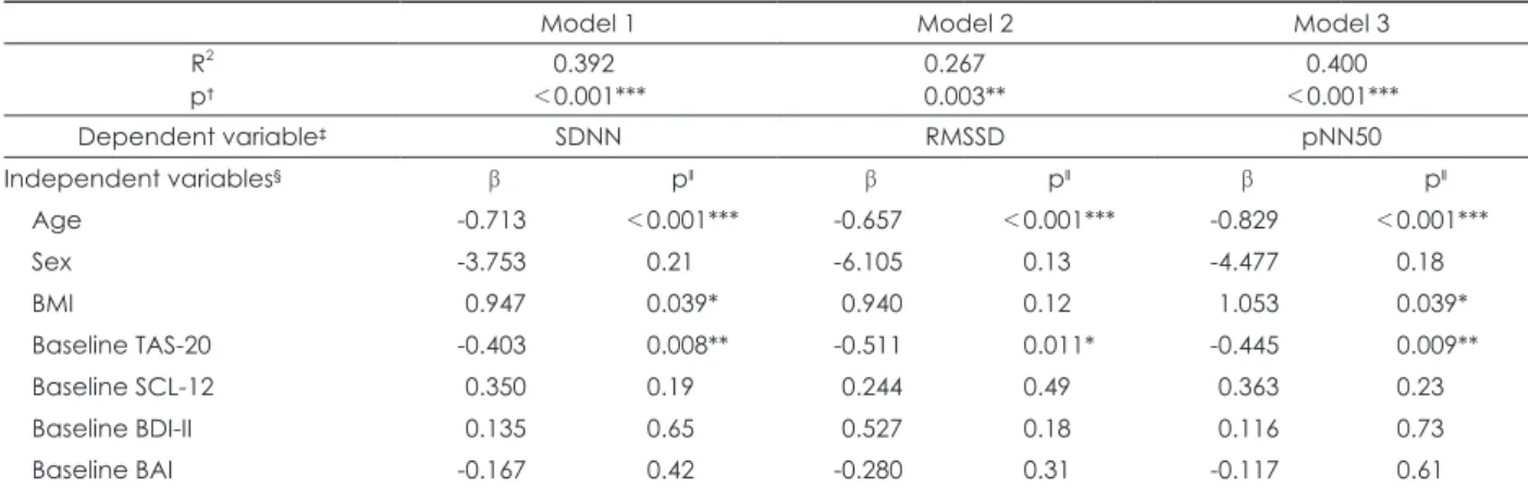
ble 2 also shows how the TAS-20 score predicted SDNN, RMSSD, and the pNN50 ; a high TAS-20 score was related to low SDNN, RMSSD, and pNN50 values in the patients with SSD.
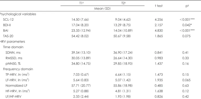
2. Longitudinal analyses of patients with SSD
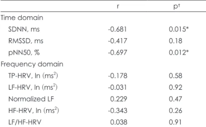
Table 3 shows the longitudinal changes in psychological and HRV variables in the SSD patients. After 6 months of treat- ment, the patients had significantly reduced levels of somatic symptoms, anxiety, and depression, but there were no signifi- cant differences in the severity of alexithymia or HRV param- eters (Table 3). In correlation analyses, there was no associa- tion between the clinical outcome and baseline HRV in patients with SSD. In subgroup analyses of the alexithymia group, how- ever, the change in somatic symptoms was correlated with the baseline SDNN and pNN50 values. A low ΔSCL-12 value (Δ=
Post–Pre), which corresponds to a good clinical outcome, was associated with high baseline SDNN and pNN50 values after adjusting for age, sex, and BMI (Table 4).
DISCUSSION
This is the first prospective cohort study to investigate the association between HRV and SSD. The results were as fol- lows. First, patients with SSD have different patterns of HRV compared with healthy controls, especially in terms of SDNN and pNN50. In a correlation analysis, the severity of alexi- thymia was negatively associated with SDNN, pNN50, and RMSSD values. Second, in the longitudinal analyses, a change in somatic symptoms was negatively correlated with the base- line SDNN and pNN50 values in patients with alexithymia.
Table 2. Multiple linear regression models predicting HRV parameters in all participants (n=75)
Model 1 Model 2 Model 3
R2
p† 0.392
<0.001*** 0.267
0.003** 0.400
<0.001***
Dependent variable‡ SDNN RMSSD pNN50
Independent variables§ β pǁ β pǁ β pǁ
Age -0.713 <0.001*** -0.657 <0.001*** -0.829 <0.001***
Sex -3.753 0.21 -6.105 0.13 -4.477 0.18
BMI 0.947 0.039* 0.940 0.12 1.053 0.039*
Baseline TAS-20 -0.403 0.008** -0.511 0.011* -0.445 0.009**
Baseline SCL-12 0.350 0.19 0.244 0.49 0.363 0.23
Baseline BDI-II 0.135 0.65 0.527 0.18 0.116 0.73
Baseline BAI -0.167 0.42 -0.280 0.31 -0.117 0.61
* : p<0.05, ** : p<0.01, *** : p<0.001, †: p values are computed for modeling using multiple linear regression analysis (enter meth- od), ‡: Dependent variables are SDNN, RMSSD and pNN50, § : Independent variables are age, sex, BMI, TAS-20, SCL-12, BDI-II, BAI,
∥ : p values are computed for each independent variables using multiple linear regression analysis (enter method). SDNN : stan- dard deviation of normal to normal R-R intervals, RMSSD : root mean square of successive differences between consecutive R-R in- tervals, pNN50 : proportions of adjacent R-R intervals differing by >50 milliseconds, TAS-20 : Toronto Alexithymia Scale-20, SCL-12 :
In the baseline analyses, patients with SSD had lower SDNN and pNN50 values. SDNN provides a measure of the total HRV, reflecting periodic and random sources of variability.28) Sympathetic nervous system (SNS) and parasympathetic ner- vous system (PNS) activity both affect SDNN ; however, in short-term recordings obtained at rest, parasympathetic-me- diated RSA was the major contributor to SDNN.28) In com- parison, pNN50 provides an index of PNS activity, and is more reliable than the SDNN in short-term HRV recordings.28) Be- cause both SDNN and pNN50 reflect PNS activity, our study shows lower PNS activity of patients with SSD compared with healthy controls. Many studies have also revealed that patients with somatic symptoms have lower parasympathetic activi-
ty.10,11) A recent study found task-related HRV abnormalities
in patients with SSD, especially in SDNN and pNN50.14) Our study supports these findings regarding the possibility of au- tonomic imbalance in SSD, reflected especially in PNS activ- ity. However, similar HRV patterns were observed in patients with depressive and anxiety disorder. In patients with depres- sive disorder, RMSSD, SDNN, and HF-HRV were reduced and the LF/HF ratio increased.7,8) Also, the severity of depres- sion was inversely associated with HRV in patients with de- pressive disorder, especially with the RMSSD, SDNN, and
HF-HRV values.7,8) Likewise, patients with anxiety disorder exhibited lower RMSSD, SDNN, pNN50, and HF-HRV val- ues than did healthy controls.9) Thus low SDNN and pNN50 values may reflect the pathophysiological mechanisms but are not specific markers of SSD.
Next question is which psychological variables correlate these phenomena. We found negative associations between the severity of alexithymia and HRV parameters, especially SDNN, pNN50, and RMSSD. There were no effects of the level of anxiety and depression on HRV parameters. Actual- ly, a previous study found that alexithymia was negatively correlated with HF-HRV, one of the index of PNS activity, in healthy young males.19) Also, alexithymia is known to be re- lated to hypothalamus-pituitary-adrenal (HPA) axis func- tion.31) For these reasons, alexithymia is thought to reflect ANS dysfunction, including HPA axis compromise.19,31) Our find- ings also suggest that alexithymia is associated with PNS ac- tivity. Although alexithymia is a core feature of patients with SSD, several studies have suggested that correlations between alexithymia and somatic symptoms were modulated by de- pression ; alexithymia was not a significant factor when de- pression was taken into account.38,39) For these reasons, alexi- thymia is considered to share pathophysiology with depression
Table 3. Longitudinal change of psychological variables and HRV variables after 6 months in SSD (n=24)
T1† T2†
t test p‡
Mean (SD) Psychological variables
SCL-12 14.50 (7.66) 9.04 (4.62) 4.256 <0.001***
BDI-II 17.04 (8.20) 13.29 (8.75) 2.157 0.042*
BAI 23.33 (12.94) 14.04 (10.89) 4.830 <0.001***
TAS-20 54.42 (8.02) 50.67 (9.58) 1.865 0.075
HRV parameters Time domain
SDNN, ms 39.54 (13.10) 36.90 (17.24) 0.841 0.41
RMSSD, ms 30.05 (13.89) 26.64 (14.30) 0.983 0.33
pNN50, % 34.80 (14.75) 29.85 (18.93) 1.437 0.16
Frequency domain
TP-HRV, ln (ms2) 7.03 (0.67) 6.64 (1.15) 1.473 0.15
LF-HRV, ln (ms2) 5.64 (0.83) 5.07 (1.42) 1.935 0.065
Normalized LF 57.71 (20.77) 55.86 (18.98) 0.485 0.63
HF-HRV, ln (ms2) 5.27 (0.88) 4.81 (1.31) 1.638 0.12
LF/HF-HRV 2.33 (2.44) 1.93 (1.98) 0.826 0.42
* : p<0.05, ** : p<0.01, *** : p<0.001, †: T1 indicates baseline and T2 indicates 6 months later, ‡: p values are calculated with paired t test. SSD : somatic symptom disorder, BMI : body mass index, SCL-12 : the somatic symptom subscales of Symptom Check- list-90-Revision, BDI-II : Beck Depression Inventory-II, BAI : Beck Anxiety Inventory, TAS-20 : Toronto Alexithymia Scale-20, SDNN : stan- dard deviation of normal to normal R-R intervals, RMSSD : root mean square of successive differences between consecutive R-R intervals, pNN50 : proportions of adjacent R-R intervals differing by >50 milliseconds, TP-HRV : total power of heart rate variability, LF- HRV : low-frequency power of heart rate variability, Normalized LF : normalized low-frequency power of heart rate variability, HF- HRV : high-frequency power of heart rate variability, LF/HF HRV : ratio of low-frequency power to high frequency power of heart rate variability
in patients with SSD.38,39) However, in the present study, alexi- thymia modulated HRV in patients with SSD after adjusting for depression. These finding raise the possibility that an in- dependent neuropathology links SSD and alexithymia. Sev- eral neuroanatomical studies also supported the relevance of alexithymia to HRV in SSD patients. Previous studies found that the extent of alexithymia was associated with specific brain structures, including the cingulate cortex, insula, pre- frontal cortex, and amygdala.32) In functional brain imaging studies, the activities in the anterior cingulate cortex, insula, and prefrontal cortex were related to alexithymia evident dur- ing socioemotional processing.32) Other studies suggested that vagal-mediated HRV parameters, such as the RMSSD and HF- HRV were correlated with the activities of the cingulate and prefrontal cortices.33) Furthermore, patients with somatic symp- toms exhibited significant changes in functional interconnec- tivity among the prefrontal and cingulate cortices, insula, and the supramarginal and occipital gyri.34,35) These neuroana- tomical and functional studies and our current study suggest that patients with SSD have low vagal-medicated HRV, medi- ated by the level of alexithymia.
Our longitudinal analyses examined the relationship be- tween HRV and clinical outcome. We did not observe corre- lations between the baseline HRV and clinical outcome in patients with SSD. However, subgroup analyses suggested
that high baseline SDNN and pNN50 values were associated with good clinical outcomes in patients with alexithymia. As previously mentioned, high SDNN and pNN50 values reflect PNS function. ANS dysfunction is thought to be involved in the pathophysiology of SSD.3) Therefore, the clinical outcome of SSD would be expected to be correlated with the extent of ANS dysfunction. Our results raise the possibility that clini- cal outcomes of SSD are related to ANS dysfunction, espe- cially vagal-mediated HRV. Although our longitudinal analy- ses show statistically significant finding, the sample size was too small to reveal any robust correlation between HRV and clinical outcomes in SSD. Accordingly, our longitudinal re- sults should be viewed as preliminary ; further studies are needed. Nevertheless, our study suggests the possibility that baseline HRV is associated with clinical outcomes of SSD.
Alexithymia is a core feature of patients with SSD. A pre- vious study found that alexithymia was associated with the number of somatic symptoms and various coping skills in patients with somatic symptoms.36) Also, a high prevalence of alexithymia has been reported in patients with somatic symp- toms, and the severity of such symptoms and treatment out- comes were thought to be related to alexithymia.36,37) In the present study, patients with SSD have lower HRV parameter, mediated alexithymia. Moreover, some HRV parameters were associated with clinical outcomes in SSD patients with high levels of alexithymia. Our findings suggest that alexithymia was related to certain HRV parameters that reflect PNS func- tions, suggesting that SSD patients with high level alexithymia exhibit more PNS dysfunction that their clinical outcomes are associated with the extent of such dysfunctions. Different neu- rophysiological pathways may be active in SSD patients with and without alexithymia. Thus, clinical assessment of alexi- thymia in patients with SSD may allow us to better understand the nature and clinical course of the illness.
Our other notable findings were as follows. First, we found no interval change in either alexithymia or HRV. Interesting- ly, in patients with autism spectrum disorder characterized by high alexithymia, low vagal-mediated HRV was associat- ed with changes in the prefrontal and limbic brain regions.40,41) It is possible that alexithymia and vagal-mediated HRV both predispose to SSD. Second, although both pNN50 and HF- HRV are indices of PNS activity, they yielded inconsistent results.28) One possible explanation is that pNN50 is affected by hormonal changes or systemic conditions rather than by PNS activity alone, whereas HF-HRV is modulated by RSA.28) Third, previous studies revealed that depression and anxiety levels were correlated clinically with HRV.7-9) Although, in
Table 4. Partial correlation analyses between baseline HRV pa- rameters and post-pre changes (Δ) of SCL-12 in SSD with alexi- thymia (n=15)
r p†
Time domain
SDNN, ms -0.681 0.015*
RMSSD, ms -0.417 0.18
pNN50, % -0.697 0.012*
Frequency domain
TP-HRV, ln (ms2) -0.178 0.58
LF-HRV, ln (ms2) -0.031 0.92
Normalized LF 0.229 0.47
HF-HRV, ln (ms2) -0.343 0.26
LF/HF-HRV 0.038 0.91
* : p<0.05, ** : p<0.01, *** : p<0.001, †: p values are calcu- lated with partial correlation analysis after adjusting for age, sex and BMI. SSD : somatic symptom disorder, SCL-12 : the somatic symptom subscale of Symptom Checklist-90-Revision, SDNN : standard deviation of normal to normal R-R intervals, RMSSD : root mean square of successive differences between consecu- tive R-R intervals, pNN50 : proportions of adjacent R-R intervals differing by >50 milliseconds, TP-HRV : total power of heart rate variability, LF-HRV : low-frequency power of heart rate variabil- ity, Normalized LF : normalized low-frequency power of heart rate variability, HF-HRV : high-frequency power of heart rate vari- ability, LF/HF HRV : ratio of low-frequency power to high frequen- cy power of heart rate variability
the present study, patients with SSD exhibited more depres- sion and anxiety than controls, these differences were not re- lated to HRV. As mentioned above, we excluded patients clini- cally diagnosed with depressive and anxiety disorder. Thus, our findings suggest that the HRV pattern of SSD patients is caused by a mechanism that is different from patients with de- pressive and anxiety disorders. Finally, our findings consis- tently showed that HRV was inversely related to age. Similar results were observed in a previous study.30) This is especial- ly the case for middle age.
This study has several limitations. First, the results of lon- gitudinal analyses had limited power due to the small sample size. Further studies with more patients are needed. Second, HRV is modulated by multiple physiological factors, includ- ing age, sex, BMI, food intake, circadian rhythm, sleep, exer- cise.29) Although we adjusted for some confounders, many oth- ers may remain. In addition, as HRV is affected by ethnicity, it is difficult to generalize our results to other racial and eth- nic groups.42) Third, all patients received individualized treat- ment, which obviously influenced HRV ; unfortunately, we did not control the process of treatment. Also, the sample size was too small to allow for treatment-stratified analyses. Addition- ally, HRV may be influenced by psychotropic agents includ- ing antipsychotics and antidepressant, so the results should be cautiously interpreted.43) Forth, we performed HRV analyses without adjusting for multiple comparisons because of the high correlations among HRV parameters. Although this may cre- ate a risk of type I error, over-adjustment would increase the risk of type II error. Finally, alexithymia has both cognitive and affective dimensions involving different brain regions and functions.32) The TAS-20 evaluates the cognitive dimension of alexithymia. Additional study of the affective dimension is required.
In conclusion, patients with SSD showed HRV patterns un- like those of controls, and these were correlated with the lev- el of alexithymia. Thus, ANS dysfunction is involved in the pathophysiology of SSD. A longitudinal study with a larger sample size is needed to confirm the association between clini- cal outcomes and HRV. Also, biofeedback targeting HRV would increase our understanding of the role played by HRV in SSD patients.
Supplementary Materials
The online-only Data Supplement is available with this article at https://doi.org/10.22722/KJPM.2020.28.1.89.
Acknowledgments
The study was supported a National Research Foundation of Ko- rea (NRF) grant funded by the Korean government (no. NRF-2017
R1C1B2012733).
Conflicts of Interest
The authors have no financial conflicts of interest.
REFERENCES
(1) Association AP. Diagnostic and statistical manual of mental disorders (DSM-5®): American Psychiatric Pub;2013.
(2) Rief W, Martin A. How to use the new DSM-5 somatic symp- tom disorder diagnosis in research and practice: a critical eval- uation and a proposal for modifications. Annual Review of Clinical Psychology 2014;10.
(3) Rief W, Barsky AJ. Psychobiological perspectives on somato- form disorders. Psychoneuroendocrinology 2005;30:996-1002.
(4) Laederach-Hofmann K, Rüddel H, Mussgay L. Pathologi- cal baroreceptor sensitivity in patients suffering from somati- zation disorders: do they correlate with symptoms? Biological Psychology 2008;79:243-249.
(5) Zygmunt A, Stanczyk J. Methods of evaluation of autonomic nervous system function. Arch Med Sci 2010;6:11-18.
(6) Clamor A, Lincoln TM, Thayer JF, Koenig J. Resting vagal activity in schizophrenia: meta-analysis of heart rate variabil- ity as a potential endophenotype. The British Journal of Psy- chiatry 2016;208:9-16.
(7) Bassett D. A literature review of heart rate variability in de- pressive and bipolar disorders. Aust N Z J Psychiatry 2016;50:
511-519.
(8) Kidwell M, Ellenbroek BA. Heart and soul: heart rate vari- ability and major depression. Behav Pharmacol 2018;29:152- 164.
(9) Chalmers JA, Quintana DS, Abbott MJ, Kemp AH. Anxiety Disorders are Associated with Reduced Heart Rate Variabili- ty. A Meta-Analysis. Front Psychiatry 2014;5:80.
(10) Tak LM, Riese H, de Bock GH, Manoharan A, Kok IC, Rosmalen JG. As good as it gets? A meta-analysis and system- atic review of methodological quality of heart rate variability studies in functional somatic disorders. Biol Psychol 2009;82:
101-110.
(11) Tak LM, Janssens KA, Dietrich A, Slaets JP, Rosmalen JG.
Age-specific associations between cardiac vagal activity and functional somatic symptoms: a population-based study. Psy- chother Psychosom 2010;79:179-187.
(12) Liu Q, Wang EM, Yan XJ, Chen SL. Autonomic functioning in irritable bowel syndrome measured by heart rate variability:
a meta-analysis. J Dig Dis 2013;14:638-646.
(13) Huang WL, Liao SC, Yang CC, Kuo TB, Chen TT, Chen IM, Gau SS. Measures of Heart Rate Variability in Individu- als With Somatic Symptom Disorder. Psychosom Med 2017;
79:34-42.
(14) Lee D, Kim SJ, Cheon J, Hwang EH, Jung YC, Kang JI.
Characteristics of Autonomic Activity and Reactivity During Rest and Emotional Processing and Their Clinical Correlations in Somatic Symptom Disorder. Psychosom Med 2018;80:690-
697.
(15) Huang WL, Liao SC, Tu YK, Yang CCH, Kuo TBJ, Gau SS. Autonomic reactivity during reading of a somatic distress script in patients with somatic symptom disorder. J Psychosom Res 2019;123:109729.
(16) Duddu V, Isaac MK, Chaturvedi SK. Alexithymia in somato- form and depressive disorders. J Psychosom Res 2003;54:435- 438.
(17) Ozturk A, Kilic A, Deveci E, Kirpinar I. Investigation of fa- cial emotion recognition, alexithymia, and levels of anxiety and depression in patients with somatic symptoms and related dis- orders. Neuropsychiatr Dis Treat 2016;12:1047-1053.
(18) Nakao M, Takeuchi T. Alexithymia and Somatosensory Am- plification Link Perceived Psychosocial Stress and Somatic Symptoms in Outpatients with Psychosomatic Illness. J Clin Med 2018;7:112.
(19) Lischke A, Pahnke R, Mau-Moeller A, Behrens M, Grabe HJ, Freyberger HJ, Hamm AO, Weippert M. Inter-individ- ual Differences in Heart Rate Variability Are Associated with Inter-individual Differences in Empathy and Alexithymia.
Front Psychol 2018;9:229.
(20) First MB, Williams JB, Karg RS, Spitzer RL. User’s Guide for the SCID-5-CV: Structured Clinical Interview for DSM-5 Disorders, Clinician Version: American Psychiatric Associa- tion;2016.
(21) Kim K, Kim J, Won H. Korean manual of symptom check- list-90-revision. Seoul: Jung Ang Juk Sung Publisher 1984:8-10.
(22) Zijlema WL, Stolk RP, Löwe B, Rief W, White PD, Rosmalen JG. How to assess common somatic symptoms in large-scale studies: a systematic review of questionnaires. Journal of Psy- chosomatic Research 2013;74:459-468.
(23) Beck AT, Steer RA, Ball R, Ranieri WF. Comparison of Beck Depression Inventories-IA and-II in psychiatric outpatients.
Journal of Personality Assessment 1996;67:588-597.
(24) Beck AT, Epstein N, Brown G, Steer RA. An inventory for measuring clinical anxiety: psychometric properties. J Con- sult Clin Psychol 1988;56:893-897.
(25) Bagby RM, Parker JD, Taylor GJ. The twenty-item Toronto Alexithymia Scale--I. Item selection and cross-validation of the factor structure. J Psychosom Res 1994;38:23-32.
(26) Taylor GJ, Bagby RM, Parker JD. Disorders of affect regula- tion: Alexithymia in medical and psychiatric illness: Cam- bridge University Press;1999.
(27) Reyes del Paso GA, Langewitz W, Mulder LJ, Van Roon A, Duschek S. The utility of low frequency heart rate variability as an index of sympathetic cardiac tone: a review with empha- sis on a reanalysis of previous studies. Psychophysiology 2013;
50:477-487.
(28) Shaffer F, Ginsberg JP. An Overview of Heart Rate Variabil- ity Metrics and Norms. Front Public Health 2017;5:258.
(29) Laborde S, Mosley E, Thayer JF. Heart Rate Variability and
Cardiac Vagal Tone in Psychophysiological Research-Recom- mendations for Experiment Planning, Data Analysis, and Data Reporting. Front Psychol 2017;8:213.
(30) Kim GM, Woo JM. Determinants for heart rate variability in a normal Korean population. J Korean Med Sci 2011;26:1293- 1298.
(31) Hua J, Le Scanff C, Larue J, José F, Martin J-C, Devillers L, Filaire E. Global stress response during a social stress test:
impact of alexithymia and its subfactors. Psychoneuroendo- crinology 2014;50:53-61.
(32) Goerlich KS. The Multifaceted Nature of Alexithymia-A Neu- roscientific Perspective. Frontiers in Psychology 2018;9.
(33) Winkelmann T, Thayer JF, Pohlack S, Nees F, Grimm O, Flor H. Structural brain correlates of heart rate variability in a healthy young adult population. Brain Struct Funct 2017;222:
1061-1068.
(34) Kim SM, Hong JS, Min KJ, Han DH. Brain Functional Con- nectivity in Patients With Somatic Symptom Disorder. Psycho- som Med 2019;81:313-318.
(35) Li Q, Xiao Y, Li Y, Li L, Lu N, Xu Z, Mou X, Mao S, Wang W, Yuan Y. Altered regional brain function in the treatment- naive patients with somatic symptom disorder: a resting-state fMRI study. Brain Behav 2016;6:e00521.
(36) Tominaga T, Choi H, Nagoshi Y, Wada Y, Fukui K. Rela- tionship between alexithymia and coping strategies in patients with somatoform disorder. Neuropsychiatric Disease and Treat- ment 2014;10:55.
(37) Kano M, Endo Y, Fukudo S. Association between alexi- thymia and functional gastrointestinal disorders. Frontiers in Psychology 2018;9:599.
(38) Probst T, Sattel H, Gündel H, Henningsen P, Kruse J, Schneider G, Lahmann C. Moderating effects of Alexithymia on associations between the therapeutic alliance and the out- come of brief psychodynamic-interpersonal psychotherapy for Multisomatoform disorder. Frontiers in Psychiatry 2017;8:261.
(39) Saariaho AS, Saariaho TH, Mattila AK, Karukivi MR, Jou- kamaa MI. Alexithymia and depression in a chronic pain pa- tient sample. Gen Hosp Psychiatry 2013;35:239-245.
(40) Thapa R, Alvares GA, Zaidi TA, Thomas EE, Hickie IB, Park SH, Guastella AJ. Reduced heart rate variability in adults with autism spectrum disorder. Autism Research;2019.
(41) Bernhardt BC, Valk SL, Silani G, Bird G, Frith U, Singer T.
Selective disruption of sociocognitive structural brain net- works in autism and alexithymia. Cereb Cortex 2014;24:3258- 3267.
(42) Ernst G. Hidden Signals-The History and Methods of Heart Rate Variability. Front Public Health 2017;5:265.
(43) Alvares GA, Quintana DS, Hickie IB, Guastella AJ. Auto- nomic nervous system dysfunction in psychiatric disorders and the impact of psychotropic medications: a systematic re- view and meta-analysis. J Psychiatry Neurosci 2016;41:89-104.