∙ Received: February 29, 2012. Accepted: April 6, 2012.
∙ Corresponding author: Woo Young Jung
Department of Nuclear Medicine, Seoul Asan Medical Center, 88, Olympic-ro 43-gil, Songpa-gu, Seoul 138-736, Korea Tel: +82-2-3010-5421, Fax: +82-2-3010-4588 E-mail: [email protected]
Original Article 전신 PET/CT 검사에서 환자의 팔 위치에 따른 CT 유효선량의 비교 연구
서울아산병원 핵의학과
성지혜 ․ 박순기 ․ 김정선 ․ 박승용 ․ 정우영
A Comparative Study on the CT Effective Dose by the Position of Patient’s Arm
Ji Hye Seong, Soon Ki Park, Jung Sun Kim Seung Yong Park and Woo Young Jung Department of Nuclear Medicine, Asan Medical Center, Seoul, Korea
Purpose: In the whole body PET/CT scan, it is natural to lift the patient’s arm for its quality improvement. However, when the lesion is located in head and neck, the arms should be located lower. This study was designed to compare the CT effective dose for each arm position applying Automatic Exposure Control (AEC). Materials and Methods:
45 patients who had 18F-FDG whole body PET/CT scan were studied with Biograph Truepoint 40 (SIEMENS, GERMANY), Biograph Sensation 16 (SIEMENS, GERMANY), Discovery STe 8 (GE healthcare, USA). The CT effective dose of 15 patients for each equipment was measured and comparatively analyzed in both arm-lifted position and lower-arm position. ImPACT v1.0 program was used as the method of measurement for CT effective dose. For the statistics analysis, Paired t-test which paired with SPSS 18.0 statistic program was applied. Results: In the case of arm-lifted, it was measured as 6.33±0.93 mSv for Biograph Sensation 16, 8.01±1.34 mSv for Biograph Truepoint 40, and 9.69±2.32 mSv for Discovery STe 8. When arms are located lower position, it was measure as 6.97±0.76 mSv, 8.95±1.85 mSv, 13.07±2.87 mSv for each. CT effective dose according to the arm position was 9.2% for Biograph Truepoint 40, 10.5% for Biograph Sensation 16, and 25.9% for Discovery Ste 8. The statistics analysis showed the meaningful difference (p<0.05). Conclusion: For the whole body PET/CT case, CT effective dose applying AEC was decreased the radiation exposure of the patients when the arm was lifted for 15.2% of average value. The patient who has no lesion in head and neck would decrease the artifact occurrence in objective part and lower the CT effective dose. Also, for the patient who had lesion in head and neck, the artifact in objective part can be lower by putting the arms down, the fact that CT effective dose increases should be concerned in its whole body PET/CT scan. (Korean J Nucl Med Technol 2012;16(1):44-49)
Key Words : PET/CT, Effective dose, AEC (Automatic Exposure Control)
INTRODUCTION
Recently, generalization with improvement on the radiology field and the crisis of Fukushima nuclear disaster, the public interest in radiation exposure is increasing. PET, admitted its excellent usefulness in the diagnosis of cancer, has been
developed in PET/CT system which fused functional imaging and anatomic imaging nowadays.1,2) These developments lead to increase of exposure dose compared with PET by introducing multi slice CT to PET/CT. MDCT has its strong point for shorter evaluation time and higher resolution, however, the resolution improvement needs thinner and lower fragments and it is related with the increasement of dose.3,4) According to these facts, Automatic Exposure Control (AEC) system was designed with MDCT for better quality of imaging and lower radiation dose in PET/CT scan.5,6) AEC system, investigating the best tube current by
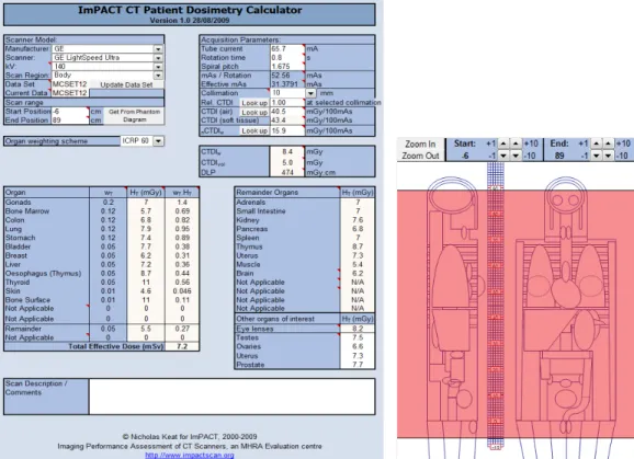
Fig. 1. Enter the parameters which are vendor, scanner type, kV, mA, rotation cycle, pitch, collimation and scan range you can get the effective dose(ImPACT v1.0).
thickness of the subject for lower radiation dose of patients, is used in most of PET/CT scans. It is reported as lowering 20~40% approximate radiation dose measured in patient body.7) Also, in Asan Medical Center, the whole body PET/CT scan is applied in lifting the patient’s arms above their head for decreasing the occurrence of beam hardening artifact. If the objective part includes Head and neck, melanoma, region of arm so that the patient cannot raise their arms, the arms are located in lower position or FOV.8,9) In this study, CT effective dose applying AEC system according to the position of patient’s arm in whole body PET/CT scan was compared.
MATERIALS AND METHOD
1. Materials
From December 2008 to July 2011, 45 persons (Male : 25, Female : 20, Average age : 51±14) who had whole body
18F-FDG PET/CT scan in the same equipment were studied for its difference between the arm position in nuclear
medicine department of Asan Medical Center.
2. Method and Dose Analysis
The imaging of all patients covered from skull vertex to upper thigh. Biograph Truepoint 40 (SIEMENS, Bio 40, Germany), Biograph Sensation 16 (SIEMENS, Bio 16, Germany) and Discovery STe 8 (GE Healthcare, Milwaukee, Mi, USA, DSTe 8) were used. The 45 patients’ data for raising their arm and lowering the arm were collected in each Bio 40, Bio 16, DSTe 8. Based on the data, 50% of CT dose were reduced and computed its average value. The measurement of CT effective dose were made by ImPACT v1.0 (NRPB, UK) program which based on Monte Carlo data of NRPB (Fig. 1).
3. CT Exposure Condition
PET/CT scan condition for whole body is tube voltage 120 kVp, 100 ref. mAs (CARE Dose4D), rotation time 0.5 sec, collimation 24×2.2, pitch 0.8, Slice thickness 5.0 mm, FOV 500 mm, Matrix 168×168 for Bio 40. For Bio 16, tube
Table 1. Scan parameter at Bio 40, Bio 16 and DSTe 8.
Parameter Bio 40 Bio 16 DSTe 8
AEC CARE Dose4D CARE Dose4D AutomA 3D
kVp 120 120 140
Rotation time (sec) 0.5 0.5 0.8
Collimation (mm) 24 x 1.2 16 x 0.75 10
Spiral pitch 0.8 1.25 1.675
Slice thickness (mm) 5.0 5.0 3.75
FOV (axial) 500 500 500
Matrix 168 × 168 128 × 128 128 × 128
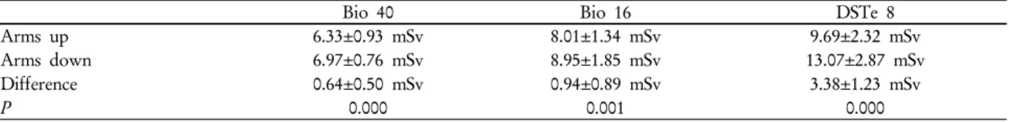
Table. 2. Estimated effective dose according to patient’s arm position of BIO40, BIO16 and DSTe8 for CT in PET/CT scans using ImPACT v1.0. (Unit: mSv)
Bio 40 Bio 16 DSTe 8
Arms up 6.33±0.93 mSv 8.01±1.34 mSv 9.69±2.32 mSv
Arms down 6.97±0.76 mSv 8.95±1.85 mSv 13.07±2.87 mSv
Difference 0.64±0.50 mSv 0.94±0.89 mSv 3.38±1.23 mSv
P 0.000 0.001 0.000
*P < 0.05.
Fig. 2. The difference of CT effective dose according to patient’s arm position.
voltage 120 kVp, 100 ref. mAs (CARE Dose4D), rotation time 0.5 sec, collimation 16×0.75, pitch 1.25, Slice thickness 5.0 mm, FOV 500 mm, Matrix 128×128 was setted up. In DSTe 8, the patients were scanned under the setting of tube voltage 140 kVp, 25~210 mA (AutomA 3D), rotation time 0.8 sec, collimation 10, pitch 1.675, Slice thickness 3.75 mm, FOV 500 mm, Matrix 128×128. For application of AEC, Care Dose4D (SIEMENS) and AutomA 3D (GE) were used (Table 1).
4. Statistic Analysis
The result of experiment were represented in average±
standard deviation and Pearson correlation coefficient was calculated in the correlation analysis and dispersion between the weight of patients and CT effective dose. For comparing CT effective dose in both arm-lifted and lower arm position in each equipment, bivariate coefficient of correlation was detected in correlation analysis. The statistical significance was setted for the result of p value in data was calculated below 0.05, and the statistic analysis for measured CT effective dose were analyzed by SPSS 18.0.
5. Understanding of terms AEC (Automatic Exposure Control)
The imaging setting method that arbitrarily controls the best tube current for setting the useful imaging in diagnosis by the thickness of the subject and reduces its radiation exposure as much as it can. In other words, it automatically controls the tube current by shape of the patient (thickness change), size, difference of absorbed radiation dose so that it can keep the quality of image and reduce the dose.10)
RESULTS
Table 2 presented the average±standard deviation for CT effective dose of both arm-lifted scan and lower arm scan in each equipment. In the result of reducing CT effective dose by using the dose measure program in each Bio 40, Bio 16, and DSTe 8. When the patient lifted arms, 6.33±0.93 mSv for Bio 40, 8.01±1.34 mSv for Bio 16, and 69±2.32 mSv for DSTe 8 were calculated and 6.97±0.76 mSv, 8.95±1.85 mSv, 13.07 ±2.87
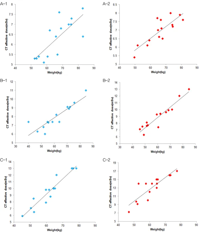
Fig. 3. Correlation between CT effective dose and weight shows positive correlation. (A) Siemens Bio 40, (B) Siemens Bio 16 and (C) GE DSTe 8, when the patient’s arms over their head(1) and arms down(2).
mSv for each in lower arm position. According to the position of patients’ arms, CT effective dose were distinguished as 9.2% in Bio 40, 10.5% for Bio 16, and 25.9% for DSTe 8 (Fig. 2). The reduced CT effective dose from each equipment has statistically significant
(p<0.05) according to position of patients’ arms.
Besides, Fig. 3 presented the correlation analysis of CT effective dose by the weight in both arm-lifted and lower arm scan. The coefficient of correlations (r²) for each case were
C-1 C-2
A-1 A-2
B-1 B-2
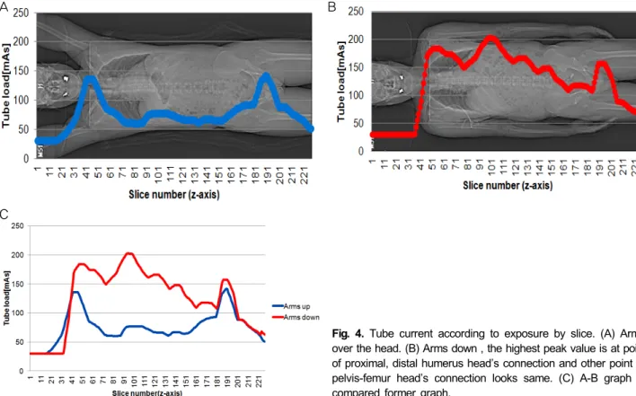
Fig. 4. Tube current according to exposure by slice. (A) Arms over the head. (B) Arms down , the highest peak value is at point of proximal, distal humerus head’s connection and other point of pelvis-femur head’s connection looks same. (C) A-B graph is compared former graph.
calculated as 0.777, 0.835 for Bio 40, 0.889, 0.946 for Bio 16, 0.949, 0.868 for DSTe 8. All instruments had strong positive correlation.
DISCUSSION
CT effective dose for the position of patients’ arm applied AEC presented the largest difference in DSTe 8 for 25.9%
while Bio 40 and Bio 16 calculated as 9.2% and 10.5%. The difference between these effective dose could be considered as the difference of AEC, mAs value which used in Siemens equipment and reference noise value which used in GE. In the table, AutomA 3D of GE is constructed in each longitudinal part and angular part (Table 3).11-13) Care Dose4D of Siemens is tube current automatic exposure control system which fused two parts mentioned above.14-16). One time
18F-FDG FDG injection for whole body PET/CT scan presented 11mSv of exposure dose for adult. It can be said that patient exposed to 17~24 mSv radiation for one time PET/CT scan, though the value can be different in patient weight. Figure 4 is dispersion graph of tube current investigated by Slice. In the graph of lower arm case, it shows
the high peak for the connection of Proximal and Distal humerus head. For the connection of Pelvis and femur head, which does not include arms, the value presented similarity.
Figure 4-C explained the comparison between two cases of Figure 4-A,B, and it presented big difference when the arms are included in FOV. CT effective dose according to the inclusion of arms for FOV was calculated in 0.64 mSv for Bio 40, 0.94 mSv for Bio 16, and 3.38 mSv for DSTe 8 and it is considered as the effective dose of distal forearm.
However, though the study focused to the same patient who had both arm-lifted scan and lower arm scan, the scan did not operate in same time and the change of weight was occurred during the period. Also, a few error could be possibly occurred despite of same setting for FOV. Due to its limitation of the reference mAs standard in AEC system used in this case, which defined an adult for 70~80 kg and an pediatric for 20~30 kg which based on the average of Western, it would be better if the study about mAs for Korean body type is progressed.17)
CONCLUSION
A B
C
Through this study, 15.2% of patient effective dose reduction was ascertained when arms were lifted during the scan. Also the arm-lifted position during whole body PET/CT let beam hardening artifact occurrence to decrease and increase the quality of image.
On the other hand, when target region is located in head and neck, arms should be putting down for its scan for lower artifact occurrence. For melanoma or region of arm, the scan should be made including the arms in FOV. PET scan in the past used to be operated in lower arm position for the comfort of patient due to its long scanning time, however, whole body PET/CT scan nowadays shortens its scanning time for 30 minutes. Therefore lifting arms of patient would be helpful for lower radiation exposure dose. For this, setting up the patient posture under the consideration of objective of scan, characteristic of instrument, and condition of patient.
요 약
최근 영상 의학 분야의 발전으로 인한 보편화와 일본의 후 쿠시마 원전사태가 일어나면서 방사선 노출에 대한 대중들의
관심이 높아지게 되었다. 이러한 방사선 피폭에 대한 대중들
의 관심으로 현재 거의 대부분의 PET/CT검사에서 사용되고 있는 AEC (Automatic Exposure Control) 시스템은 환자의 피 폭선량 감소를 위해 피사체의 두께에 따라 최적의 관전류를 조사한다. 또한, 서울아산병원에서는 전신 PET/CT 검사 시 선속 경화 인공물(Beam hardening artifact)의 발생을 줄이기 위해 환자의 팔을 머리 위로 올리고 검사를 시행하고 있다. 본 연구에서는 전신 PET/CT검사 시 환자의 팔 위치에 따라 AEC 시스템을 적용한 CT 유효선량의 차이를 비교하고자 한다. 2008 년 12월부터 2011년 7월까지 서울아산병원 핵의학과를 내원하 여 동일 장비에서 팔을 올렸을 경우와 팔을 내렸을 경우의 전신 18F‐FDG PET/CT 검사를 시행한 환자 45명을 연구의 대상으로 하였고, 실험 장비는 Biograph Truepoint 40 (Bio 40), Biograph Sensation 16 (Bio 16), Discovery STe 8 (DSTe 8)을 사용하였 다. 각 장비당 15명의 동일 환자를 대상으로, 팔을 올리고 검 사하였을 경우와 팔을 내리고 검사하였을 경우의 CT 유효선
량을 산출하여 비교 분석하였다. CT 유효선량의 측정 방법은
ImPACT v1.0 프로그램을 사용하였다. 이 연구를 통해 팔을 올 리고 검사한 경우에 팔을 내리고 검사한 경우보다 총 15.2%의 환자 유효선량이 감소된 것을 확인할 수 있었다. 전신 PET/CT 검사 시 환자의 팔을 올린 자세는 선속 경화 인공물 발생을 줄
이고, 영상의 질을 향상시킬 수 있을 것이라 생각된다.
REFERENCES
1. Townsend DW, Beyer T: A combined PET/CT scanner: the path to true image fusion. The British J Radiol 75:S24-S30 (2002).
2. Beyer T, Townsend DW, Burn T, et al: A combined PET/CT scanner for clinical oncology. J Nucl Med 41:1369-1379 (2000).
3. Dawson P: Patient dose in multi-slice CT: Why is it increasing and dose it matter? Br J Radiol 77:S10-S13 (2004).
4. Yates SJ, Pike LC, Goldstone KE: Effect of multi-slice scanners on patient dose from routine CT examinations in East Anglia.
Br J Radiol 77:472-478 (2004).
5. Iball GR, Brettle DS, Moore AC: Assessment of tube current modulation in pelvic CT. Br J Radiol 79:62-70 (2006).
6. Rizzo S, Kalra M, Schmidt B, et al: Comparison of angular and combined automatic tube current modulation techniques with constant tube current CT of the abdomen and pelvis. Am J Roentgenol 186:673-679 (2006).
6. McCollough CH. Automatic exposure control in CT: are we done yet? Radiology 2005;237;755-6.
7. Thomas Beyer, MD. Acquisition Protocol Considerations for Combined PET/CT Imaging. The Journal of Nuclear Medicine 2004; 45:29S-30S.
7. Dominique Delbeke. Procedure Guideline for Tumor Imaging with 18F-FDG PET/CT 1.0*, 2006. Available at:
http://www.snm.org/guidelines. Approved February 11, 2006 8. McCollough CH, Bruesewitx MR, Kofler JM Jr: CT dose re-
duction and dose management tools: Overview of available options. RadioGraphics 26:503-512 (2006)
9. Kalra MK, Rizzo SM, Novelline RA. Reducing radiation dose in emergency computed tomography with automatic exposure control techniques. Emerg Radiol 2005;11:267-74.
10. Kalra MK, Maher MM, Toth TL, Schmidt B, Westerman BL Morgan HT, et al. Techniques and applications of automatic tube current modulation for CT. Radiology 2004;233:649-57.
11. Kalra MK, Naz N, Rizzo SM, Blake MA. Computed tomog- raphy radiation dose optimization: scanning protocols and clin- ical applications of automatic exposure control. Curr Probl Diagn Radiol 2005;34:171-81.
12. Keat N. CT scanner automatic exposure control systems.
MHRA evaluation report 0516 (February 2005). London:
Medicines and Healthcare Regulatory Agency; 2005.
13. McCollough CH, Bruesewitz MR, Kofler JM Jr. CT dose re- duction and dose management tools: overview of available options. Radiographics 2006;26;503-12.
14. General Electronic. Brochure: LightSpeed 2369740-142 (Swedish text). General Electronic Company 2004;6:7-16.
15. Lewis M, Keat N, Edyvean S: Report 06013: 32 to 64 slice CT scanner comparison report version 14. London, England:
ImPACT, 2006. Available at: http://www.impactscan. org/re- ports/Report06013. Accessed June 16, 2008.