코골이 환자에서 무호흡-저호흡지수, 수면설문지,
구인두부 소견 및 두부 규격 방사선 계측학적 변수들의 평가
계명대학교 의과대학 신경과학교실, 이비인후과학교실*, 가정의학과교실†, 예방의학교실‡, 치과학교실§,
경북대학교 치과대학 구강악안면외과학교실∥
김현아 조용원 이 형 이주화 안병훈* 서영성† 이미영‡ 황상희§ 권대근∥
An Evaluation of Apnea-Hypopnea Index, Sleep Questionnaires, Oropharyngeal Findings and Cephalometric Parameters in Patients with Snoring
Hyun-Ah Kim, M.D., Yong-Won Cho, M.D., Hyung Lee, M.D., Joo-Hwa Lee, Byung-Hoon Ahn, M.D.*, Young-Sung Suh, M.D.
†, Mi-Young Lee, M.D.
‡, Sang-Hee Hwang, D.D.S.
§, Tae-Geon Kwon, D.D.S.
∥Departments of Neurology, Otolaryngology * , Family Medicine
†, Preventive Medicine
‡and Dentistry
§, Keimyung University College of Medicine, Daegu;
Department of Oral & Maxillofacial Surgery, Kyungpook National University College of Dentistry
∥, Daegu, Korea
Background: Although obstructive sleep apnea (OSA) is a common disorder, it often goes undiagnosed due to
limited availability of the polysomnography (PSG) and a lack of interest in this condition. The aim of this study was to investigate the relationship between severities of obstructive sleep apnea, sleep questionnaires, oropharyngeal findings and cephalometric parameters in patients whom snore.Methods: Fifty-seven (46 males) patients presenting snoring or other symptoms of OSA were evaluated
retrospectively at the sleep disorder clinic in Keimyung University, Dongsan Medical Center were included in this study. All patients completed an overnight polysomnography, several sleep questionnaires and oropharyngic and cephalometric examinations. The sleep questionnaires included the Stanford Sleepiness Scale (SSS), Epworth Sleepiness Scale (ESS), Pittsburgh Sleep Quality Index (PSQI), and Beck Depression Inventory (BDI). The oropharyngeal examinations included tonsil grade and modified Mallampati grade.Results: There were altogether 42 (38 male) patients diagnosed with OSA. The mean age of the patients was 42.4
±12.8 years, the body mass index (BMI) was 26.1±2.7 kg/m2 and the apnea-hypopnea index (AHI) was 28.1 3±
0.3. Sex difference (male), BMI, tonsil grade and some parameters of the cephalometric examination had a significant positive correlation with the AHI. However, the AHI correlated poorly with the sleep questionnaires.
Conclusions: Although tonsil grade, modified Mallampati grade and some parameters of the cephalometric
examination can be utilized as a useful method to evaluate OSA, the AHI correlated poorly with self-reported sleep questionnaires. These findings suggest that the severity of sleep apnea should be quantified with both physiologic and subjective measures.J Korean Neurol Assoc 23(2):215-221, 2005
Key Words: Obstructive sleep apnea, Questionnaires, Cephalometry, Polysomnography
Received June 14, 2004 Accepted October 8, 2004
*Yong-Won Cho, M.D.
Department of Neurology, Keimyung University College of Medicine
194 Dongsan-dong, Jung-gu, Daegu, 700-712 Korea Tel: +82-53-250-7831 Fax: +82-53-250-7840 E-mail: [email protected]
서 론
폐쇄성 수면무호흡증은 성인 남성에서 0.3~1%의 유병률을
지닌 비교적 흔한 질환이다.
1-2폐쇄성 수면무호흡증은 수면 동
안 반복적으로 상부 기도에 협착이 일어나고 이로 인하여 공기 의 흐름이 막혀, 혈중 산소포화도가 떨어지고 미세각성이 일어 나서 수면이 분절되는 질환으로, 대개 심한 코골이와 주간 졸음 이 동반된다.
3수면다원검사가 수면무호흡증을 진단하고 심한 정도를 평가하는 표준검사방법으로 알려져
4,5있으나 검사 방법 이 불편하고 비싸며 수면무호흡증에 대한 사회적 관심 부족 등 으로 인해 진단되지 않은 채 지내는 환자가 많다.
6폐쇄성 수면무호흡증은 수면 시 시끄러운 소리를 내는 습관 으로 생각할 뿐 질병으로 인식되지 않았으나, 폐쇄성 수면무호 흡증이 집중력과 지적 능력의 저하, 주간 두통이나 고혈압, 부 정맥, 야간 심허혈, 심근경색, 뇌졸중 등의 뇌-심혈관계 질환과 연관성이 있다는 사실
7-9이 밝혀지면서 최근에 점차 관심이 높 아지고 있다.
수면무호흡증의 정도는 환자가 느끼는 임상 증상과 검사실 검사 소견을 모두 고려해야 하나 현재는 수면다원검사만으로 판단되고 있으며 수면다원검사상의 이상 소견과 임상 증상과의 관련성에 대해 아직 논란이 많다.
10또한 우리 나라에서는 수면 다원검사가 아직까지 널리 보편화되지 않아 한정된 수면클리닉 에서만 검사할 수 있으므로 관련 연구가 미비한 실정이다. 따라 서 심한 코골이를 주소로 내원한 환자에서 수면다원검사를 하 기에 앞서 환자의 자각 주간 증상과 이학적 검사 소견 등을 종 합하여 수면다원검사 결과와 비교 분석해 보는 것은 유용하다 고 생각된다.
따라서, 본 저자들은 코골이를 주소로 내원한 환자에서 자각 주간 증상, 구인두의 이학적 검사 소견 등과 폐쇄성 수면무호흡 증의 수면다원검사 소견과의 관계를 알아보고자 본 연구를 진 행하였다.
대상과 방법
1. 대상
본 연구는 코골이를 포함하여 수면무호흡증이 의심되는 증상 으로 계명대학교 동산의료원 수면장애클리닉을 방문한 환자들 중 수면 설문지, 이비인후과적 진찰, 두부 규격 방사선 계측 (cephalometry) 및 수면다원검사를 모두 실시한 환자를 대상으 로 하였다. 수면다원검사에서 중추성 수면무호흡증이 나온 환 자는 대상에서 제외하였다. 조사 기간은 2002년 9월부터 2004 년 3월까지였다.
2. 방법
1) 수면설문지 문항의 작성
수면설문지는 나이와 성별 외에 체질량 지수(body mass index, kg/m
2), 과거력, 흡연, 알코올 및 카페인 섭취, 수면 유 도제 복용 등의 인구 통계학적 내용이 포함되었다. 수면에 관한 척도로 주간수면과다를 평가하기 위해 Standard Sleepiness Scale (SSS)
11과 Epworth Sleepiness Scale (ESS)
12을 사용하 였고, 수면의 질을 평가하기 위해 Pittsburg Sleep Quality Index (PSQI)
13를 사용하였다. 각 척도에서 SSS가 4점 이상, 혹은 ESS가 10점 이상인 경우는 주간 졸림으로 정의하였고, PSQI가 5점보다 크면 수면의 질이 나쁜 것으로 정의하였다.
11-13또한, 대상 환자들의 우울증 정도를 평가하기 위해 한국판 Beck Depression Inventory (K-BDI)
14를 이용하였다.
수면설문지는 외래 진료를 하기 위해 방문한 날 혹은 수면다 원검사를 하기 위해 내원한 당일 저녁에 작성하였고 설명이 필 요한 경우는 숙달된 검사자가 도와주었다.
2) 이비인후과적 진찰 및 두부 규격 방사선 계측
대상 환자들은 본원 수면클리닉 이비인후과 전문의에게 의뢰 하여 비과적 문제 여부를 평가하고 편도 등급(tonsil grade)과 Modified Mallampati grades (MMP)
15를 측정하였다. MMP는 혀를 내밀지 않은 상태에서 혀와 연구개 등 구조물의 위치를 평 가하여 기도가 어느 정도 좁아져 있는지를 알아보는 방법이다.
15두부 규격 방사선 사진은 핀란드 Planmaca사의 Proline 으 로 치과에서 촬영하였다. 방법은 환자가 직립한 상태에서 귀- 막대(ear-rod)를 외이도에 삽입한 후 방사선 조사 직전 침을 한번 삼키게 하고 인후부가 자연스러운 상태가 되었을 때 치 아가 맞물린 상태(P1)와 하악을 전방으로 내민 상태(P2)로 두 번 촬영하였다. 현상한 후 투사(tracing) 용지에 투사하고 다 시 V-Ceph 2.0 program (CyberMed Co, Korea)에 연결된 Intous 2 digitizer (Wacom Co, USA)로 계측점을 입력하였다.
각각의 연조직 및 경조직 계측 항목은 Fig. 1에 나타나 있다.
15Posterior airway space (PAS)와 Oropharyngeal airway width 2 (Oph2)는 하부 구-인두부 기도의 폭경을 나타내는 것 이고 연구개의 너비는 soft palate width (SPW), 길이는 posterior nasal spine (PNS) to tip of soft palate contour (PNS-P), 위치는 angle from ANS to PNS to P (ANS-PNS-P angle)로 비교해 볼 수 있다. 또한 shortest distance from hyoid bone to mandibular plane (MP-H)은 하악 평면과 설 골의 위치를 나타낸다.
3) 수면다원검사
수면다원검사는 미국 Glass-Telefactor사의 Aurora기종을
non-OSA (N:15, 26.3%)
OSA (N:42, 73.7%) Age (years)
Sex
Male (%)*
Female (%)
Body mass index (kg/m
2) Smoking + (%)
Alcohol + (%) Caffeine + (%)
41.3±15.8 8 (53.3) 7 (46.7) 25.8±2.9 4 (28.6) 7 (50.6) 12 (85.7)
42.7±11.7 38 (90.5) 4 (9.5) 26.2±2.6 18 (46.2) 24 (60.0) 34 (82.9) OSA; obstructive sleep apnea, *p<0.05
Table 1. General characteristics of study subjects
non-OSA OSA
Oropharyngeal findings Modified Mallampati grade Tonsil grade*
2.3±0.8 2.1±0.3
2.6±0.7 2.4±0.5 Cephalometric parameters
PAS PNS-P ANS-PNS-P SPW Oph2*
MP-H
9.2±3.7 42.9±4.1 119.8±22.8 12.1±2.1 8.8±4.5 18.8±5.7
11.9±4.9 44.9±5.6 125.2±5.5 12.8±2.2 11.9±4.4 20.6±6.5 OSA; obstructive sleep apnea, PAS (mm); linear measurement between the base of tongue and posterior pharyngeal wall along the line B-Go, PNS-P (mm); posterior nasal spine (PNS) to tip of soft palate contour, PNS-PNS-P angle (˚); angle from ANS to PNS to P, SPW (mm); soft palate width, Oph2 (mm); oropharyngeal airway width 2, MP-H (mm); shortest distance fom hypid bone to mandibular plane, *p<0.05
Table 3. Oropharyngeal parameters
non-OSA OSA
Sleep questionaries
Stanford Sleepiness Scales Epworth Sleepiness Scales Subjective Sleep quality PSQI
Beck depression index
3.8±1.7 8.1±5.3 1.4±0.9 3.3±2.6 8.3±5.2
3.2±1.5 9.2±4.5 1.6±0.9 3.2±3.2 9.9±7.7 Sleep architectures
Total resting time (minutes) Total sleep time (minutes) Sleep efficiency (%) Sleep stage 1 (%)*
Sleep stage 2 (%)*
Sleep stage 3 (%) Sleep stage 4 (%) REM sleep (%)
439±60.1 346.3±60.4 80.3±11.5 16.6±6.6 52.3±10.5 5.8±3.0 8.5±7.0 16.6±5.2
438.6±55.6 369.5±65.5 85.5±10.8 25.8±10.3 44.4±10.3 5.5±3.7 7.4±6.2 16.4±6.4 OSA; obstructive sleep apnea, PSQI; pittsburgh sleep quality index.
*p<0.05.
Table 2. Clinical variables 이용하여 하룻밤 동안 수면검사실에서 실시하였다. 수면다원검
사의 판독은 현재 표준화되어 사용하고 있는 Rechschaffen과 Kale의 방법으로 하였다.
16무호흡은 10초 이상 공기 흐름이 기 저선에 비해 50% 이상 감소하는 경우로, 저호흡은 10초 이상 공기 흐름이 50%까지 감소하지 않지만 산소 불포화도가 3% 이 상이거나 각성이 동반된 경우로 정의하였다.
17시간 당 무호흡저호흡지수(apnea-hypopnea index; AHI) 가 5회 이상일 때 수면무호흡증으로 정의하였고
18-22이들 그룹 을 시간 당 무호흡 및 저호흡지수가 5회 미만인 비수면무호흡군 과 나누어 자료를 분석하였다. 또한 AHI가 5 이상 15 이하일 때 경한 군으로, 15보다 클 때를 중한 군으로 나누어 비교하였다.
4) 통계 분석
비수면무호흡군과 폐쇄성 수면무호흡군 간 각 변수의 비교에 는 t-test를 이용하였고 변수들과 AHI와의 관련성을 파악하기 위해 상관분석을 사용하였다. 폐쇄성 수면무호흡증에 영향을 미치는 변수를 알아보기 위해 단순변수분석에서 경계치 이상의 통계학적 유의성이 있거나( p <0.1), 혼란변수로 알려진 인자를 독립변수로 하고 무호흡의 유무를 종속변수로 하여 로지스틱 회귀분석을 하였다. 통계 분석에는 SPSS 통계 프로그램 11.0 version (SPSS Inc, Chicago, IL, USA)을 이용하였다.
결 과
모든 검사를 실시한 대상 환자는 57명으로 나이는 14세에서 69세까지 평균 42.4±12.8세였다. 남자가 46명(80.7%)이고 여 자는 11명(19.3%)이었다. 평균 체질량지수는 26.1±2.7kg/m
2이었다. 흡연자는 22명(38.6%), 음주자는 31명(54.4%) 그리고 카페인이 든 음료를 즐겨 마시는 사람은 46명(80.7%)이었다.
수면다원검사 결과 시간 당 무호흡저호흡지수가 5 미만인 비 수면무호흡군은 15명(26.3%)이었고 시간 당 무호흡저호흡지수
5 이상인 폐쇄성 수면무호흡군이 42명(73.7%)이었다. 비수면
무호흡군과 폐쇄성 수면무호흡군으로 나누어 비교해 보았을
때, 폐쇄성 수면무호흡군에서 통계학적으로 남성이 여성보다
더 많았으나 체질량지수와 흡연, 음주, 카페인 복용은 두 군 간
에 유의한 차이가 없었다(Table 1). 두 군 간의 임상적 특징의
비교에서 수면설문지를 통한 수면척도는 두 군 간에 유의한 차
이가 없었으나 구인두부의 이학적 검사에서 편도 등급이 폐쇄
성 수면무호흡군에서 높았고 두부 규격 방사선 계측에서 Oph2
가 폐쇄성 수면무호흡군에서 더 길었다( p <0.05). 또한 수면의
구조는 폐쇄성 수면무호흡군에서 깊은 수면인 비렘수면 3,4기
Variables β S.E O.R p Value Age
Sex (male) Body mass index Modified Mallampati grade Tonsil grade
PAS PNS-P Oph2 MP-H
0.034 2.814 -0.054 1.171 2.586 -0.178 -0.043 0.327 -0.004
0.042 1.087 0.213 0.699 1.882 0.253 0.128 0.26 0.062
1.035 16.683 0.947 3.226 13.274 0.837 0.958 1.386 0.996
0.408 0.01 0.799 0.094 0.169 0.481 0.738 0.209 0.953 PAS (mm); linear measurement between the base of tongue and posterior pharyngeal wall along the line B-Go, PNS-P (mm); posterior nasal spine (PNS) to tip of soft palate contour, Oph2 (mm); oropharyngeal airway width 2, MP-H (mm); shortest distance fom hypid bone to mandibular plane, S.E; standard error, OR; odds ratio
Table 5. Logistic regression analysis for obstructive sleep apnea
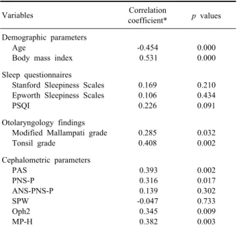
Variables Correlation
coefficient* p values Demographic parameters
Age
Body mass index
-0.454 0.531
0.000 0.000 Sleep questionnaires
Stanford Sleepiness Scales Epworth Sleepiness Scales PSQI
0.169 0.106 0.226
0.210 0.434 0.091 Otolaryngology findings
Modified Mallampati grade Tonsil grade
0.285 0.408
0.032 0.002 Cephalometric parameters
PAS PNS-P ANS-PNS-P SPW Oph2 MP-H
0.393 0.316 0.139 -0.047
0.345 0.382
0.002 0.017 0.302 0.733 0.009 0.003 PSQI; Pittsburgh Sleep Quality Index, PAS (mm); linear measure- ment between the base of tongue and posterior pharyngeal wall along the line B-Go, PNS-P (mm); posterior nasal spine (PNS) to tip of soft palate contour, Oph2 (mm); oropharyngeal airway width 2, MP-H (mm); shortest distance fom hypid bone to mandibular plane, *Pearson's correlation
Table 4. Correlation analysis of the apnea-hypopnea index
Figure 1. Cephalometric landmarks.
S; Sella, N; Nasion, A point; Supspinale, B point; supra- mentale, PNS-P (mm); soft palate length = posterior nasal spine (PNS) to tip of soft palate contour (P), ANS-PNS-P angle(°);
angle from anterior nasal spine (ANS) to PNS to P, SPW (mm); soft palate width, Oph2 (mm); Oropharyngeal airway width 2 = distance from tongue base to soft palate to posterior pharyngeal wall at the level of the tip of soft palate along a line parellel to ANS-PNS, PAS (mm); linear measurement between the base of tongue and posterior pharyngeal wall along the line B-Go (Gonial angle), MP-H (mm); shortest distance from hyoid bone to mandibular plane.
가 적고, 얕은 수면인 비렘수면 1단계가 통계적으로 유의하게 많았다(Table 2, 3).
폐쇄성 수면무호흡군을 AHI 15 이하의 경한 군과 16 이상의 중한 군으로 나누어 비교하였을 때는 중한 군에서 나이는 더 젊 고 체질량계수와 Oph2가 유의하게 높았다( p <0.05).
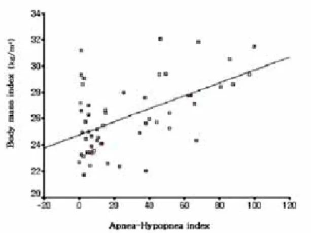
무호흡-저호흡지수와 관련이 있는 변수를 알아보기 위한 상 관분석에서 나이는 음의 상관 관계를 보였고 체질량지수, 편도 등급, MMP, PAS, PNS-P, Oph2, MP-H 등은 양의 상관 관계
를 보였다( p <0.05). 따라서 나이가 젊을수록, 체질량지수가 높 을수록, 또 편도나 혀가 클수록, 연구개 길이가 길고 기도의 전 후 폭경이 클수록 AHI가 높았다(Fig. 2). 그러나 자가 수면설문 지의 척도들과는 상관성이 없었다(Table 4).
폐쇄성 수면무호흡증에 영향을 미치는 변수를 알아보기 위해
로지스틱 회귀분석을 한 결과 여자에 비해 남자가 17배 정도 유
의하게 영향을 미치는 것으로 나타났으며 MMP는 약 3배의 영
향을 미치는 것으로 나왔으나 통계적으로 경계치 유의성을 보
였다(Table 5).
Figure 2. Correlation coefficient between body mass index and
apnea-hypopnea index in the Pearson correlation test (n=57).고 찰
코골이를 주소로 내원한 환자들에서 수면다원검사를 기준으 로 수면무호흡증을 진단 하였을 때 약 74%가 시간당 무호흡저 호흡지수가 5 이상으로 폐쇄성 수면무호흡증이었고 26%가 시 간 당 무호흡저호흡지수가 5 미만이었다. 그러나 두 군 간의 인 구 통계학적 및 임상적 특성을 비교하였을 때 성별과 편도 등급 에서만 유의한 차이를 보였다. 이러한 결과는 여성에 비해 남성
에서,
21,22또 편도가 클수록 수면무호흡증이 많다는 것을 시사
하여 다른 연구 결과와 비슷하였다.
15,23한편, 비수면무호흡군과 폐쇄성 수면무호흡군 간의 자가 수 면척도를 통한 비교에서는 두 군 사이에 유의한 차이가 없었다.
무호흡저호흡지수와 주간 졸리움에 관한 상관 분석에서도 본 연구에서는 관련이 없는 것으로 나왔다. 이는 Chervin RD 등
24의 연구 결과와는 유사하나 Johns 등
25은 연구 결과와는 상반되 었다. 두 군 사이에 수면 구조를 비교해 보았을 때 폐쇄성 수면 무호흡군에서 얕은 수면이 많고 깊은 수면이 적어 주간 졸리움 이 더 많을 것이라는 기대와 달랐다. 이는 주간 졸리움이 야간 수면 구조의 변화 외에 다른 요인들, 즉 우울증, 불안, 스트레 스, 수면 위생 및 건강 상태 등과 복합적으로 관련되어 나타나 기 때문일 것으로 추정된다. 이상의 결과만으로 볼 때 폐쇄성 수면무호흡증 환자에 있어 주간 졸리움의 임상 증상으로 수면 다원검사에 의한 수면무호흡증의 심한 정도를 예측하거나, 수 면다원검사에 의한 무호흡저호흡지수로 주간 졸리움의 임상적 증상을 예측할 수는 없다는 것을 알 수 있다. 그러나 본 연구의 제한점으로 수면클리닉을 방문한 환자들을 대상으로 하였다는 점과 수면 중 각성 지수의 비교 분석을 하지 않은 점 및 식도 압력계를 사용하지 않아 상기도 저항증후군을 감별하지 못한
점 등이 있다. 수면클리닉을 방문한 환자들은 비록 수면다원검 사에서 이상 소견이 보이지 않더라도 수면과 관련된 증상을 가 지고 내원하므로 수면다원검사에 이상이 있는 사람들과의 임상 증상의 비교는 상대적으로 차이가 적을 것으로 기대된다. 또한 일반인을 대상으로 무호흡저호흡지수와 주간 졸리움 척도와의 상관 관계를 연구하였을 때 관련이 있다는 연구
23도 있어 수면 설문지 결과와 무호흡저호흡지수와의 관계에 대해서는 향후 더 많은 사람들을 대상으로 수면 중 각성 지수 및 상기도 저항 증 후군의 감별 등을 고려하여 추가적인 연구가 필요할 것으로 생 각된다.
비만이 수면무호흡증의 위험인자이며
26반대로 수면무호흡증 이 비만에 기여하는 것으로 알려져 있다.
27,28본 연구에서는 비 수면무호흡군과 폐쇄성 수면무호흡군 간의 체질량지수에서는 차이가 없었으나 수면무호흡증이 경한 군과 심한 군으로 나누 어 비교해 보았을 때는 유의한 차이가 있는 것으로 나타났다.
이는 수면무호흡증 환자에서 체질량지수가 높을수록 무호흡증 의 정도가 심할 것으로 기대되어, 체중이 어떤 방법으로든 폐쇄 성 수면무호흡증에 기여하는 것으로 생각된다.
폐쇄성 수면무호흡증의 진단과 치료에 있어 상부 기도의 해 부학적 평가는 필수적인 검사이다. 왜냐하면 폐쇄성 수면무호 흡증에 기여하는 요인들이 복잡하며 하나로 설명되지 않는 경 우가 많아 상부기도의 전체적인 평가 없이는 치료에 따른 예후 를 예측하기가 어렵기 때문이다.
29편도의 크기 및 혀와 구강 내 구조와 수면무호흡증의 연관성에 대한 보고들이 많이 있다.
30,31MMP는 구강 내에서의 혀의 위치와 경구개, 연구개와 혀의 관 계를 단계화한 것으로 수면무호흡증이 심할수록 단계가 높다고 알려져 있다.
15본 연구에서도 편도 단계와 MMP가 무호흡저호 흡지수와 유의한 연관성을 가지는 것으로 조사되었다. 두부 규 격 방사선 계측에 의한 해부학적 평가에서 연구개 조직이 두꺼 울수록, 연구개 길이가 길수록 더 수면무호흡 지수가 높은 것으 로 알려져 있다.
31즉 연구개의 해부학적 구조가 수면무호흡에 영향을 미친다고 알려져 있으며 이는 우리 결과와 유사하였다.
한편 설후방 기도 부위의 전후 폭경은 좁을수록 무호흡저호흡
지수가 높을 것이라는 일반적인 가정
32,33과 달리 본 연구에서는
무호흡저호흡지수가 높은 환자일수록 오히려 기도 부위의 전후
폭경이 증가하는 양상이 통계적으로 유의하게 나타났다. 이러
한 소견은 로지스틱 회귀분석에서 편도 등급이 높은 영향을 미
친다는 결과와 함께 분석할 때 수면무호흡이 심한 환자가 편도
등급이 유의하게 더 크고 이것이 좌우 폭경에 영향을 미친다는
것을 알 수 있다. 편도가 커져서 기도를 막고 있는 환자의 경우
보상적으로 기도 자체를 앞뒤로 확장시켜 기도를 확보하려는
경향을 나타내는 것으로 보인다. 따라서 수면무호흡증 환자의
해부학적 평가를 할 때 전면에서 관찰하는 이비인후과적 검진 소견과 측면에서 평가하는 두부 규격 방사선계측 결과를 함께 종합하여야 하겠다. 그러나 이번 연구에서는 대상 환자수가 적 어 성별에 따른 차이를 비교하지 못하였고, 또 식도 압력을 측 정하지 못해 비수면무호흡증후군 환자 중 혀와 후방기도강 문 제에 의한 상기도 저항 증후군을 나타내는 환자를 배제하지 못 한 제한점이 있다.
결론적으로, 본 연구에서 수면다원검사에 의한 폐쇄성 수면 무호흡증의 정도는 구인두부의 이학적검사 소견과 두부 규격 방사선 계측 결과와 연관성이 있었으나 수면설문지를 통한 임 상 증상과는 연관성이 없었다. 이러한 결과는 수면 클리닉에서 수면무호흡증이 의심되는 환자의 생리적인 심각성과 주관적 증 상의 정도를 평가 비교하는 데 주의를 요한다. 또한 수면무호흡 증 환자의 평가에 있어 이비인후과적 검진과 더불어 두부 규격 방사선 계측의 필요성을 다시 한번 확인하였고 수면무호흡 환 자의 진단과 향후 치료에 있어 여러 과의 협진 체계가 필요하다 고 생각된다.
REFERENCES
1. Lavie P. Incidence of sleep apnea in presumably healthy working population: a significant relationship with excessive daytime sleepiness. Sleep 1983;6:312-318.
2. Gislason T, Almqvist M, Eriksson G, Taube A, Boman G.
Prevalence of sleep apnea syndrome among Swedish men-an epidemiological study. J Clin Epidemiol 1988;41:571-576.
3. Redline S, Strohl KP. Recognition and consequences of obstructive sleep apnea hyponea syndrome. Clin Chest Med 1998:19:1-19.
4. Coleman RM, Roffwarg HP, Kennedy SJ, Guilleminault C, Cinque F, Cohn MA, et al. Sleep-wake disorders based on a polysomnographic diagnosis. A national cooperative study. JAMA 1982;247:997-1003.
5. Roth T, Hartse KM, Zorick F, Conway W. Multiple naps and the evaluation of daytime sleepiness in patients with upper airway sleep apnea. Sleep 1980;3:425-439.
6. Baumel MJ, Maislin G, Pack AI. Population and occupational screening for obstructive sleep apnea: are we there yet? Am J
Respir Crit Care Med 1997;155:9-14.
7. Bassiri AG, Guilleminault C. Clinical features and evaluation of obstructive sleep apnea-hypopnea syndrome. In: Kryger MH, Roth T, Dement WC. Principles and Practice of Sleep Medicine. 3rd ed.
Philadelphia: Saunders. 2000;869-878.
8. Strollo PJ Jr, Rogers RM. Obstructive sleep apnea. N Engl J Med 1996;334:99-104.
9. Schafer H, Koehler U, Ploch T, Peter JH. Sleep-related myocardial ischemia and sleep structure in patients with obstructive sleep apnea and coronary heart disease. Chest 1997;111:387-393.
10. Rivlin J, Hoffstein V, Kalbfleisch J, McNicholas W, Zamel N, Bryan AC. Upper airway morphology in patients with idiopathic obstructive sleep apnea. Am Rev Respir Dis 1984;129:355-360.
11. Hoddes E, Zarcone V, Smythe H, Phillips R, Dement WC.
Qualitification of sleepiness: a new approach. Psychophysiology 1973;10:431-436.
12. Johns MW. A new method for measuring daytime sleepiness: the Epworth sleepiness scale. Sleep 1991;14:540-545.
13. Buysse DJ, Reynolds CF 3rd, Monk TH, Berman SR, Kupfer DJ.
The Pittsburgh Sleep Quality Index: a new instrument for psychiatric practice and research. Psychiatry Res 1989;28:193-213.
14. Bastien CH, Vallieres A, Morin CM. Validation of the Insomnia Severity Index as an outcome measure for insomnia research. Sleep
Med 2001;2:297-307.
15. Friedman M, Tanyeri H, La Rosa M, Landsberg R, Vaidyanathan K, Pieri S, et al. Clinical predictors of obstructive sleep apnea.
Laryngoscope 1999;109:1901-1907.
16. Rechtschaffen A, Kales AD. A manual of standardized terminology,
techniques and scoring system for sleep stages of human subjects. Los
Angeles: Brain information Service/Brain Research Institute, 1968.17. Keenan BS. Respiratory monitoring during sleep: polysomnography.
In: Guilleminault C. Sleeping and Waking Disorders: Indications and
Techniques. Menlo Park: Addison-Wesley, 1982;183-212.
18. Young T, Peppard P, Palta M, Hla KM, Finn L, Morgan B, et al. Population-based study of sleep-disordered breathing as a risk factor for hypertension. Arch Intern Med 1997;157:1746-1752.
19. Young T, Blustein F, Finn L, Palta M. Sleep-disordered breathing and motor vehicle accidencts in a population-based sample of employed adults. Sleep 1997;20:608-613.
20. Young T. Finn L, Hla KM, Morgan B, Palta M. Snoring as part of a dos-response relationship between sleep-disordered breathing and blood pressure. Sleep 1996;19:202-205.
21. Maislin G, Pack AI, Kribbs NB, Smith PL, Schwartz AR, Kline LR, et al. A survey screen for prediction of apnea. Sleep 1995;18:158-166.
22. Viner S, Szalai JP, Hoffstein V. Are history and physical examination a good screening test for sleep apnea? Ann Int Med 1991;115:356-359.
23. Richardson MA, Seid AB, Cotton RT, Benton C, Kramer M.
Evaluation of tonsils and adenoids in Sleep Apnea Syndrome.
Laryngoscope 1980;90:1106-1110.
24. Chervin RD, Aldrich MS. The Epworth Sleepiness Scale may not reflect objective measures of sleepiness or sleep apnea. Neurology 1999;52:125-131.
25. Johns MW. Daytime sleepiness, snoring, and obstructive sleep apnea. The Epworth Sleepiness Scale. Chest 1993;103:30-36.
26. Deegan PC, McNicholas WT. Predictive value of clinical features for the obstructive sleep apnea syndrome. Eur Respir J 1996;
9:117-124.
27. Inoue K. Takano H. Yoshikawa T. Interleukin-6, obstructive sleep apnea, and obesity. Chest 2003;124:1621-1622; author reply 1622-1623.
28. Kurtz D. Krieger J. Kowalski J. Hoff E. Mangin P. Nyctero- hemeral variations in plasma growth hormone (GH) levels and sleep apnea syndromes: their relationship with obesity. Rev
Electroencephalogr Neurophysiol Clin 1980;10:366-375.
29. Kwon TG, Cho YW, Ahn BH, Suh YS. Cephalometric predictors of obstructive sleep apnea. J Kor Oral Maxillofac Surg 2000;29:338- 345.
30. Bradlly TD, Brown IG, Grossman RF, Zamel N, Martinez D,
Phillipson EA, et al. Pharyngeal size in snorers, nonsnorers, and patients with obstructive sleep apnea. N Engl J Med 1986;
315:1327-1331.
31. Zucconi M, Ferini-Strambi L, Palazzi S, Curci C, Cucchi E, Smirne S. Craniofacial cephalometric evaluation in habitual snorers with and without obstructive sleep apnea. Otolaryngol Head Neck Surg 1993;109:1007-1013.
32. Jamieson A, Guilleminault C, Partinen M, Quera-Salva MA.
Obstructive sleep apnea patients have craniomandibular abnor- malities. Sleep 1986;9:469-477.
33. Battagel JM, L’ Estrange PR. The cephalometric morphology of patients with obstructive sleep apnoea. Eur J Orthod 1996;18:557- 569.